. 2024 Mar;36(10):e2300320.
doi: 10.1002/adma.202300320. Epub 2023 Jul 8.

Nanozyme-Based Robotics Approach for Targeting Fungal Infection

Affiliations

Nanozyme-Based Robotics Approach for Targeting Fungal Infection

Min Jun Oh et al. Adv Mater. 2024 Mar.

Abstract

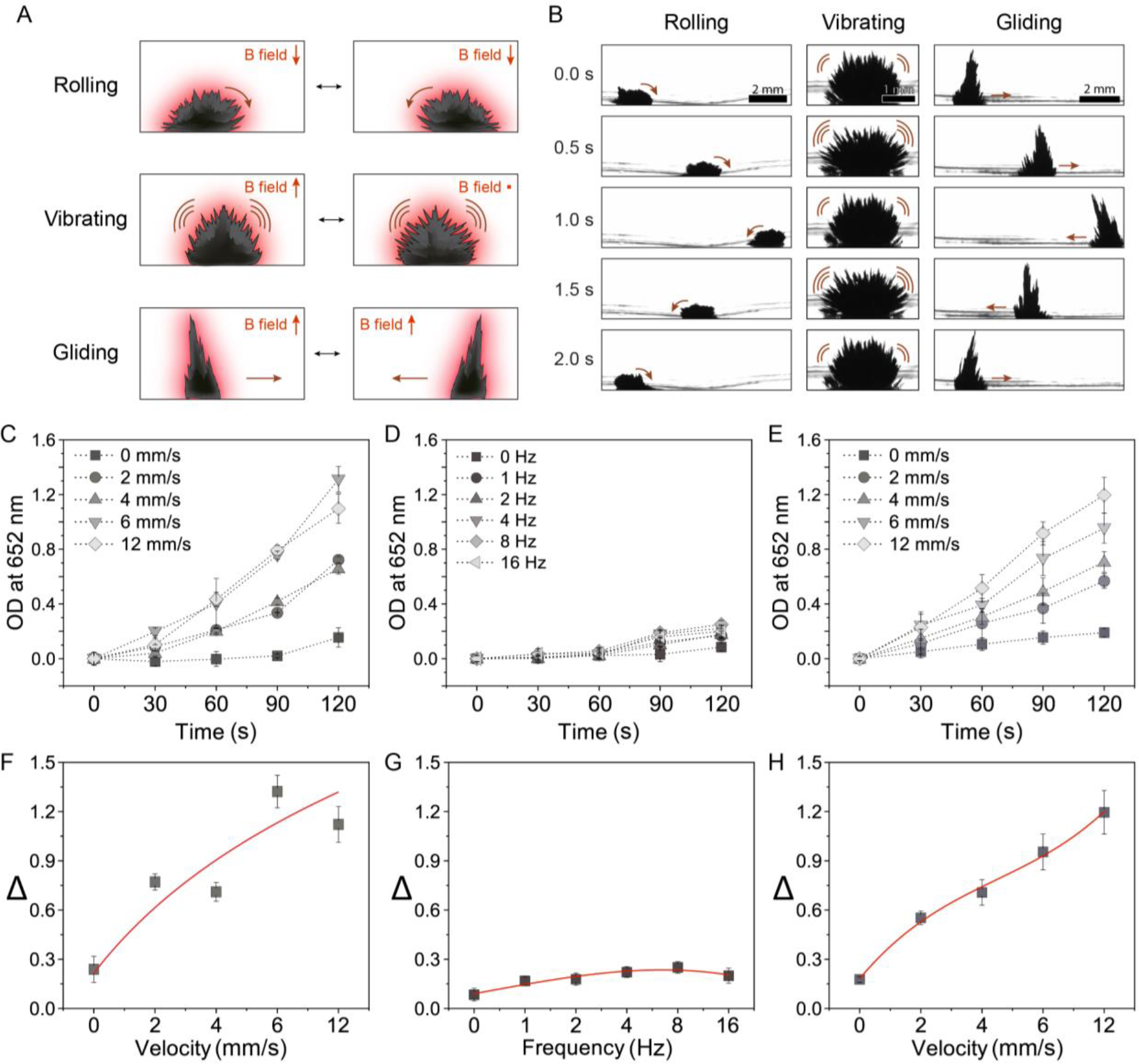
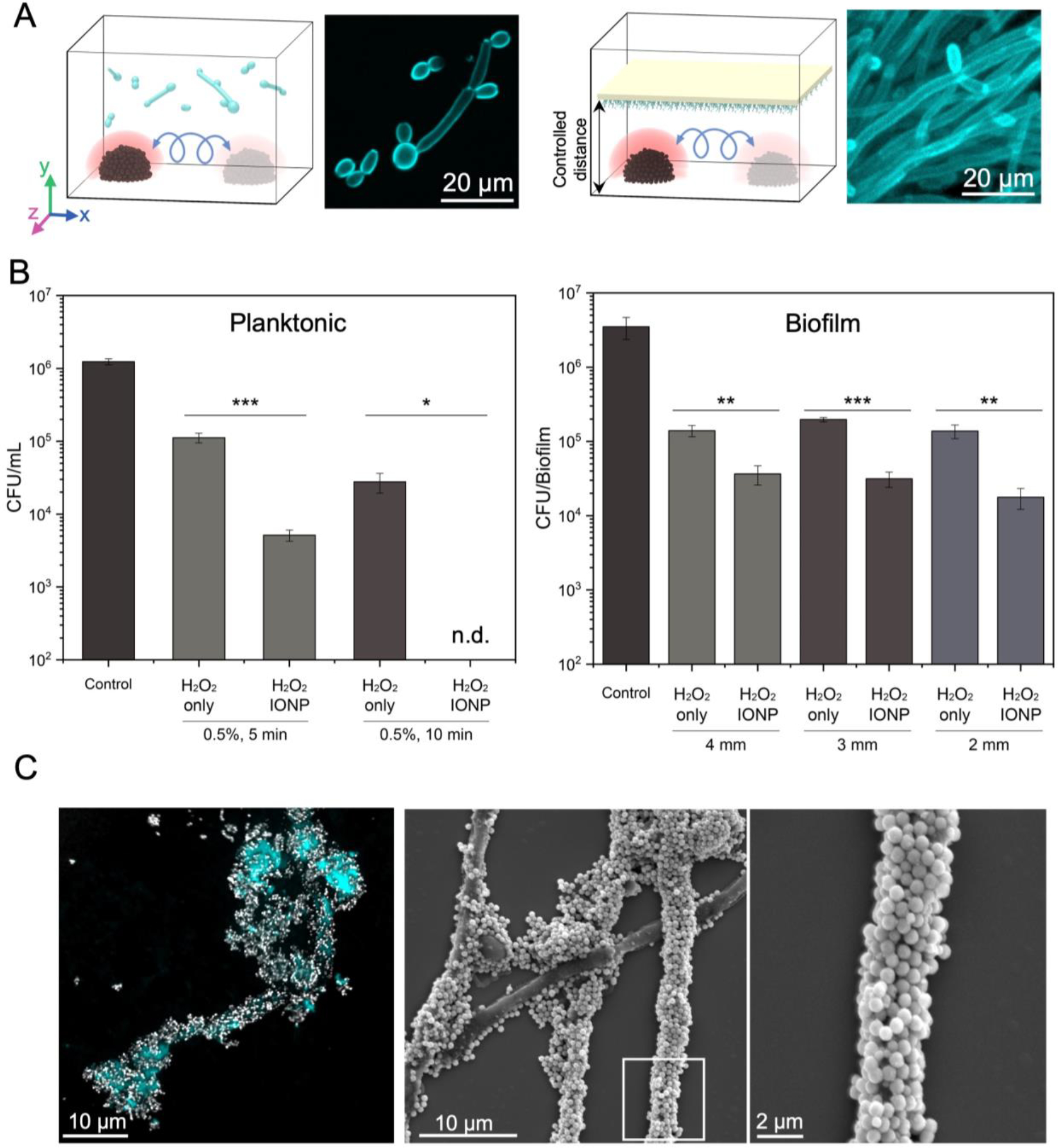
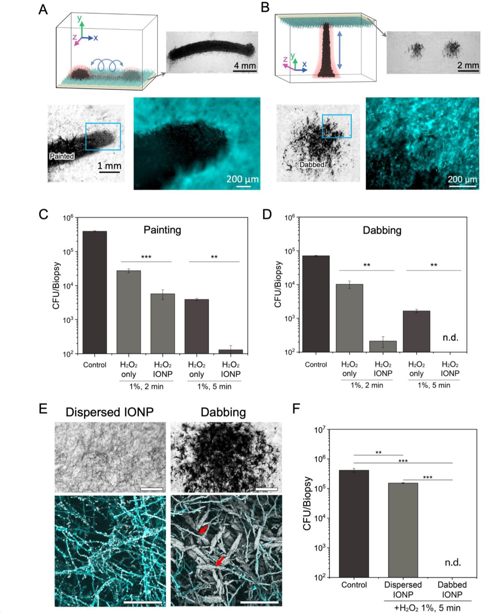
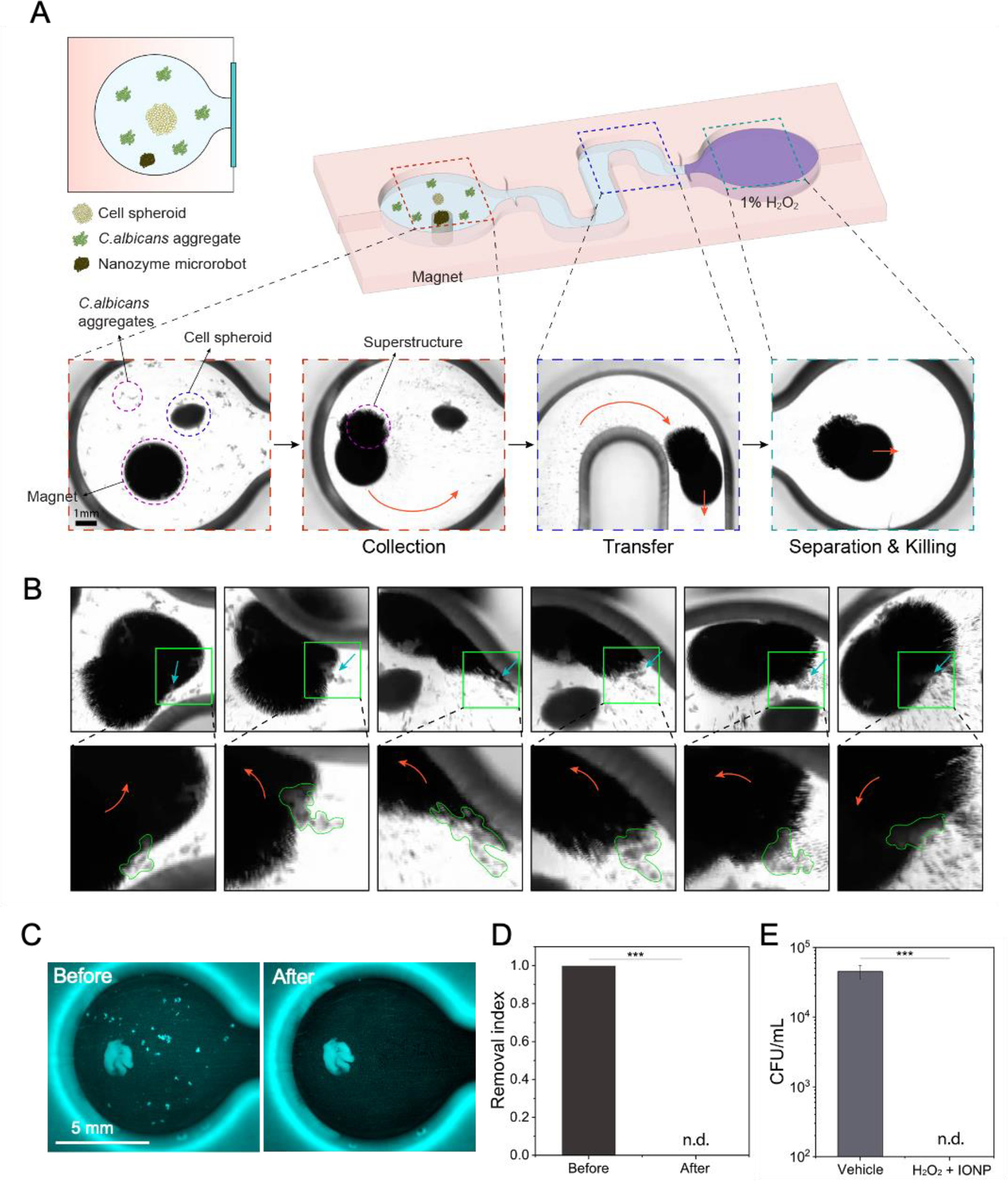
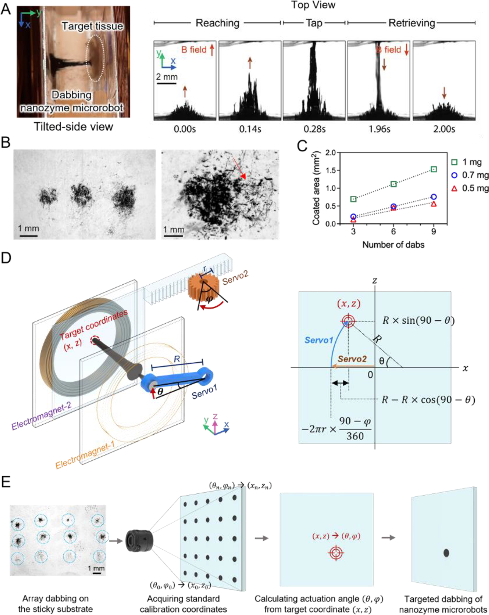
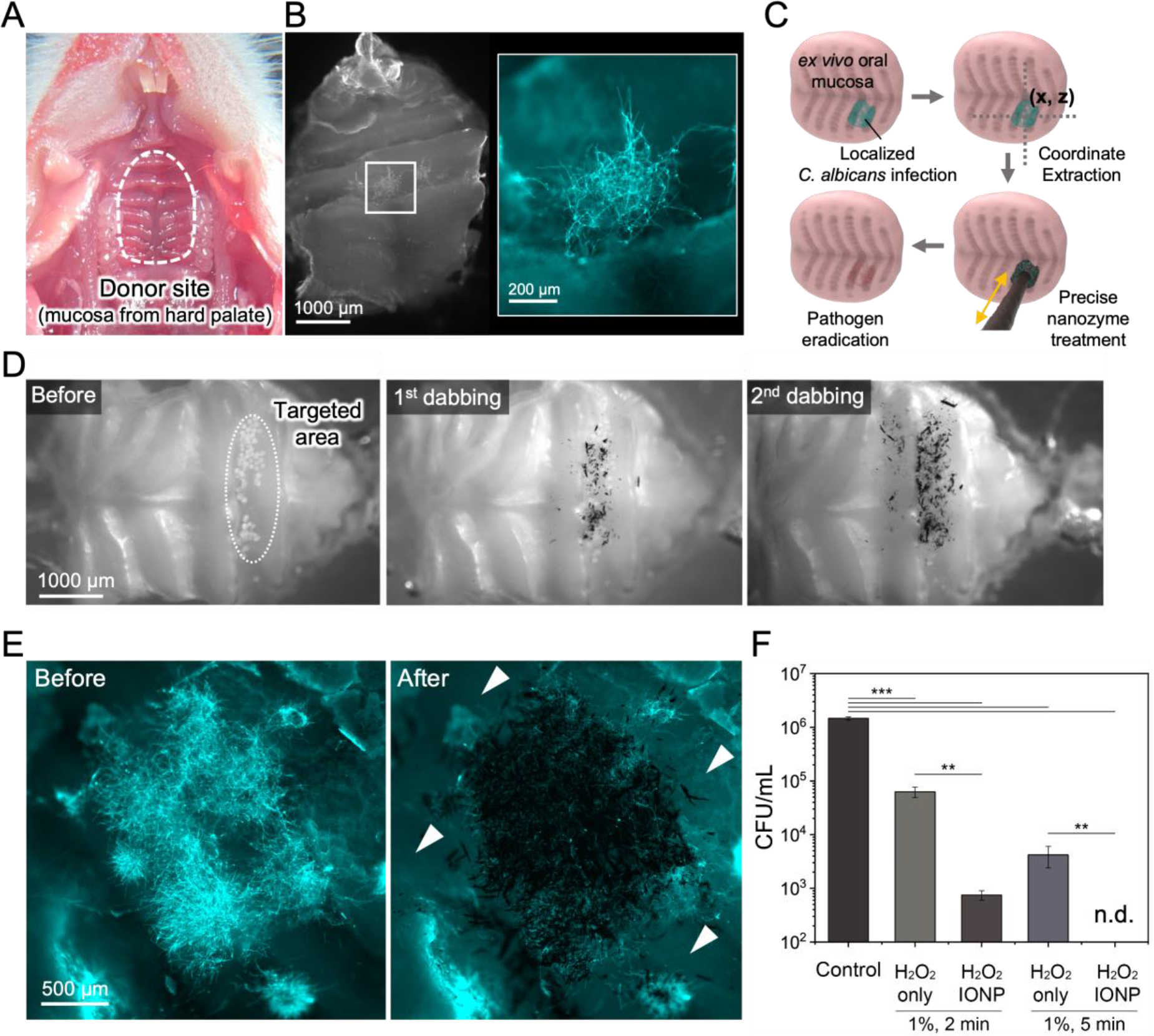
Fungal pathogens have been designated by the World Health Organization as microbial threats of the highest priority for global health. It remains a major challenge to improve antifungal efficacy at the site of infection while avoiding off-target effects, fungal spreading, and drug tolerance. Here, a nanozyme-based microrobotic platform is developed that directs localized catalysis to the infection site with microscale precision to achieve targeted and rapid fungal killing. Using electromagnetic field frequency modulation and fine-scale spatiotemporal control, structured iron oxide nanozyme assemblies are formed that display tunable dynamic shape transformation and catalysis activation. The catalytic activity varies depending on the motion, velocity, and shape providing controllable reactive oxygen species (ROS) generation. Unexpectedly, nanozyme assemblies bind avidly to fungal (Candida albicans) surfaces to enable concentrated accumulation and targeted ROS-mediated killing in situ. By exploiting these tunable properties and selective binding to fungi, localized antifungal activity is achieved using in vivo-like cell spheroid and animal tissue infection models. Structured nanozyme assemblies are directed to Candida-infected sites using programmable algorithms to perform precisely guided spatial targeting and on-site catalysis resulting in fungal eradication within 10 min. This nanozyme-based microrobotics approach provides a uniquely effective and targeted therapeutic modality for pathogen elimination at the infection site.

Keywords: Candida albicans; assemblies; biofilms; iron oxide; microrobots; mucosal.


Figures

Figure 1.
Figure 1.. Assembly, control, and functional properties of robotic nanozyme assemblies and their mode of action.
(A) On-site assembly of individual nanozymes into catalytically active superstructures. The motion dynamics, morphology, and the location of catalysis of the structured assemblies can be controlled creating nanozyme microrobots for targeting fungal infection. (B) Electromagnet cores guide the nanozyme microrobot assemblies with controllable morphology, position, and motion using programmed algorithms. (C) Programmable dynamic motions via magnetic field modulation enable controlled catalytic activities and targeted treatment.
Figure 2.
Figure 2.. Catalytic properties of the nanozyme microrobots.
(A) Catalytic activity in situ generated by motion dynamics. The TMB assay demonstrates the generation of ROS on-site from H2O2 by the catalytically active (peroxidase-like) nanozyme microrobots. (B) The location of the catalysis is controlled by the shape and motion of the nanozyme assemblies. (1 mg mL−1 IONPs). Catalytic activity dynamics of (C) rolling, (D) vibrating, and (E) gliding motions. (F) Velocity dependent catalysis of rolling. (G) Frequency dependent catalysis of vibrating. (H) Velocity dependent catalysis of gliding. The rolling motion shows efficient catalytic activity. Data are mean ± standard deviation (n=3).
Figure 3.
Figure 3.. Fungal killing and binding by nanozymes.
(A) Experimental design for killing planktonic fungal cells (left) and biofilms (right) using convective mixing. The rolling nanozyme microrobots create convective ROS mixing and dispersion against planktonic cells or biofilm surfaces at fixed distances. (B) Cell viability counts show killing of planktonic cells and biofilms of C. albicans at specific distances (2, 3 and 4 mm from the outer surface of the nanozyme microrobot) via catalytic activation of 0.5% H2O2. (C) Confocal images and SEM micrographs demonstrate the binding of nanozymes on C. albicans cells. Data are mean ± standard deviation; * P < 0.05, ** P< 0.01, *** P < 0.001 by one-way analysis of variance with Tukey’s multiple-comparison test (n=12), and n.d. stands for not detectable.
Figure 4.
Figure 4.. Precision-guided delivery of nanozyme for site-specific catalytic killing of fungal biofilms.
(A) Experimental design for precise delivery of nanozymes to fungal biofilms using a ‘painting nanozyme microrobot’. (B) Experimental design for targeting specific areas with submillimeter precision using a ‘dabbing nanozyme microrobot’. (C) Cell viability counts show eradication of C. albicans cells within painted areas by the painting microrobot. (D) Cell viability counts show complete killing of C. albicans cells within dabbed areas by the dabbing microrobot. (E) Coating efficiency of dispersed IONP treatment and IONP dabbing. Bright field microscopy (upper, scale bar: 500 μm) and confocal microscopy images (lower, scale bar:100 μm) show clear differences in IONP coating density on C. albicans biofilms. IONP dabbing delivers a greater amount of IONPs to the target area than dispersed IONP treatment. Hyphae of C. albicans are heavily coated with IONPs (red arrows). (F) Cell viability counts show limited killing of C. albicans cells by dispersed IONP treatment. Data are mean ± standard deviation; ** P< 0.01, *** P < 0.001 by one-way analysis of variance with Tukey’s multiple-comparison test (n=12 for panel C, n=8 for panel D and n=8 for panel F), and n.d. stands for not detectable.
Figure 5.
Figure 5.. Precision capturing and killing of fungal aggregates using nanozyme-microrobotics strategy.
(A) A schematic of the experimental platform for testing C. albicans capture and killing in the presence of cell spheroid using nanozyme microrobots. (B) Fungal binding, dragging, and engulfing by the nanozyme microrobot. Close-up images show the fungal aggregates marked by green lines. (C) Before and after fluorescence imaging shows fungal aggregates effectively removed without binding or disturbing the cell spheroid by a magnetically controlled nanozyme microrobot. (D) Quantitative image analyses show complete removal of fungal aggregates. (E) Cell viability counts show eradication of the targeted and captured fungal aggregates via catalytic activation of H2O2. Data are mean ± standard deviation; *** P < 0.001 by Student’s t-test (n=4), and n.d. stands for not detectable.
Figure 6.
Figure 6.. Precision programming and automation for the dabbing nanozyme microrobot.
(A) The shape of the dabbing superstructure extending and tapping the targeted surface as visualized via a stereoscope. (B) Controllable dabbing and precision targeting provide localized delivery of tunable amounts of nanozyme. C. albicans coated with nanozymes on the targeted area (right image). (C) The area of biofilm coated with nanozymes depends on the IONP concentration. (D) Targeting location is determined by converting servo motor rotation positions to x-z coordinates. (E) A nanozyme dabbing test array aids fine-tuning of the positioning accuracy.
Figure 7.
Figure 7.. Precision targeting and antifungal killing on murine mucosa using dabbing nanozyme-microrobots.
(A) Explant of murine oral mucosal tissue harvested from the palate. (B) Focal C. albicans infection developed on the oral mucosa characterized by localized hyphal accumulation. (C) A schematic diagram of the coordinate extraction and precision-guided treatment using the dabbing nanozyme microrobot. (D) Bright field images showing sequential nanozyme dabbing. (E) Fluorescence imaging shows high spatial precision for targeting the site of fungal infection. (F) Cell viability counts show effective killing of targeted fungal cells on mucosa via catalytic activation of H2O2 on site. Data are mean ± standard deviation; ** P< 0.01, *** P < 0.001 by one-way analysis of variance with Tukey’s multiple-comparison test (n=8), and n.d. stands for not detectable.

References

    1. Vallabhaneni S, Mody RK, Walker T, Chiller T, Infect Dis Clin North Am. 2016, 30, 1–11. - PubMed
    1. Brown GD, Denning DW, Gow NAR, Levitz SM, Netea MG, White TC, Sci. Transl. Med. 2012, 4, 165rv13. - PubMed
    1. WHO, WHO fungal priority pathogens list to guide research, development and public health action, 2022, ISBN; 978-92-4-006024-1.
    1. Perfect JR, Nat. Rev. Drug Discovery 2017, 16, 603–616. - PMC - PubMed
    1. Sousa F, Ferreira D, Reis S, Costa P, Pharmaceuticals 2020, 13, 248. - PMC - PubMed