. 2023 Jun;22(6):100563.
doi: 10.1016/j.mcpro.2023.100563. Epub 2023 May 3.

Sensitive, High-Throughput HLA-I and HLA-II Immunopeptidomics Using Parallel Accumulation-Serial Fragmentation Mass Spectrometry

Sensitive, High-Throughput HLA-I and HLA-II Immunopeptidomics Using Parallel Accumulation-Serial Fragmentation Mass Spectrometry

Kshiti Meera Phulphagar et al. Mol Cell Proteomics. 2023 Jun.

Abstract

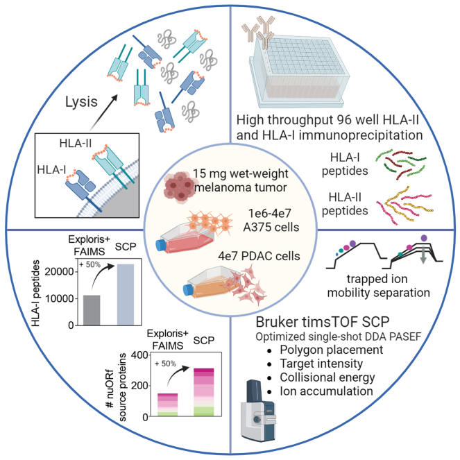
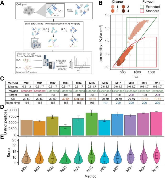
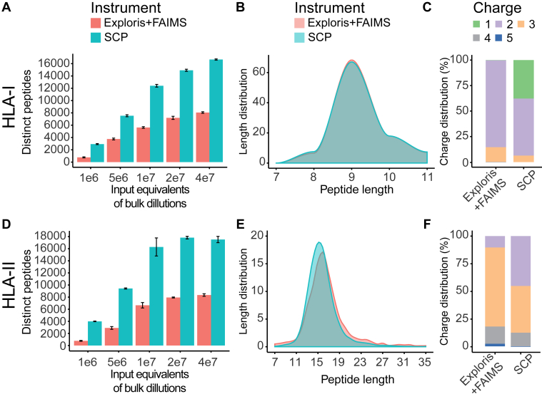
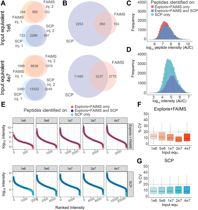
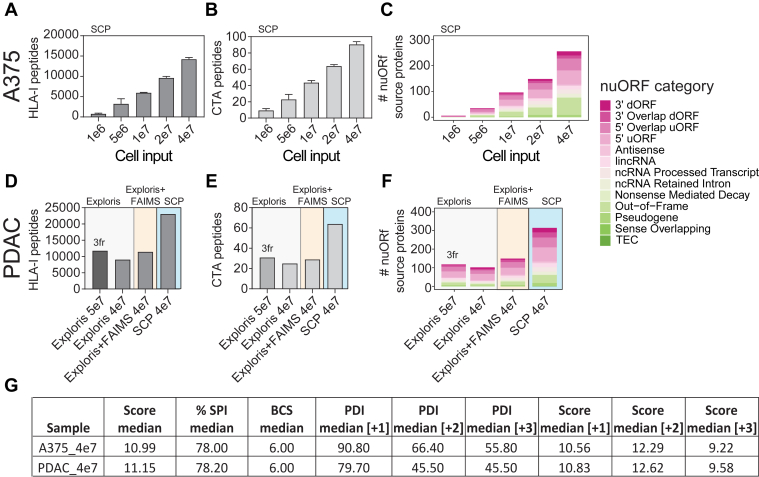
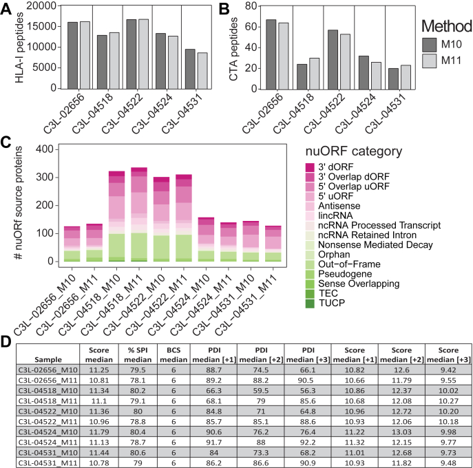
Comprehensive and in-depth identification of the human leukocyte antigen class I (HLA-I) and class II (HLA-II) tumor immunopeptidome can inform the development of cancer immunotherapies. Mass spectrometry (MS) is a powerful technology for direct identification of HLA peptides from patient-derived tumor samples or cell lines. However, achieving sufficient coverage to detect rare and clinically relevant antigens requires highly sensitive MS-based acquisition methods and large amounts of sample. While immunopeptidome depth can be increased by off-line fractionation prior to MS, its use is impractical when analyzing limited amounts of primary tissue biopsies. To address this challenge, we developed and applied a high-throughput, sensitive, and single-shot MS-based immunopeptidomics workflow that leverages trapped ion mobility time-of-flight MS on the Bruker timsTOF single-cell proteomics system (SCP). We demonstrate greater than twofold improved coverage of HLA immunopeptidomes relative to prior methods with up to 15,000 distinct HLA-I and HLA-II peptides from 4e7 cells. Our optimized single-shot MS acquisition method on the timsTOF SCP maintains high coverage, eliminates the need for off-line fractionation, and reduces input requirements to as few as 1e6 A375 cells for >800 distinct HLA-I peptides. This depth is sufficient to identify HLA-I peptides derived from cancer-testis antigen and noncanonical proteins. We also apply our optimized single-shot SCP acquisition methods to tumor-derived samples, enabling sensitive, high-throughput, and reproducible immunopeptidome profiling with detection of clinically relevant peptides from less than 4e7 cells or 15 mg wet weight tissue.

Keywords: HLA-I and II immunopeptidomics; high-throughput acquisition; sensitive single-shot MS analysis; trapped ion mobility.


Conflict of interest statement

Conflict of interest S. A. C. is a member of the scientific advisory boards of Kymera, PTM BioLabs, Seer, and PrognomIQ. A.S.V.J. is an employee of Bruker. All other authors declare no competing interests.

Figures

None
Graphical abstract
Fig. 1
Fig. 1
Human leukocyte antigen (HLA) sample preparation overview and timsTOF single-cell proteomics (SCP) acquisition method optimization for analysis of HLA-I peptides enables inclusion of singly charged peptides.A, schematic overview of HLA sample preparation and mass spectrometry (MS) acquisition schemes. Serial HLA-I and HLA-II enrichment, acid elution of peptides from HLA complexes, and peptide desalting performed in a semiautomated 96-well format, followed by single-shot data-dependent acquisition (DDA) analysis of purified HLA-I and HLA-II peptides using our standard workflow with the Exploris + FAIMS or the timsTOF SCP. B, HLA-I peptide spectrum matches (PSMs) identified on the timsTOF SCP using the standard precursor filter (“polygon”) in pink and the extended polygon in green across m/z and ion mobility (IM) dimensions. Colors indicate precursor charge state. C, timsTOF SCP parameter overview, including IM range, polygon placement, target intensity value (“target”), collision energy (CE) slope, accumulation, and ramp time for M00–M10. D, number of unique HLA-I peptides from 1e7 cell equivalents from bulk sample preparation postfiltering in duplicates with M00–M10 on the timsTOF SCP. Median and standard deviation are shown. E, corresponding score distributions of HLA-I peptides for methods indicated in D. FAIMS, field asymmetric waveform ion mobility spectrometry.
Fig. 2
Fig. 2
Single-shot acquisition of HLA-I and HLA-II peptides on the timsTOF single-cell proteomics (SCP) increases identifications greater than twofold compared with Exploris + FAIMS.A, unique HLA-I peptides identified by single injections on Exploris + FAIMS (red) and timsTOF SCP (blue) from bulk digest at indicated input equivalents. Mean and standard deviation is shown. B, peptide length distribution across HLA-I peptides identified on the Exploris + FAIMS (red) and the timsTOF SCP (blue) from bulk digest at indicated input equivalents from bulk sample preparation. C, HLA-I peptide charge states for Exploris + FAIMS or timsTOF SCP. D, unique HLA-II peptides identified by single injections on Exploris + FAIMS (red) and timsTOF SCP and indicated input equivalents (blue) from bulk sample preparation. Mean and standard deviation is shown. E, HLA-II peptide length distribution for Exploris + FAIMS (red) and the timsTOF SCP (blue). F, charge state distribution for HLA-II peptides on Exploris + FAIMS or timsTOF SCP. FAIMS, field asymmetric waveform ion mobility spectrometry; HLA, human leukocyte antigen.
Fig. 3
Fig. 3
HLA-I peptide identifications on the timsTOF single-cell proteomics (SCP) show high reproducibility and extended dynamic range.A, overlap of HLA-I peptides across multiple injections on the Exploris + FAIMS or the timsTOF SCP at 1e6 or 4e7 A375 cell input equivalents from bulk sample preparation. B, overlap of HLA-I peptides between the Exploris + FAIMS and the timsTOF SCP at 1e6 (upper panel) or 4e7 (lower panel) A375 cells. C, Log10 intensity (MS1 AUC) distribution of HLA-I peptides uniquely identified on the Exploris + FAIMS (pink) or both instruments (dark blue). D, Log10 intensity (MS1 AUC) distribution of HLA-I peptides uniquely identified on the timsTOF SCP (light blue) or both instruments (dark blue). E, ranked peptide intensity at different cell input equivalents from bulk sample preparation on the Exploris + FAIMS (upper panel) or the timsTOF SCP (lower panel). Respective intensity of peptides uniquely identified on the Exploris + FAIMS (pink), the timsTOF SCP (light blue), or both instruments (dark blue). F, percent CV of peptide intensity between technical replicates at different cell input equivalents from bulk sample preparation on Exploris + FAIMS. Dashed line indicates 20% CV. G, % CV of peptide intensity between technical replicates identified on the timsTOF SCP at indicated input equivalents from bulk sample preparation. Dashed line indicates 20% CV. AUC, area under the curve; FAIMS, field asymmetric waveform ion mobility spectrometry; HLA, human leukocyte antigen; MS, mass spectrometry.
Fig. 4
Fig. 4
Single-shot acquisition of HLA-I peptides from low amounts of tumor-derived cell lines enables detection of cancer-testis antigen (CTA) and novel unannotated ORFs (nuORFs)–derived peptides on the timsTOF single-cell proteomics (SCP).A, unique HLA-I peptides directly enriched from 1 to 40 million A375 cells as indicated by single-shot injections on timsTOF SCP. B, peptides detected in A that map to CTA source proteins from CTdatabase (44). C, unique nuORF source proteins contributing to HLA-I immunopeptidome of 1 to 40 million A375 cells (6). D, unique HLA-I peptides identified from 40 to 50 million pancreatic ductal adenocarcinoma (PDAC) line in three offline StageTip fractions on the Exploris (Exploris, 3fr) or single-shot injections on Exploris ± FAIMS or the timsTOF SCP. E, peptides detected in D that map to CTA source proteins from CTdatabase (44). F, unique nuORF source proteins identified in a patient-derived PDAC cell line with the acquisition schemes indicated in D. G, quality metrics for HLA-I peptides from 4e7 A375 or PDAC samples calculated by Spectrum Mill including median score, %SPI, BCS, and median PDI by charge state on the SCP. %SPI, percent scored peak intensity; BCS, backbone cleavage score; HLA, human leukocyte antigen; PDAC, pancreatic ductal adenocarcinoma; PDI, percent precursor dissociation intensity.
Fig. 5
Fig. 5
Single-shot DDA-PASEF of low-input primary melanoma tumors on the timsTOF single-cell proteomics (SCP) enables deep profiling of the HLA-I immunopeptidome and detection of cancer-testis antigen (CTA) and novel unannotated ORF (nuORF)–derived peptides.A, unique HLA-I peptides enriched from 15 mg wet weight primary melanoma tumors (estimated <1e7 cells) identified by single-shot injections on timsTOF SCP with methods M10–M11. B, peptides detected in A that map CTA source proteins from CTdatabase (44). C, unique nuORF source proteins represented in HLA-I immunopeptidome of the respective melanoma tumors (6). D, quality metrics of HLA-I peptides from primary melanoma tumors including median score, %SPI, BCS, median PDI, and scores by charge state using M10 or M11 on the SCP. %SPI, percent scored peak intensity; BCS, backbone cleavage score; DDA, data-dependent acquisition; HLA, human leukocyte antigen; PASEF, parallel accumulation-serial fragmentation; PDI, percent precursor dissociation intensity.

Update of

References

    1. Waldman A.D., Fritz J.M., Lenardo M.J. A guide to cancer immunotherapy: from T cell basic science to clinical practice. Nat. Rev. Immunol. 2020;20:651–668. - PMC - PubMed
    1. Ott P.A., Hu Z., Keskin D.B., Shukla S.A., Sun J., Bozym D.J., et al. An immunogenic personal neoantigen vaccine for patients with melanoma. Nature. 2017;547:217–221. - PMC - PubMed
    1. Sahin U., Derhovanessian E., Miller M., Kloke B.-P., Simon P., Löwer M., et al. Personalized RNA mutanome vaccines mobilize poly-specific therapeutic immunity against cancer. Nature. 2017;547:222–226. - PubMed
    1. Guedan S., Ruella M., June C.H. Emerging cellular therapies for cancer. Annu. Rev. Immunol. 2019;37:145–171. - PMC - PubMed
    1. Chong C., Coukos G., Bassani-Sternberg M. Identification of tumor antigens with immunopeptidomics. Nat. Biotechnol. 2022;40:175–188. - PubMed

Publication types

Substances