Case Reports
. 2023 May 4;16(5):e253499.
doi: 10.1136/bcr-2022-253499.

Aggressive giant cell lesion of mandible-confusing to common: true neoplasm versus reactive lesion

Affiliations
Case Reports

Aggressive giant cell lesion of mandible-confusing to common: true neoplasm versus reactive lesion

Santha Devy Arumugam et al. BMJ Case Rep. .

Abstract

Destructive lesions in the craniofacial region especially in the jawbones, if associated with giant cells, include a spectrum of lesions that pose difficulty in diagnosis. The nature of such a lesion in the jawbones is questionable about whether it is a reactive/benign lesion or aggressive/non-aggressive. Clinical, radiological and histopathological correlation may be a reliable indicator to differentiate between the qualities of the lesion, which directly accounts for effective and individual planning of the treatment. Here we present a case of a woman in her late 20s with an unusual destructive lesion of the mandible.

Keywords: Dentistry and oral medicine; Radiology (diagnostics); Surgical diagnostic tests.


Conflict of interest statement

Competing interests: None declared.

Figures

Figure 1
Figure 1
Diffuse intraoral swelling of size 5×4 cm anteroposteriorly from 31 to 37 tooth region with buccal and lingual cortical plate expansion.
Figure 2
Figure 2
Orthopantamogram showing a well-defined multilocular radiolucency involving the periapical region of right lower central incisor extending to the mesial aspect of left lower I molar tooth is evident well above the inferior alveolar canal; divergence of roots noted in relation to left lower canine and II premolar teeth, marked root resorption, extending to the middle third of the root noted in left lower I premolar region and apical root resorption in left lower II premolar region.
Figure 3
Figure 3
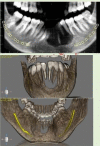
Cone-beam CT (CBCT) panoramic image and CBCT reconstructed (three-dimensional format) anterior and posterior view showing well-defined radiolucent areas with complete loss of alveolar bone from 35 region to 44 teeth region and floating of 34, 33, 32, 31, 41, 42 teeth. There is evidence of inferior alveolar nerve on both sides of mandible.
Figure 4
Figure 4
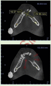
Cone-beam CT axial view of mandible towards apical region shows complete bone loss in buccal and lingual cortex with expansion.
Figure 5
Figure 5
(A–D) Photomicrograph showing lesional areas, highly cellular with proliferation of ovoid to spindle-shaped stromal cells along with presence of plenty of mononucleated and multinucleated giant cells (A, B). Lesional area is surrounded by fibrous capsule; haemorrhagic areas, extravasated red blood cells and bone were noticed (A) (H&E 10×). Highly proliferative stromal tissue with blood vessels and inflammatory cells (H&E 40×) (C). Immunopositivity for CD68 on multinucleated giant cells and individual mononuclear cells (immunohistochemistry 10×) (D).
Figure 6
Figure 6
Surgical picture showing en bloc resection of mandible with tooth-bearing alveolus, and part of basal bone from 42 to 35 was removed.
Figure 7
Figure 7
(A–D) Photomicrograph showing the dense fibrous tissue with areas of spindle-shaped mesenchymal cells and areas of osteoid formation and areas of fibrohistiocytic appearance (A). Stromal cells appear to be highly active with blood vessels and inflammatory cells (B). Multinucleated giant cells in lumen of blood vessels (C) (H&E 40×), and areas of bony resorption noticed (D) (H&E 40×).
Figure 8
Figure 8
Postoperative panoramic image showing partially edentulous arch with loss of 35, 34, 33, 32, 31, 41, 42 teeth; along with this, bone plating was done in the mandible anterior region.

References

    1. Gupta G, Athanikar S, Pai V, et al. . Giant cells in dermatology. Indian J Dermatol 2014;59:481. 10.4103/0019-5154.139887 - DOI - PMC - PubMed
    1. Brodbeck WG, Anderson JM. Giant cell formation and function. Current Opinion in Hematology 2009;16:53–7. 10.1097/MOH.0b013e32831ac52e - DOI - PMC - PubMed
    1. Ranjan V, Chakrabarty S, Arora P, et al. . Classifying giant cell lesions: a review. J Indian Acad Oral Med Radiol 2018;30:297. 10.4103/jiaomr.jiaomr_81_18 - DOI
    1. Brooks PJ, Glogauer M, McCulloch CA. An overview of the derivation and function of multinucleated giant cells and their role in pathologic processes. Am J Pathol 2019;189:1145–58. 10.1016/j.ajpath.2019.02.006 - DOI - PubMed
    1. McNally AK, Anderson JM. Beta1 and beta2 integrins mediate adhesion during macrophage fusion and multinucleated foreign body giant cell formation. Am J Pathol 2002;160:621–30. 10.1016/s0002-9440(10)64882-1 - DOI - PMC - PubMed

Publication types