. 2023 May 4;14(1):2561.
doi: 10.1038/s41467-023-38311-0.

Genomic mutation landscape of skin cancers from DNA repair-deficient xeroderma pigmentosum patients

Affiliations

Genomic mutation landscape of skin cancers from DNA repair-deficient xeroderma pigmentosum patients

Andrey A Yurchenko et al. Nat Commun. .

Abstract

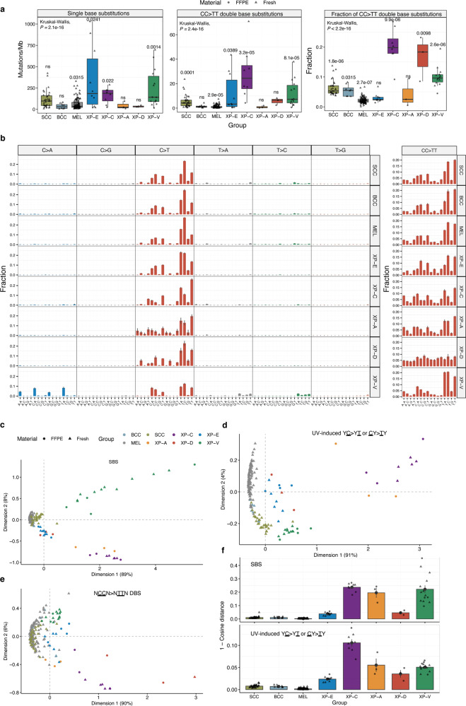
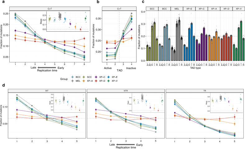
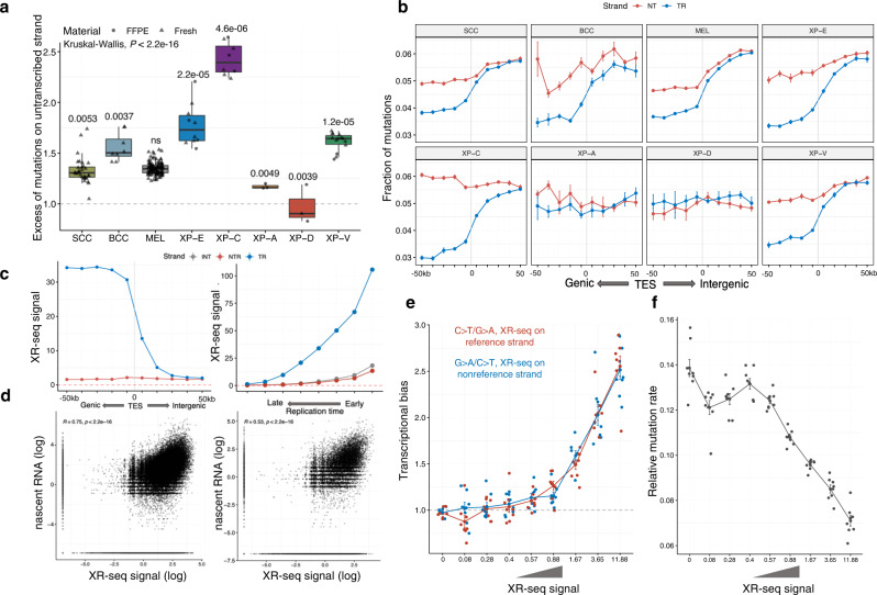
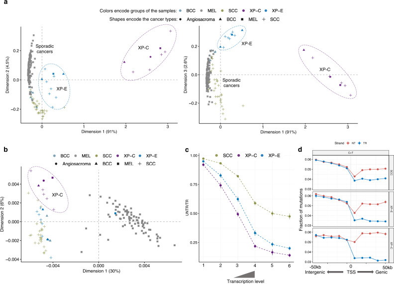
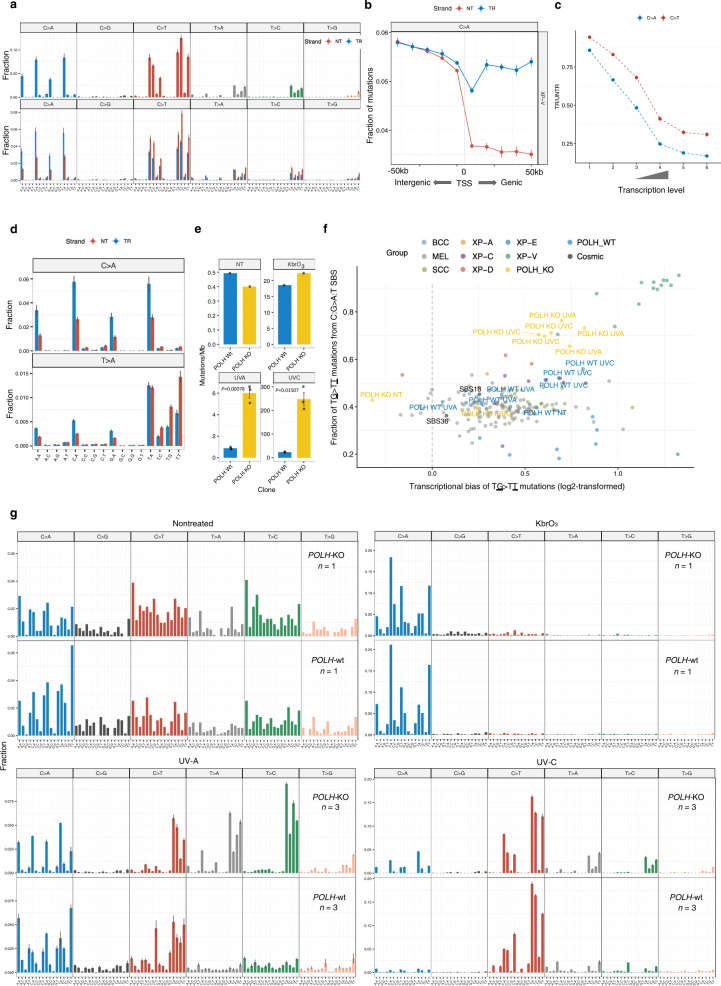
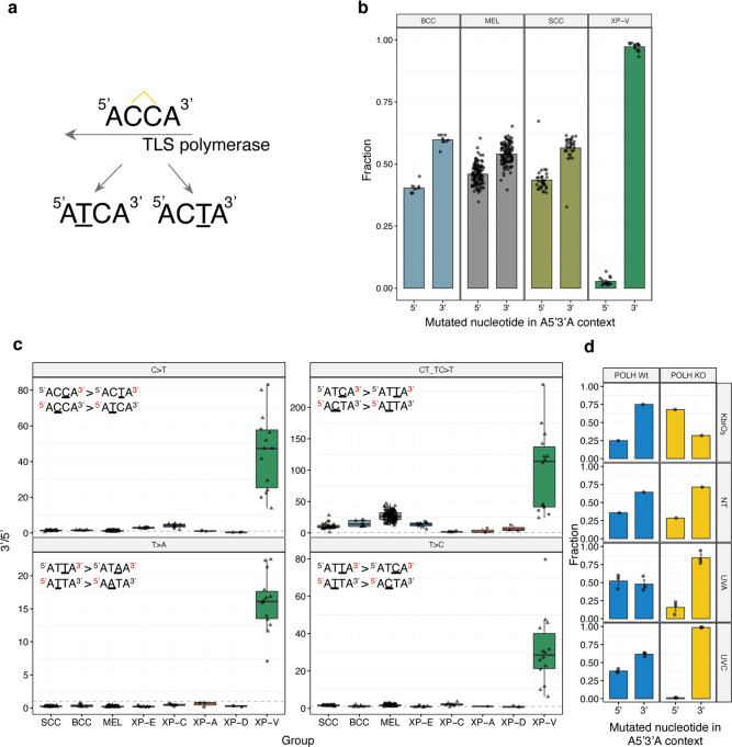
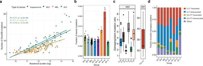
Xeroderma pigmentosum (XP) is a genetic disorder caused by mutations in genes of the Nucleotide Excision Repair (NER) pathway (groups A-G) or in Translesion Synthesis DNA polymerase η (V). XP is associated with an increased skin cancer risk, reaching, for some groups, several thousand-fold compared to the general population. Here, we analyze 38 skin cancer genomes from five XP groups. We find that the activity of NER determines heterogeneity of the mutation rates across skin cancer genomes and that transcription-coupled NER extends beyond the gene boundaries reducing the intergenic mutation rate. Mutational profile in XP-V tumors and experiments with POLH knockout cell line reveal the role of polymerase η in the error-free bypass of (i) rare TpG and TpA DNA lesions, (ii) 3' nucleotides in pyrimidine dimers, and (iii) TpT photodimers. Our study unravels the genetic basis of skin cancer risk in XP and provides insights into the mechanisms reducing UV-induced mutagenesis in the general population.


Conflict of interest statement

The authors declare no competing interests.

Figures

Fig. 1
Fig. 1. Mutation landscape of the studied cancers.
a Tumor mutation burden of all single base substitutions (SBS; left panel), double base substitutions (CC > TT; middle panel) per group and a proportion of CC > TT DBS relative to C > T SBS in pyrimidine context (right panel). All the cancer types were combined together per XP group. P-values from nonparametric ANOVA are indicated (Kruskal-Wallis test for global P-value estimation and Mann–Whitney U test, two-sided for individual groups P-values). Boxes depict the interquartile range (25–75% percentile), lines—the median, whiskers—1.5× the IQR below the first quartile and above the third quartile. Source data are provided as a Source Data file. b Trinucleotide-context mutation profiles of SBS (left panel) and tetranucleotide-context mutation profiles of CC > TT DBS (right panel) per group. Data are presented as mean values +/− SEM. c Multidimensional scaling (MDS) plot based on the Cosine similarity distance between the SBS trinucleotide-context mutation profiles of the samples. d MDS plot based on the Cosine similarity distance between the trinucleotide-context mutation profiles of the samples using only C > T mutations with an adjacent pyrimidine (YC > YT or CY > TY), the typical UV mutation context. e MDS plot based on the Cosine similarity distance between the tetranucleotide-context mutation profiles of the samples using only CC > TT double base substitutions. f Mean Cosine dissimilarity (1-Cosine distance) between original and reconstructed trinucleotide-context mutation profiles using only SBS7a/b/c/d COSMIC mutation signatures for all SBS (upper panel) and C > T mutations with adjacent pyrimidine only (lower panel). Data are presented as mean values +/− SEM. Source data are provided as a Source Data file. Sample size for all the panels (tumors): n = 31 for SCC, n = 8 for BCC, n = 113 for MEL, n = 10 for XP-E, n = 8 for XP-C, n = 3 for XP-A, n = 3 for XP-D and n = 14 for XP-V.
Fig. 2
Fig. 2. Genomic topography of mutagenesis in the skin cancers (SEM intervals are indicated).
a Fraction of C > T mutations from pyrimidine dimers in genomic regions grouped in 8 equal size bins by replication timing (RT) for XP groups and sporadic skin cancers. The box contains the slope values from linear regressions across 8 RT bins. Data are presented as mean values +/− SEM. Source data are provided as a Source Data file. b Fraction of C > T mutations from pyrimidine dimers per group in 1 Mb regions centered at the boundary between active and inactive topologically associated domains (split into two bins each). Data are presented as mean values +/− SEM. Source data are provided as a Source Data file. c Fraction of C > T mutations from pyrimidine dimers per group across different chromatin states (R – repressed, A and A2 – active, H – heterochromatin, I – inactive). Data are presented as mean values +/− SEM. Source data are provided as a Source Data file. d Fractions of C > T mutations from pyrimidine dimers in intergenic regions (left panel), on the untranscribed (middle panel) and transcribed (right panel) DNA strands of gene regions grouped in 5 equal size bins by replication timing (RT) for XP groups and sporadic skin cancers. The boxes contain the slope values from linear regressions across 5 RT bins. I intergenic regions, NTR untranscribed strand of genes, TR transcribed strand of genes. Data are presented as mean values +/−  SEM. Source data are provided as a Source Data file. Sample size for all the panels (tumors): n = 31 for SCC, n = 8 for BCC, n = 113 for MEL, n = 10 for XP-E, n = 8 for XP-C, n = 3 for XP-A, n = 3 for XP-D and n = 14 for XP-V.
Fig. 3
Fig. 3. TC-NER activity behind transcription end sites (TES) of genes.
a The transcriptional bias (TRB) per group (ratio between untranscribed and transcribed strand) for C > T mutations with adjacent pyrimidines for XP groups and sporadic skin cancers. P-values from nonparametric ANOVA are indicated. Boxes depict the interquartile range (25–75% percentile), lines—the median, whiskers—1.5× the IQR below the first quartile and above the third quartile. n = 31 for SCC, n = 8 for BCC, n = 113 for MEL, n = 10 for XP-E, n = 8 for XP-C, n = 3 for XP-A, n = 3 for XP-D and n = 14 for XP-V (tumors). Source data are provided as a Source Data file. b Fractions of C > T mutations with adjacent pyrimidines separated by strands in the TES-centered 100 kb region (binned by 10 kb intervals). Data are presented as mean values +/− SEM. n = 31 for SCC, n = 8 for BCC, n = 113 for MEL, n = 10 for XP-E, n = 8 for XP-C, n = 3 for XP-A, n = 3 for XP-D and n = 14 for XP-V (tumors). Source data are provided as a Source Data file. c DNA context-normalized XR-seq density from XP-C cell line on untranscribed and transcribed gene strands in the TES-centered 100 kb region (binned by 10 kb intervals; left panel, n = 1). DNA context-normalized XR-seq density from XP-C cell line by replication timing for the transcribed and untranscribed DNA strands of genes and intergenic regions. I intergenic regions, NTR untranscribed strand of genes, TR transcribed strand of genes (right panel, n = 1). d Correlation between XR-seq intensity from XP-C cell line and nascent RNA-seq for genic regions (left panel, n = 1) and intergenic regions 50 kb downstream of TES (right panel, n = 1). Pearson’s r correlation coefficients and P values are indicated. e Transcriptional bias of C:G > T:A mutations on intergenic regions of XP-C tumors depending on the XR-seq intensity of XP-C cell line. SEM intervals are indicated, n = 8 tumors. f Relative mutation rate of C:G > T:A mutations in intergenic regions of XP-C tumors (n = 8) depending on the XR-seq intensity in XP-C cell line. Data are presented as mean values +/− SEM.
Fig. 4
Fig. 4. Comparison of genomic mutagenesis between sporadic cancers, XP-E and XP-C groups.
a Multidimensional scaling (MDS) plot based on the Cosine similarity distance between the SBS trinucleotide-context mutation profiles of the samples (Dimensions 1 and 2—left panel, Dimensions 1 and 3—right panel). Colors encode groups of the samples and shapes encode types of cancers. n = 31 for SCC, n = 8 for BCC, n = 113 for MEL, n = 10 for XP-E and n = 8 for XP-C (tumors). b PCA plot based on the density of mutations in 2684 1Mb-long windows along the genome (only for samples with more than 50k mutations belong to sporadic, XP-C and XP-E groups). Colors encode groups of the samples and shapes encode types of cancers. n = 4 for BCC, n = 83 for MEL, n = 26 for SCC, n = 8 for XP-C and n = 10 for XP-E (tumors). c The transcriptional bias (TRB; ratio between untranscribed and transcribed strand mutation number) for C > T mutations from pyrimidine dimers in genes grouped in 6 bins by gene expression level. Only cutaneous SCC tumors were used for XP-C and XP-E groups. Data are presented as mean values +/− SEM. n = 31 for SCC, n = 5 for XP-C and n = 7 for XP-E (tumors). Source data are provided as a Source Data file. d Fractions of C > T mutations from pyrimidine dimers separated by strands in the TSS-centered 100 kb region (binned by 10 kb intervals). Blue line—untranscribed strand for purines or transcribed for pyrimidines, red line—transcribed strand for purines or untranscribed for pyrimidines. Data are presented as mean values +/− SEM. n = 31 for SCC, n = 5 for XP-C and n = 7 for XP-E (tumors). Source data are provided as a Source Data file.
Fig. 5
Fig. 5. Mutation profiles of XP-V skin cancers and POLH-KO clones.
a Trinucleotide-context mutation profile of genomic SBS (upper panel) and genic SBS (lower panel) separated by transcribed (TR) and untranscribed (NT) strands in XP-V tumors. Blue bars— untranscribed strand for purines or transcribed for pyrimidines, red bars—transcribed strand for purines or untranscribed for pyrimidines. Data are presented as mean values +/− SEM. n = 14 tumors. b Fractions of C > A mutations separated by gene strands in the TSS-centered 100 kb region of XP-V tumors (binned by 10 kb intervals). Blue—untranscribed strand for mutations from purines and transcribed strand for mutations from pyrimidines; red— transcribed strand for mutations from purines and untranscribed strand for mutations from pyrimidines. Data are presented as mean values +/− SEM. n = 14 tumors. c The transcriptional bias (ratio between transcribed and untranscribed strand) for C > A and C > T mutations per bin of gene expression level (only XP-V samples represented by BCC, n = 11 tumors). Data are presented as mean values +/− SEM. d Trinucleotide-context mutation profiles of SBS separated by strands in XP-V tumors for C > A and T > A mutations. Data are presented as mean values +/− SEM, n = 14 tumors. e Mutations per megabase in the POLH wt and POLH-KO clones in nontreated cells (NT, n = 1 per cell line independent biological replicate), treated with KbrO3 (n = 1 per cell line independent biological replicate), UV-A (n = 3 per cell line independent biological replicates) and UV-C (n = 3 independent cell clones per cell line). Welch two sample t-test, two-sided. Data are presented as mean values +/− SEM. Source data are provided as a Source Data file. f Mutational specificity of the TG > TT mutations in XP-V tumors and POLH-KO UV-A- and UV-C-treated cell lines. X-axis: log2-transformed transcriptional bias of the TG > TT mutations per genome. Y-axis: Fraction of the mutations in the TG > TT context from the total number of C:G > A:T substitutions per genome. POLH-KO and POLH-wt clones are specifically indicated with their corresponding treatment (KbrO3, UV-A and UV-C) as well as COSMIC SBS18 and SBS36 mutational signatures associated with oxidative DNA damage (black dots). g Mutation profiles of the POLH-wt and POLH-KO clones for nontreated cells (NT), treated with KbrO3, UV-A and UV-C. Data are presented as mean values +/− SEM for UV-A and UV-C experiments. Sample size is indicated on the plots (independent cell clones).
Fig. 6
Fig. 6. Dimer translesion bias in XP-V skin cancers.
a Schematic representation of the putative CC photodimer in ACCA context and resulting mutations analyzed in the panel b. b Fraction of C > T mutations from 5’ and 3’ cytosines of the dimer in the 5’ACCA3’ context per group of tumors. Data are presented as mean values +/− SEM. n = 31 for SCC, n = 8 for BCC, n = 113 for MEL and n = 14 for XP-V (tumors). Source data are provided as a Source Data file. c “Dimer translesion bias” for different sequence contexts per group of tumors. Comparison of C > T mutation frequency in CT and TC pyrimidine dimers was performed after normalization to the number of such contexts in the genome (upper right panel). Boxes depict the interquartile range (25–75% percentile), lines - the median, whiskers − 1.5× the IQR below the first quartile and above the third quartile. n = 31 for SCC, n = 8 for BCC, n = 113 for MEL, n = 10 for XP-E, n = 8 for XP-C, n = 3 for XP-A, n = 3 for XP-D and n = 14 for XP-V (tumors). Source data are provided as a Source Data file. d Fraction of C > T mutations from 5’ and 3’ cytosines of the dimer in the 5’ACCA3’ context in the RPE-1 POLH-wt and POLH-KO clones. SEM intervals are indicated. n = 1 for NT and KbrO3 and n = 3 for UV-A and UV-C clones per cell line. Source data are provided as a Source Data file.
Fig. 7
Fig. 7. Protein-damaging effect of mutation contexts.
a Correlations between tumor mutation burden and number of oncogenic and likely oncogenic mutations in the studied skin cancer samples according to the OncoKB database. Pearson’s r coefficients and P values are indicated. b Mean fraction of exonic mutations from all the mutations per sample. Data are presented as mean values +/− SEM. n = 31 for SCC, n = 8 for BCC, n = 113 for MEL, n = 10 for XP-E, n = 8 for XP-C, n = 3 for XP-A, n = 3 for XP-D and n = 14 for XP-V (tumors). Source data are provided as a Source Data file. c Protein-damaging/silent mutation ratio per substitution type in our pooled skin cancer cohort (n = 190 tumors). Damaging mutations—all non-silent exonic (missense, truncating) and splice site mutations. Boxes depict the interquartile range (25–75% percentile), lines—the median, whiskers—1.5× the IQR below the first quartile and above the third quartile. d Mean fraction of protein-damaging mutations originating from the main mutation classes split by gene strand per group.

References

    1. Lehmann AR, McGibbon D, Stefanini M. Xeroderma pigmentosum. Orphanet J. Rare Dis. 2011;6:1–6. doi: 10.1186/1750-1172-6-70. - DOI - PMC - PubMed
    1. Bradford PT, et al. Cancer and neurologic degeneration in xeroderma pigmentosum: long term follow-up characterises the role of DNA repair. J. Med. Genet. 2011;48:168–176. doi: 10.1136/jmg.2010.083022. - DOI - PMC - PubMed
    1. Yurchenko AA, et al. XPC deficiency increases risk of hematologic malignancies through mutator phenotype and characteristic mutational signature. Nat. Commun. 2020;11:5834. doi: 10.1038/s41467-020-19633-9. - DOI - PMC - PubMed
    1. Nikolaev S, Yurchenko AA, Sarasin A. Increased risk of internal tumors in DNA repair-deficient xeroderma pigmentosum patients: analysis of four international cohorts. Orphanet J. Rare Dis. 2022;17:104. doi: 10.1186/s13023-022-02203-1. - DOI - PMC - PubMed
    1. Spivak G. Nucleotide excision repair in humans. DNA Repair. 2015;36:13–18. doi: 10.1016/j.dnarep.2015.09.003. - DOI - PMC - PubMed

Publication types