. 2024 Apr;18(2):568-581.
doi: 10.1007/s12072-023-10537-6. Epub 2023 May 4.

Hepatocellular carcinoma (HCC) tumor microenvironment is more suppressive than colorectal cancer liver metastasis (CRLM) tumor microenvironment

Affiliations

Hepatocellular carcinoma (HCC) tumor microenvironment is more suppressive than colorectal cancer liver metastasis (CRLM) tumor microenvironment

Sara Santagata et al. Hepatol Int. 2024 Apr.

Abstract

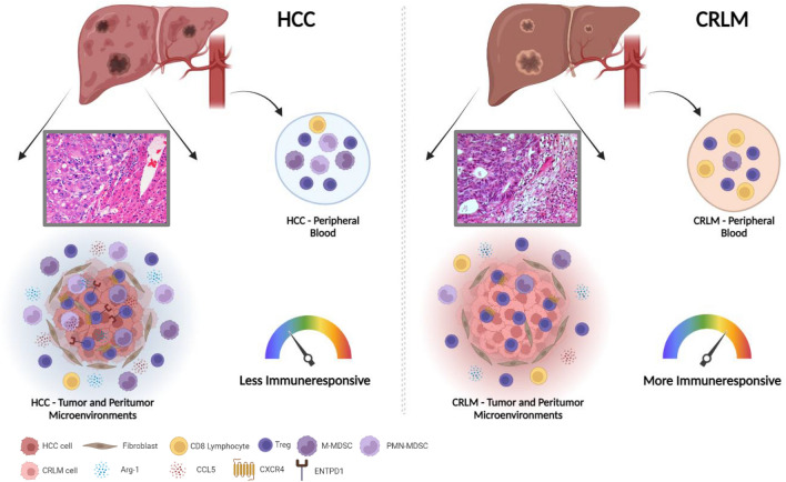
Background and purpose: While HCC is an inflammation-associated cancer, CRLM develops on permissive healthy liver microenvironment. To evaluate the immune aspects of these two different environments, peripheral blood-(PB), peritumoral-(PT) and tumoral tissues-(TT) from HCC and CRLM patients were evaluated.

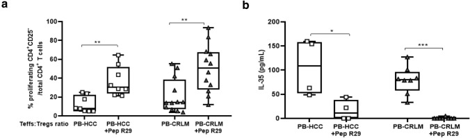
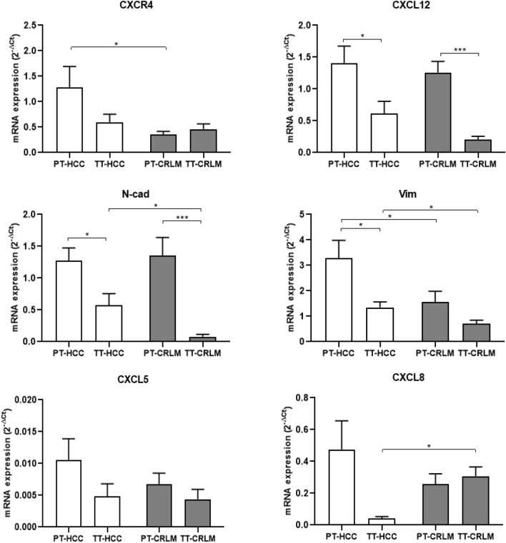
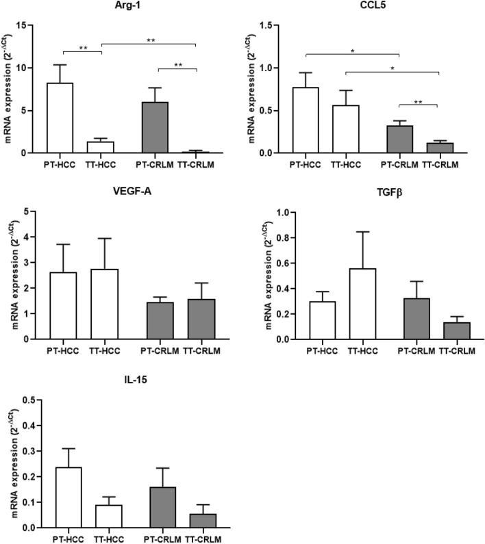
Methods: 40 HCC and 34 CRLM were enrolled and freshly TT, PT and PB were collected at the surgery. PB-, PT- and TT-derived CD4+CD25+ Tregs, M/PMN-MDSC and PB-derived CD4+CD25- T-effector cells (Teffs) were isolated and characterized. Tregs' function was also evaluated in the presence of the CXCR4 inhibitor, peptide-R29, AMD3100 or anti-PD1. RNA was extracted from PB/PT/TT tissues and tested for FOXP3, CXCL12, CXCR4, CCL5, IL-15, CXCL5, Arg-1, N-cad, Vim, CXCL8, TGFβ and VEGF-A expression.

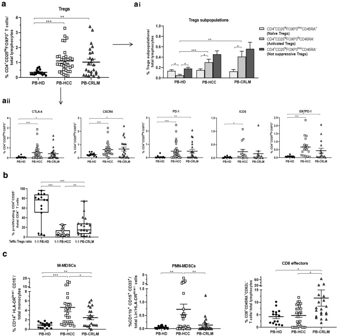
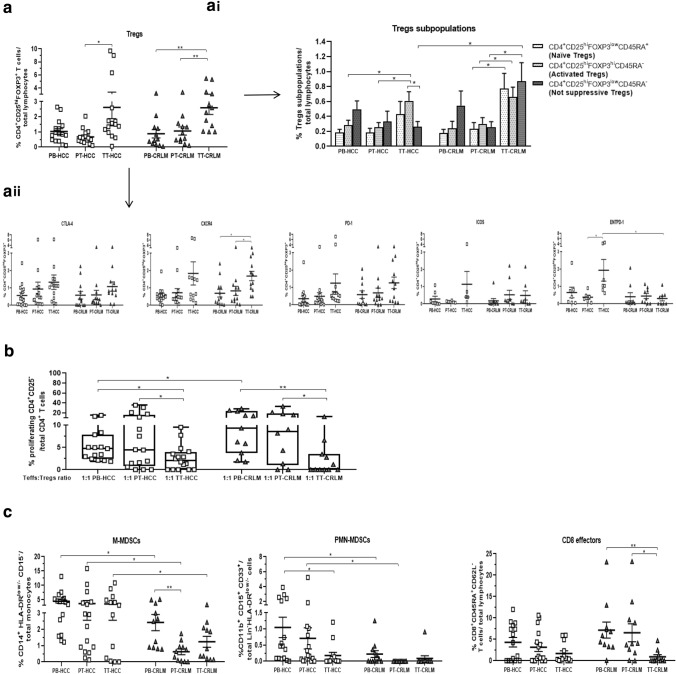
Results: In HCC/CRLM-PB, higher number of functional Tregs, CD4+CD25hiFOXP3+ was detected, although PB-HCC Tregs exert a more suppressive function as compared to CRLM Tregs. In HCC/CRLM-TT, Tregs were highly represented with activated/ENTPD-1+Tregs prevalent in HCC. As compared to CRLM, HCC overexpressed CXCR4 and N-cadherin/vimentin in a contest rich in arginase and CCL5. Monocytic MDSCs were highly represented in HCC/CRLM, while high polymorphonuclear MDSCs were detected only in HCC. Interestingly, the function of CXCR4-PB-Tregs was impaired in HCC/CRLM by the CXCR4 inhibitor R29.

Conclusion: In HCC and CRLM, peripheral blood, peritumoral and tumoral tissues Tregs are highly represented and functional. Nevertheless, HCC displays a more immunosuppressive TME due to Tregs, MDSCs, intrinsic tumor features (CXCR4, CCL5, arginase) and the contest in which it develops. As CXCR4 is overexpressed in HCC/CRLM tumor/TME cells, CXCR4 inhibitors may be considered for double hit therapy in liver cancer patients.

Keywords: CXCR4; CXCR4 inhibitors; ENTPD1; Immune cells and the microenvironment; Inflammation-associated cancer; Liver cancer; Liver metastases; Liver microenvironment; Myeloid-derived suppressor cells; Regulatory T cells.


Conflict of interest statement

The authors declare no conflicts of interest. The authors Sara Santagata, Giuseppina Rea, Daniela Castaldo, Maria Napolitano, Anna Capiluongo, Crescenzo D’Alterio, Anna Maria Trotta, Caterina Ieranò, Luigi Portella, Salvatore Di Maro, Fabiana Tatangelo,Vittorio Albino, Rita Guarino, Carmen Cutolo, Francesco Izzo, and Stefania Scala declare no conflict of interest.

Figures

Fig. 1
Fig. 1
HCC and CRLM peripheral blood (PB) Tregs are highly represented: HCC Tregs are the most active. a Phenotypic characterization of peripheral Tregs (CD4+CD25hiFoxp3+ T cells/ total lymphocytes) in 33 HCC and 24 CRLM as compared to 15 HD (HCC vs HD, p < 0.001; CRLM vs HD, p < 0.01). (ai) naïve (CD25hiFOXP3lowCD45RA+), activated (CD25hiFOXP3hiCD45RA), and not suppressive (CD25hiFOXP3lowCD45RA) Tregs in HCC, CRLM, and HD peripheral blood: (activated Tregs: HCC vs HD, p < 0.001; CRLM vs HD, p < 0.01); (HD: activated vs naïve and not suppressive Tregs, p < 0.05); (HCC and CRLM: activated vs naïve Tregs p < 0.05). (aii) Tregs activation markers in HCC and CRLM compared to HD: CTLA-4, CXCR4, PD-1, and ENTPD1 (HCC vs HD, p < 0.001); ICOS (HCC vs HD, p < 0.05); CTLA-4, CXCR4, and ENTPD1 (CRLM vs HD, p < 0.05); PD1 (CRLM vs HD, p < 0.01). b Functional PB-derived Tregs from 24 HCC, 20 CRLM patients, and 15 HD by CFSE suppression assay (PB-HCC/CRLM vs PB-HD: p < 0.001; PB-HCC vs PB-CRLM: p < 0.01). c Peripheral M-MDSCs (CD14+HLA-DRlow/−CD15) and PMN-MDSCs (CD11b+CD15+CD33+LinHLA-DRlow/−) in HCC and CRLM (M-MDSCs: HCC vs HD: p < 0.001; CRLM vs HD: p < 0.01; HCC vs CRLM: p < 0.05); (PMN-MDSCs: HCC vs HD, p < 0.01; HCC vs CRLM, p < 0.01). Peripheral CD8+ effector cells (CD8+CD45R4+CD62L.) in HCC and CRLM (CRLM vs HD, P < 0.05; HCC vs CRLM, p < 0.05)
Fig. 2
Fig. 2
Tregs are highly represented in HCC and CRLM tumor tissue (TT) as compared to peritumoral tissue (PT). ENTPD-1+Tregs and MDSCs characterized a more immunosuppressive TME in HCC. a Paired peripheral, peritumoral and tumor Tregs by flow cytometry in 16 HCC and 12 CRLM patients (TT-HCC vs PT-HCC, p < 0.05; TT-CRLM vs PT-CRLM: p < 0.01). (ai) Naïve, activated and not suppressive Tregs in HCC and CRLM patients: (activated Tregs: TT-HCC/CRLM vs PT-HCC/CRLM, p < 0.05; naïve and not suppressive: TT-CRLM vs PT-CRLM, p < 0.05); not suppressive Tregs (TT-HCC vs TT-CRLM: p < 0.05); (TT-HCC: activated Tregs vs not suppressive, p < 0.05). (aii) Tregs’ activation markers in HCC and CRLM patients: CXCR4 (TT-CRLM vs PB-/PT-CRLM: p < 0.05); ENTPD1 (TT-HCC vs PT-HCC: p < 0.05); (TT-HCC vs TT-CRLM: p < 0.05). b Functional characterization of PB-, PT-, and TT- derived Tregs from HCC and CRLM patients: (TT-HCC/CRLM vs PT-HCC/CRLM: p < 0.05); (TT-HCC/CRLM vs PB-HCC/CRLM: p < 0.01 and p < 0.05); (PB-HCC vs PB-CRLM: p < 0.05). c M-MDSCs and PMN-MDSCs in HCC and CRLM: (M-MDSCs: PB-/PT-/TT-HCC vs PB-PT-TT-CRLM: p < 0.05); (PMN-MDSCs:PB-/PT-HCC vs PB-/PT-CRLM: p < 0.05). CD8.+ effector cells in HCC and CRLM (TT-CRLM vs PT-CRLM: p < 0.05; TT-CRLM vs PB-CRLM: p < 0.01)
Fig. 3
Fig. 3
Ex vivo CXCR4 antagonism impairs PB-HCC/CRLM Tregs suppressive capability. a Functional PB-derived Tregs from 8 HCC and 12 CRLM treated for 30 min at 37 ℃ in 5% CO2 with Pep R29 (10 μM) and then added at 1:1 ratio to Teffs for 5 days (HCC: 1:1 vs 1:1 + Pep R29, p < 0.01;) (CRLM: 1:1 vs 1:1 + Pep R29, p < 0.01). b IL-35 concentration (pg/ml) in supernatant of CFSE assay from Tregs pretreated with Pep R29 (10 μM) in 4 HCC and 8 CRLM patients by ELISA: (HCC: 1:1 vs 1:1 + Pep R29, p < 0.05) (CRLM: 1:1 vs 1:1 + Pep R29, p < 0.001)
Fig. 4
Fig. 4
CXCR4 and mesenchymal markers defined HCC. Real-time PCR was performed to define CXCR4, CXCL12, N-cad, Vim, CXCL5 and CXCL8 expression in tumor and peritumoral tissue of HCC and CRLM patients. ACTB was used for normalization of target gene expression. CXCR4 (19 PT-HCC vs 23 PT-CRLM, P < 0.05); CXCL12 (20 HCC: PT vs TT, p < 0.05; 18 CRLM: PT vs TT, p < 0.001); N-cad (17 HCC: PT vs TT, p < 0.05; 21 CRLM: PT vs TT, p < 0.001) (17 TT-HCC vs 21 TT-CRLM, p < 0.05); Vim (21 HCC: PT vs TT, p < 0.05) (21 PT-HCC vs 22 PT-CRLM, p < 0.05) (21 TT-HCC vs 22 TT-CRLM, p < 0.05); CXCL8 (5 TT-HCC vs 10 TT-CRLM, p < 0.05)
Fig. 5
Fig. 5
Intrinsic tumor features modifying the tumor microenvironment. HCC-TME overexpressed ARG-1 and CCL5. Real-time PCR was performed to define Arg-1, CCL5, VEGF-A, TGFβ, and IL-15 expression in tumor and peritumoral tissue of HCC and CRLM patients. ACTB was used as normalization of target gene expression. Arg-1 (20 HCC: PT vs TT P < 0.01) (22 CRLM: PT vs TT, p < 0.01) (20 TT-HCC vs 22 TT-CRLM, p < 0.01); CCL5 (20 PT-HCC vs 22 PT-CRLM, p < 0.05) (22 CRLM: PT vs TT, p < 0.01) (20 TT-HCC vs 22 TT-CRLM p < 0.05)
Fig. 6
Fig. 6
Proposed landscape of the tumor microenvironment in HCC and CRLM

References

    1. Sung H, Ferlay J, Siegel RL, Laversanne M, Soerjomataram I, Jemal A, et al. Global cancer statistics 2020: GLOBOCAN estimates of incidence and mortality worldwide for 36 cancers in 185 countries. CA Cancer J Clin. 2021;71(3):209–249. doi: 10.3322/caac.21660. - DOI - PubMed
    1. Akinyemiju T, Abera S, Ahmed M, Alam N, Alemayohu MA, Global Burden of Disease Liver Cancer C et al. The burden of primary liver cancer and underlying etiologies from 1990 to 2015 at the Global, Regional, and National Level: results from the Global Burden of Disease Study 2015. JAMA Oncol. 2017;3(12):1683–1691. doi: 10.1001/jamaoncol.2017.3055. - DOI - PMC - PubMed
    1. Ringelhan M, Pfister D, O'Connor T, Pikarsky E, Heikenwalder M. The immunology of hepatocellular carcinoma. Nat Immunol. 2018;19(3):222–232. doi: 10.1038/s41590-018-0044-z. - DOI - PubMed
    1. Yuan D, Huang S, Berger E, Liu L, Gross N, Heinzmann F, et al. Kupffer cell-derived tnf triggers cholangiocellular tumorigenesis through JNK due to chronic mitochondrial dysfunction and ROS. Cancer Cell. 2017;31(6):771–789 e776. doi: 10.1016/j.ccell.2017.05.006. - DOI - PMC - PubMed
    1. Volponi C, Gazzillo A, Bonavita E. The tumor microenvironment of hepatocellular carcinoma: untying an intricate immunological network. Cancers. 2022;14(24):6151. doi: 10.3390/cancers14246151. - DOI - PMC - PubMed

MeSH terms