Review
. 2023 May 5;42(1):114.
doi: 10.1186/s13046-023-02691-4.

Tumor microenvironmental modification by the current target therapy for head and neck squamous cell carcinoma

Affiliations
Review

Tumor microenvironmental modification by the current target therapy for head and neck squamous cell carcinoma

Kohei Okuyama et al. J Exp Clin Cancer Res. .

Abstract

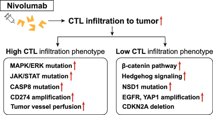
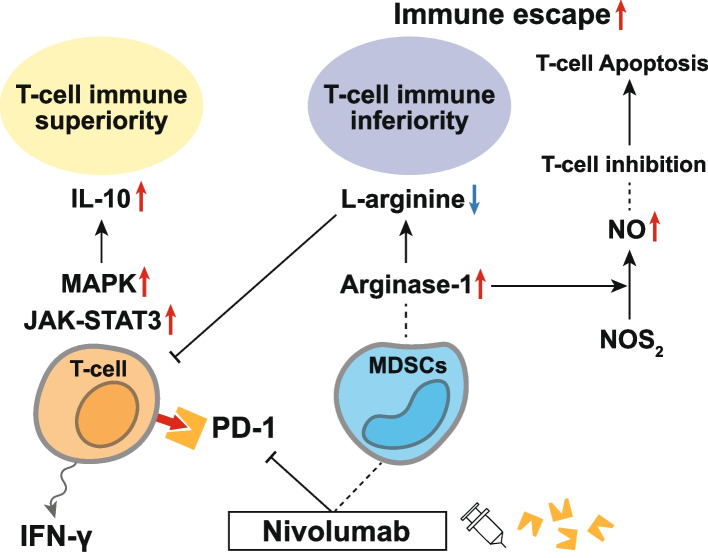
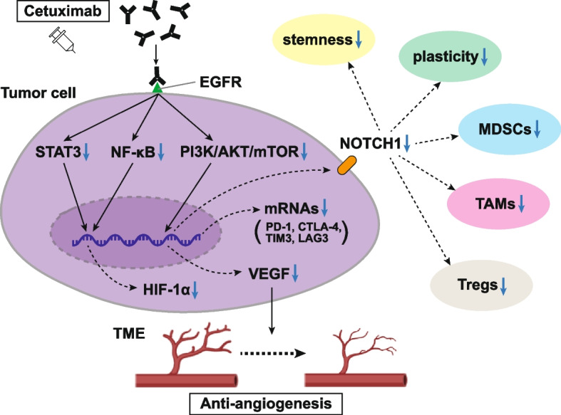
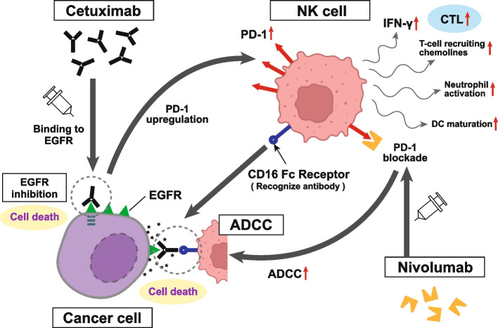
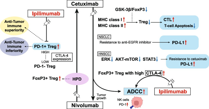
Current clinical and observational evidence supports the EXTREME regimen as one of the standards of care for patients with recurrent or metastatic head and neck squamous cell carcinoma (HNSCC) followed by the administration of immune checkpoint inhibitors (ICIs). In addition to the inhibition of the epidermal growth factor receptor (EGFR) pathway, cetuximab-mediated EGFR blockade has been shown to modulate tumor microenvironment (TME) characteristics, such as antibody-dependent cellular cytotoxicity (ADCC) activity, cytotoxic T-lymphocyte (CTL) infiltration into the tumor, anti-angiogenesis activity, and cytokine secretion via associated natural killer (NK) cells, etc.. On the other hand, there are reports that nivolumab affects the TME via Programmed cell death 1 (PD-1) inhibition, Interleukin-10 upregulation via T-cells, myeloid-derived suppressor cell-mediated immune escape induction, and tumor vessel perfusion by promoting CD8 + T-cell accumulation and Interferon-γ production in treatment-sensitive tumor cells. Actually, nivolumab administration can give T cells in the TME both immune superiority and inferiority. HNSCC treatment using cetuximab increases the frequency of FoxP3 + intratumoral effector regulatory T cells (Tregs) expressing CTL associated antigen (CTLA)-4, and targeting CTLA-4 + Tregs using ipilimumab restores the cytolytic function of NK cells, which mediate ADCC activity. Treg-mediated immune suppression also contributes to clinical response to cetuximab treatment, suggesting the possibility of the addition of ipilimumab or the use of other Treg ablation strategies to promote antitumor immunity. Moreover, also in hyper progression disease (HPD), intratumoral frequency of FoxP3 + effector Tregs expressing CTLA-4 is increased. Therefore, combination treatment with cetuximab plus anti-CTLA-4 antibody ipilimumab for HNSCC and this combination therapy after nivolumab administration for HPD may be expected to result in a higher tumor-control response. Based on the above evidence, we here suggest the efficacy of using these therapeutic strategies for patients with local-advanced, recurrent, and metastatic HNSCC and patients who do not respond well to nivolumab administration.

Keywords: CTLA-4; Cetuximab; Head and neck squamous cell carcinoma; Immune checkpoint inhibitors; Nivolumab; PD-1; Tumor microenvironment.


Conflict of interest statement

The authors declare no competing interests.

Figures

Fig. 1
Fig. 1
Differences in the responses of high- and low-CTL tumors to nivolumab administration
Fig. 2
Fig. 2
Immune superiority and inferiority of T-cells in the TME following nivolumab administration. IL-10 is one of the most important immunoregulatory cytokines that regulate T-cell responses by modulating multiple signaling pathways. Nivolumab induces potent IL-10 secretion in T cells via the activation of the MAPK pathway, in which T cells can activate T cell growth and metabolism, possibly leading to T-cell immune superiority. Based on an in vitro study, nivolumab treatment significantly reduces PD-1 levels in all T-cell populations, suggesting that IL-10 may confer a heterogeneous T-cell response to nivolumab. The engagement of IL-10 with its receptor also activates multiple signaling pathways, particularly the JAK-STAT3 pathway, indicating that nivolumab triggers several biological pathways in the TME. MDSCs exert their immunomodulatory effects via diverse mechanisms, including Arginase-1-mediated depletion of l-arginine and NO production via NOS2. Arginase-1 starves L-arginine in the TME, thus limiting T-cell proliferation. In addition to L-arginine depletion, NO production transforms the TME to promote immune evasion and prolonged NO exposure leads to T-cell apoptosis, causing T-cells to show TME immune inferiority
Fig. 3
Fig. 3
Neovascularization suppression and TME modulation by cetuximab. Treatment with cetuximab suppresses tumor-induced neovascularization in HNSCC. Increased EGFR expression shows a correlation with increased HIF-1α level and microvessel density. The previously demonstrated inhibition of VEGF by cetuximab occurs at the transcriptional level in response to reduced levels of HIF-1α. Moreover, cetuximab has been shown to attenuate the secretion of proangiogenic factors in tumor cells, such as VEGF and IL-8. Cetuximab inhibits tumor-induced angiogenesis by downregulating HIF-1α and Notch1, resulting in reduced angiogenesis and tumor shrinkage. The Notch signaling pathway plays an important role in blood vessel formation and remodeling. The inhibition of Notch signaling also reduces the number of MDSCs, TAMs, and Tregs within the tumor and inhibits the mRNA and target protein expression of the most relevant immune checkpoint molecules: PD-1, CTLA4, TIM3, and LAG3
Fig. 4
Fig. 4
ADCC activity of cetuximab treatment and the effect on nivolumab administration. Pre-clinical studies have demonstrated the ability of cetuximab to stimulate ADCC and affect antitumor immunity. In vitro, cetuximab can mobilize NK cells, activate neutrophils, and stimulate DC maturation. This contributes to an extra antitumor effect in addition to simple EGFR inhibition. NK cell activation then produces IFN-γ, which promotes PD-L1 expression in tumor cells and emits T cell-recruiting chemokines that activate a high density of CTL in the TME. Cetuximab treatment also upregulates PD-1 expression in NK cells and maximizes antitumor effects. Additionally, PD-1 blockade increases cetuximab-mediated ADCC against PD-L1-high HNSCC cells without EGFR amplification, indicating that the combination of anti-EGFR antibodies with ICIs can enhance both innate and acquired antitumor immune responses
Fig. 5
Fig. 5
TME modulation by cetuximab and nivolumab and the potential breakthrough treatment strategy using ipilimumab for HPD. Inhibiting EGFR modulates the tumor immune microenvironment in several ways, including enhancing MHC class I and II expression, decreasing the suppressive function of Tregs, promoting CTL activity, and reducing T cell apoptosis. PD-1 blockade may facilitate the proliferation of highly suppressive PD-1 + effector Treg cells in HPDs, resulting in the inhibition of antitumor immunity. Highly activated tumor-infiltrating PD-1 + effector Treg cells show higher CTLA-4 expression levels than PD-1- effector Treg cells. As the PD-1 blockade significantly enhances Treg cell suppressive activity, PD-1 blockade facilitates the proliferation of highly suppressive PD-1 + effector Treg cells expressing high CTLA-4 in HPDs, resulting in immunosuppression in HNSCC. Moreover, cetuximab treatment increases the FoxP3 + intratumoral effector Tregs expressing CTLA-4, suggesting the combination with ipilimumab restores the cytolytic functions of NK cells mediating ADCC. Targeting CTLA-4 high PD-1 effector Tregs for HNSCC may show a high response to the tumor and improve survival

References

    1. de Andrade DA, Machiels JP. Treatment options for patients with recurrent or metastatic squamous cell carcinoma of the head and neck, who progress after platinum-based chemotherapy. Curr Opin Oncol. 2012;24:211–217. doi: 10.1097/CCO.0b013e3283510773. - DOI - PubMed
    1. Mendelsohn J, Baselga J. Status of epidermal growth factor receptor antagonists in the biology and treatment of cancer. J Clin Oncol. 2003;21:2787–2799. doi: 10.1200/JCO.2003.01.504. - DOI - PubMed
    1. Trivedi S, Concha-Benavente F, Srivastava RM, Jie HB, Gibson SP, Schmitt NC, et al. Immune biomarkers of anti-EGFR monoclonal antibody therapy. Ann Oncol. 2015;26:40–47. doi: 10.1093/annonc/mdu156. - DOI - PMC - PubMed
    1. Jie HB, Schuler PJ, Lee SC, Srivastava RM, Argiris A, Ferrone S, et al. CTLA-4+ regulatory T cells increased in cetuximab-treated head and neck cancer patients suppress NK cell cytotoxicity and correlate with poor prognosis. Cancer Res. 2015;75:2200–2210. doi: 10.1158/0008-5472.CAN-14-2788. - DOI - PMC - PubMed
    1. Perez R, Crombet T, de Leon J, Moreno E. A view on EGFR-targeted therapies from the oncogene-addiction perspective. Front Pharmacol. 2013;4:53. doi: 10.3389/fphar.2013.00053. - DOI - PMC - PubMed