. 2023 May 4;10(1):21.
doi: 10.1186/s40779-023-00454-y.

Elimination of methicillin-resistant Staphylococcus aureus biofilms on titanium implants via photothermally-triggered nitric oxide and immunotherapy for enhanced osseointegration

Affiliations

Elimination of methicillin-resistant Staphylococcus aureus biofilms on titanium implants via photothermally-triggered nitric oxide and immunotherapy for enhanced osseointegration

Yong-Lin Yu et al. Mil Med Res. .

Abstract

Background: Treatment of methicillin-resistant Staphylococcus aureus (MRSA) biofilm infections in implant placement surgery is limited by the lack of antimicrobial activity of titanium (Ti) implants. There is a need to explore more effective approaches for the treatment of MRSA biofilm infections.

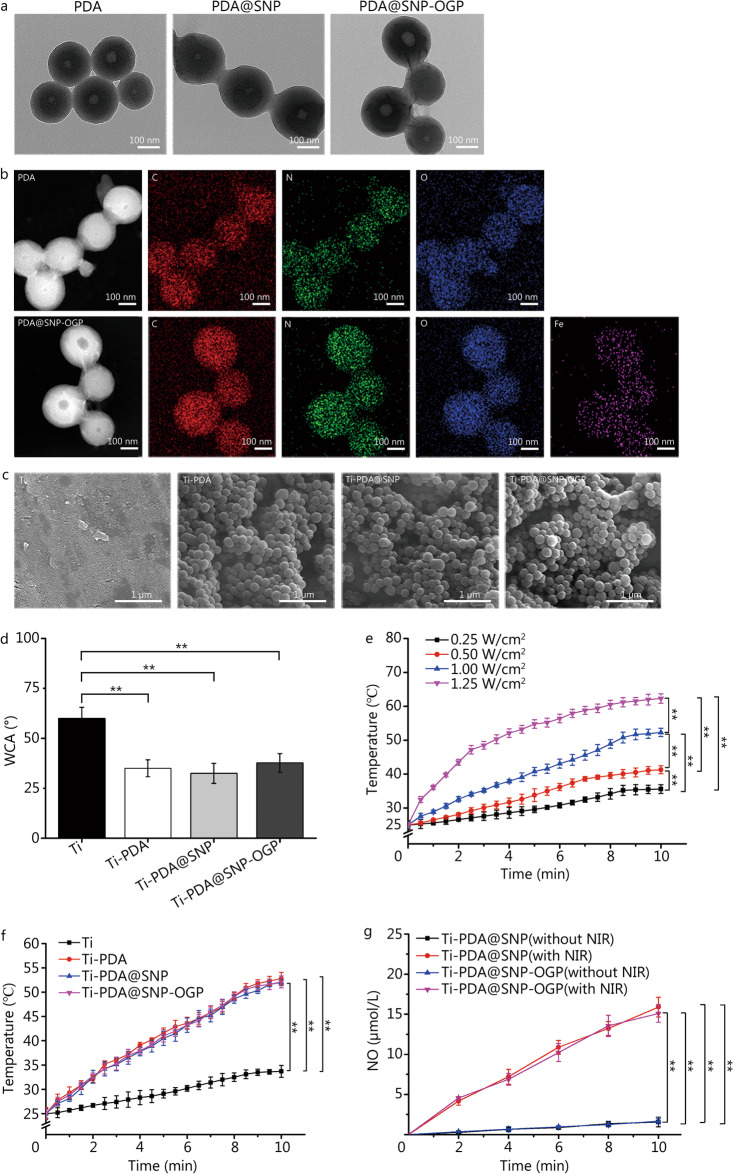
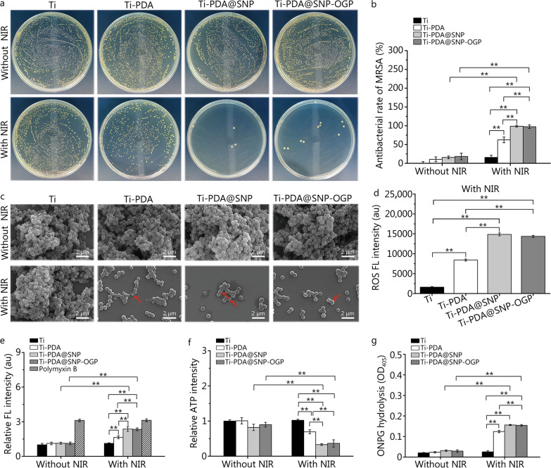
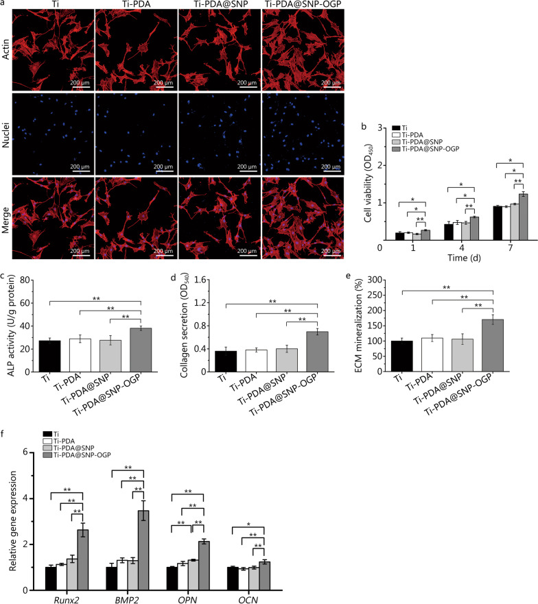
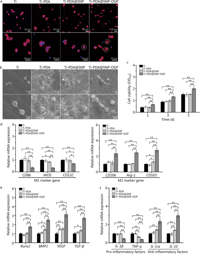
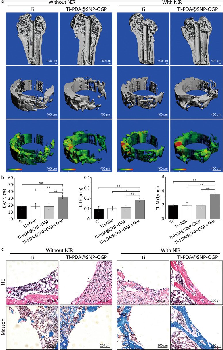
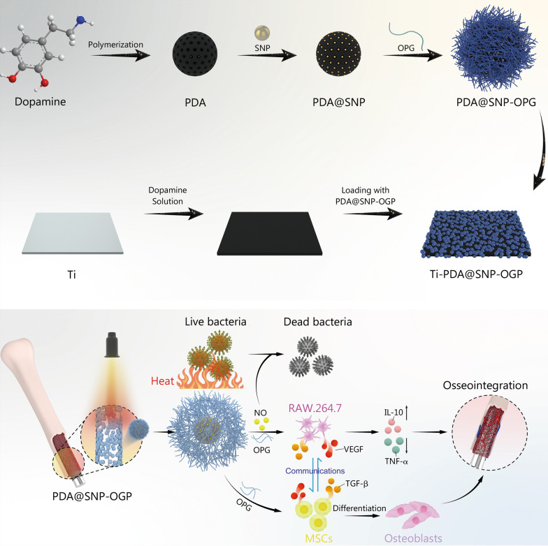
Methods: Herein, an interfacial functionalization strategy is proposed by the integration of mesoporous polydopamine nanoparticles (PDA), nitric oxide (NO) release donor sodium nitroprusside (SNP) and osteogenic growth peptide (OGP) onto Ti implants, denoted as Ti-PDA@SNP-OGP. The physical and chemical properties of Ti-PDA@SNP-OGP were assessed by scanning electron microscopy, X-ray photoelectron spectroscope, water contact angle, photothermal property and NO release behavior. The synergistic antibacterial effect and elimination of the MRSA biofilms were evaluated by 2',7'-dichlorofluorescein diacetate probe, 1-N-phenylnaphthylamine assay, adenosine triphosphate intensity, o-nitrophenyl-β-D-galactopyranoside hydrolysis activity, bicinchoninic acid leakage. Fluorescence staining, assays for alkaline phosphatase activity, collagen secretion and extracellular matrix mineralization, quantitative real‑time reverse transcription‑polymerase chain reaction, and enzyme-linked immunosorbent assay (ELISA) were used to evaluate the inflammatory response and osteogenic ability in bone marrow stromal cells (MSCs), RAW264.7 cells and their co-culture system. Giemsa staining, ELISA, micro-CT, hematoxylin and eosin, Masson's trichrome and immunohistochemistry staining were used to evaluate the eradication of MRSA biofilms, inhibition of inflammatory response, and promotion of osseointegration of Ti-PDA@SNP-OGP in vivo.

Results: Ti-PDA@SNP-OGP displayed a synergistic photothermal and NO-dependent antibacterial effect against MRSA following near-infrared light irradiation, and effectively eliminated the formed MRSA biofilms by inducing reactive oxygen species (ROS)-mediated oxidative stress, destroying bacterial membrane integrity and causing leakage of intracellular components (P < 0.01). In vitro experiments revealed that Ti-PDA@SNP-OGP not only facilitated osteogenic differentiation of MSCs, but also promoted the polarization of pro-inflammatory M1 macrophages to the anti-inflammatory M2-phenotype (P < 0.05 or P < 0.01). The favorable osteo-immune microenvironment further facilitated osteogenesis of MSCs and the anti-inflammation of RAW264.7 cells via multiple paracrine signaling pathways (P < 0.01). In vivo evaluation confirmed the aforementioned results and revealed that Ti-PDA@SNP-OGP induced ameliorative osseointegration in an MRSA-infected femoral defect implantation model (P < 0.01).

Conclusions: These findings suggest that Ti-PDA@SNP-OGP is a promising multi-functional material for the high-efficient treatment of MRSA infections in implant replacement surgeries.

Keywords: Methicillin-resistant Staphylococcus aureus; Nitric oxide; Osseointegration; Osteo-immunomodulation; Photothermal effect; Polydopamine nanoparticles; Titanium implants.


Conflict of interest statement

The authors declare no competing financial interest.

Figures

Fig. 1
Fig. 1
Characterization of Ti-PDA@SNP-OGP. a Transmission electron microscopy (TEM) images of PDA, PDA@SNP or PDA@SNP-OGP nanoparticles. Scale bar = 100 nm. b Elemental mappings of PDA and PDA@SNP-OGP nanoparticles. Scale bar = 100 nm. c Scanning electron microscopy (SEM) images of Ti, Ti-PDA, Ti-PDA@SNP or Ti-PDA@SNP-OGP. Scale bar = 1 μm. d Water contact angles (WCA) of Ti, Ti-PDA, Ti-PDA@SNP and Ti-PDA@SNP-OGP. e Heating curves of Ti-PDA@SNP-OGP using NIR irradiation with different power intensities. f Heating curves of Ti, Ti-PDA, Ti-PDA@SNP or Ti-PDA@SNP-OGP with NIR irradiation (808 nm, 1.00 W/cm2). g The cumulative concentrations of NO from Ti-PDA@SNP or Ti-PDA@SNP-OGP with or without NIR irradiation (808 nm, 1.00 W/cm2) for 10 min. **P < 0.01; Ti titanium, PDA polydopamine nanoparticles, SNP sodium nitroprusside, OGP osteogenic growth peptide, NO nitric oxide, NIR near-infrared light
Fig. 2
Fig. 2
In vitro anti-biofilm activity of Ti-PDA@SNP-OGP with or without NIR irradiation. a. Representative images of MRSA colonies in agar plates from each group with or without NIR irradiation. b Elimination efficacy of MRSA biofilms based on the results of agar plates in each group. c Scanning electron microscopy (SEM) images of MRSA biofilms from each group withor without NIR irradiation. Scale bar = 2 μm. Red arrows indicate the damaged cell membranes. d ROS FL intensity of MRSA from each group by DCFH-DA probe under NIR irradiation. e Relative FL intensity of MRSA under various conditions by NPN fluorescent probe, polymyxin B was used as the positive control. The fluorescence intensities of the experimental groups were normalized to that of pristine Ti without NIR irradiation. f Relative ATP intensity of MRSA in each group measured with a fluorescence spectrophotometer. The relative fluorescence intensity of the experimental groups was obtained by normalizing the fluorescence intensity to that of pristine Ti without NIR irradiation. g ONPG hydrolysis of MRSA from each group using a spectrophotometric microplate reader. **P < 0.01; Ti titanium, PDA polydopamine nanoparticles, SNP sodium nitroprusside, OGP osteogenic growth peptide, NIR near-infrared light, MRSA methicillin-resistant Staphylococcus aureus scanning, ROS reactive oxygen species, DCFH-DA 2′,7′-dichlorofluorescein diacetate, NPN 1-N-phenylnaphthylamine, FL fluorescence, ATP adenosine triphosphate, ONPG o-nitrophenyl-β-D-galactopyranoside
Fig. 3
Fig. 3
Cell proliferation and osteogenesis evaluation of MSCs on Ti or functionalized Ti substrate. a Fluorescence images of MSCs on Ti or functionalized Ti substrate, Hoechst 33258 (blue) and Actin (red). Scale bar = 200 μm. b Cell proliferation of MSCs on Ti or functionalized Ti substrate by CCK-8 assay. c ALP activity of MSCs on Ti or functionalized Ti substrate after incubation for 7 d. d Collagen secretion of MSCs on Ti or functionalized Ti substrate were detected and quantified by Sirius red staining. e ECM mineralization of MSCs in each sample were measured and quantified by Alizarin red staining. f mRNA expression of osteogenesis-related genes Runx2, BMP2, OPN, and OCN measured by qRT‑PCR. *P < 0.05, **P < 0.01; Ti titanium, PDA polydopamine nanoparticles, SNP sodium nitroprusside, OGP osteogenic growth peptide, MSCs marrow stromal cells, ALP alkaline phosphatase, ECM extracellular matrix, Runx2 runt-related transcription factor 2, BMP2 bone morphogenetic protein 2, OPN osteopontin, OCN osteocalcin, qRT‑PCR quantitative real‑time reverse transcription‑polymerase chain reaction
Fig. 4
Fig. 4
Effects of Ti or functionalized Ti substrate on macrophage phenotype reprogramming and anti-inflammation capacity in vitro. a Cytoskeleton staining of RAW 264.7 cells on Ti or functionalized Ti substrate after culturing for 24 h, Hoechst 33258 (blue) and Actin (red). Scale bar = 50 μm. White dotted circles represent the pseudopodium. b Scanning electron microscopy (SEM) images of RAW264.7 cells on Ti or functionalized Ti substrate after culturing for 24 h. Scale bar = 5 μm. c Cell viability of RAW264.7 cells on various samples after cultured for 1, 3, and 5 d. d mRNA expression of M1 marker genes CD86, iNOS and CD11C and M2 marker genes CD206, Arg-1 and CD163 in lipopolysaccharide (LPS)-stimulated RAW264.7 cells. e mRNA expression of Runx2, BMP2, VEGF and TGF-β genes in RAW264.7 cells. f mRNA expression of pro-inflammatory genes IL-1β and TNF-α and anti-inflammatory genes IL-1ra and IL-10 in LPS-stimulated RAW264.7 cells. *P < 0.05, **P < 0.01; Ti titanium, PDA polydopamine nanoparticles, SNP sodium nitroprusside, OGP osteogenic growth peptide, CD86 cluster of differentiation 86, iNOS inducible nitric oxide synthase, CD11C cluster of differentiation 11C, CD206 cluster of differentiation 206, Arg-1 arginase-1, CD163 cluster of differentiation 163, Runx2 runt-related transcription factor 2, BMP2 bone morphogenetic protein 2, VEGF vascular endothelial growth factor, TGF-β transforming growth factor-β, IL-1β interleukin-1β, TNF‑α tumor necrosis factor-α, IL-1ra interleukin-1ra, IL-10 interleukin-10
Fig. 5
Fig. 5
Bone regeneration in an MRSA-infected femoral defect implantation model in vivo. a Micro-CT images of new bone. Scale bar = 400 μm. b Quantitative analysis of newly-formed bone tissues based on three‑dimensional (3D) reconstruction images of micro-CT, including bone volume/total volume (BV/TV), trabecular thickness (Tb.Th), and trabecular number (Tb.N) after 4 weeks. c Representative images of HE and Masson’s trichrome staining at bone-implant interface. Scale bar = 200 μm. **P < 0.01; Ti titanium, PDA polydopamine nanoparticles, SNP sodium nitroprusside, OGP osteogenic growth peptide, HE hematoxylin and eosin
Fig. 6
Fig. 6
Schematic diagrams showing that Ti-PDA@SNP-OGP eliminates MSRA biofilms via photothermally-triggered NO and immunotherapy for enhanced osseointegration. Ti titanium, PDA polydopamine nanoparticles, SNP sodium nitroprusside, OGP osteogenic growth peptide, NO nitric oxide, VEGF vascular endothelial growth factor, TGF-β transforming growth factor-β, IL-10 interleukin-10, TNF‑α tumor necrosis factor-α

Similar articles

Cited by

References

    1. Jiao Y, Niu LN, Ma S, Li J, Tay FR, Chen JH. Quaternary ammonium-based biomedical materials: state-of-the-art, toxicological aspects and antimicrobial resistance. Prog Polym Sci. 2017;71:53–90. doi: 10.1016/j.progpolymsci.2017.03.001. - DOI - PMC - PubMed
    1. Zhao Q, Wu J, Li Y, Xu R, Zhu X, Jiao Y, et al. Promotion of bone formation and antibacterial properties of titanium coated with porous Si/Ag-doped titanium dioxide. Front Bioeng Biotechnol. 2022;10:1001514. doi: 10.3389/fbioe.2022.1001514. - DOI - PMC - PubMed
    1. Zhang QY, Yan ZB, Meng YM, Hong XY, Shao G, Ma JJ, et al. Antimicrobial peptides: mechanism of action, activity and clinical potential. Mil Med Res. 2021;8(1):48. - PMC - PubMed
    1. Yang J, Wang C, Liu X, Yin Y, Ma YH, Gao Y, et al. Gallium-carbenicillin framework coated defect-rich hollow TiO2 as a photocatalyzed oxidative stress amplifier against complex infections. Adv Funct Mater. 2020;30(43):2004861. doi: 10.1002/adfm.202004861. - DOI
    1. Jiao D, Yang S. Overcoming resistance to drugs targeting KRASG12C mutation. Innovation (Camb) 2020;1(2):100035. - PMC - PubMed

Publication types