. 2023 Oct 3;146(10):4217-4232.
doi: 10.1093/brain/awad148.

Choroid plexus mis-splicing and altered cerebrospinal fluid composition in myotonic dystrophy type 1

Affiliations

Choroid plexus mis-splicing and altered cerebrospinal fluid composition in myotonic dystrophy type 1

Curtis A Nutter et al. Brain. .

Abstract

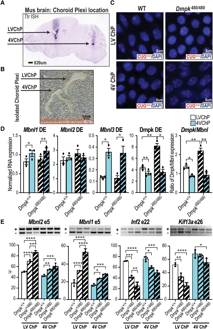
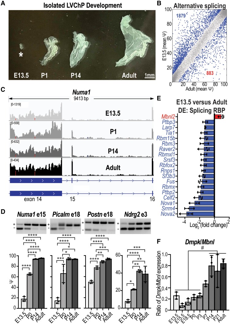
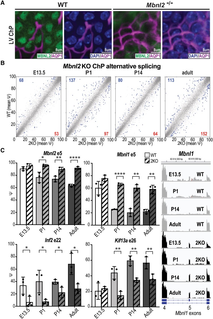
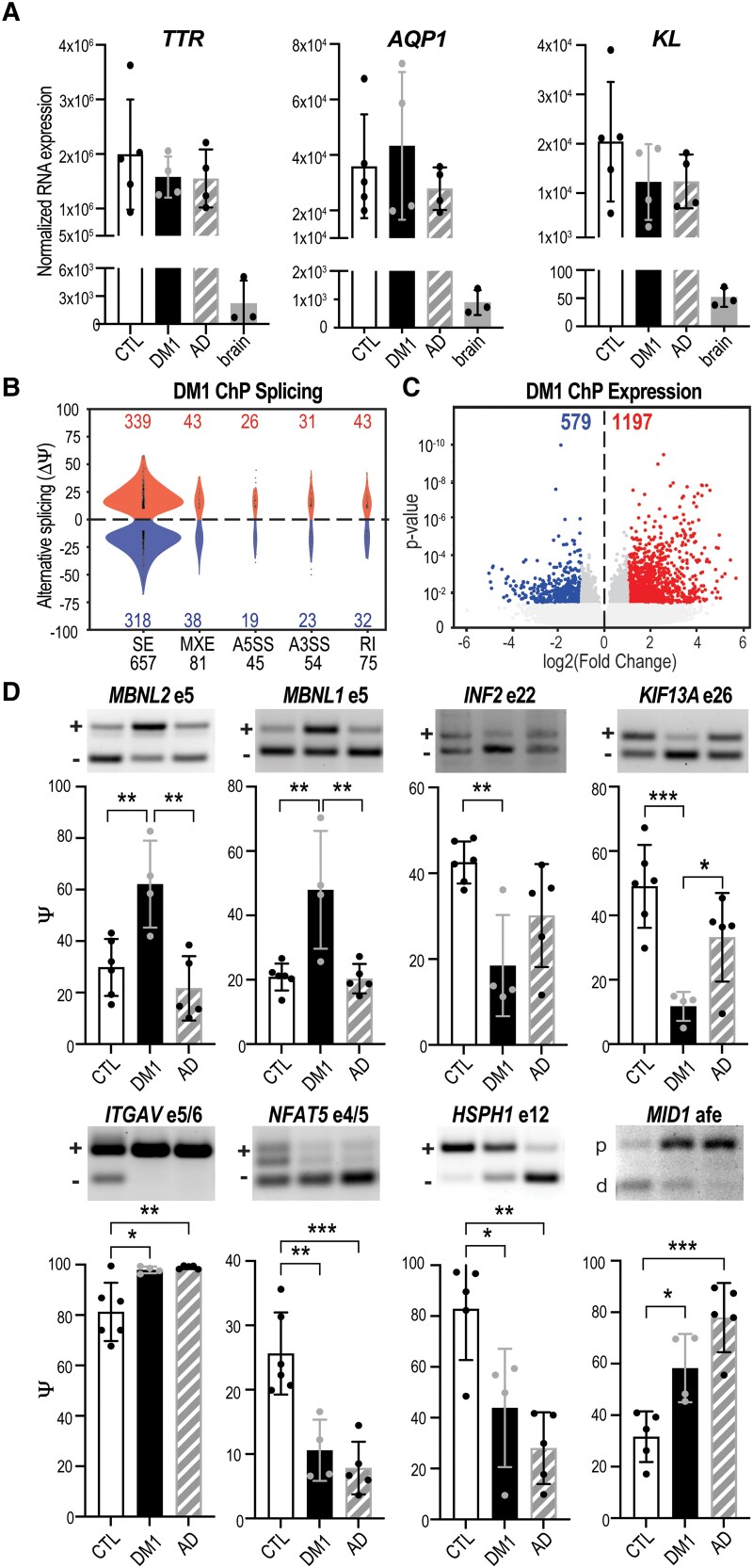
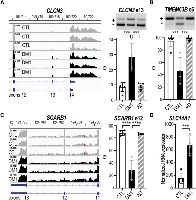
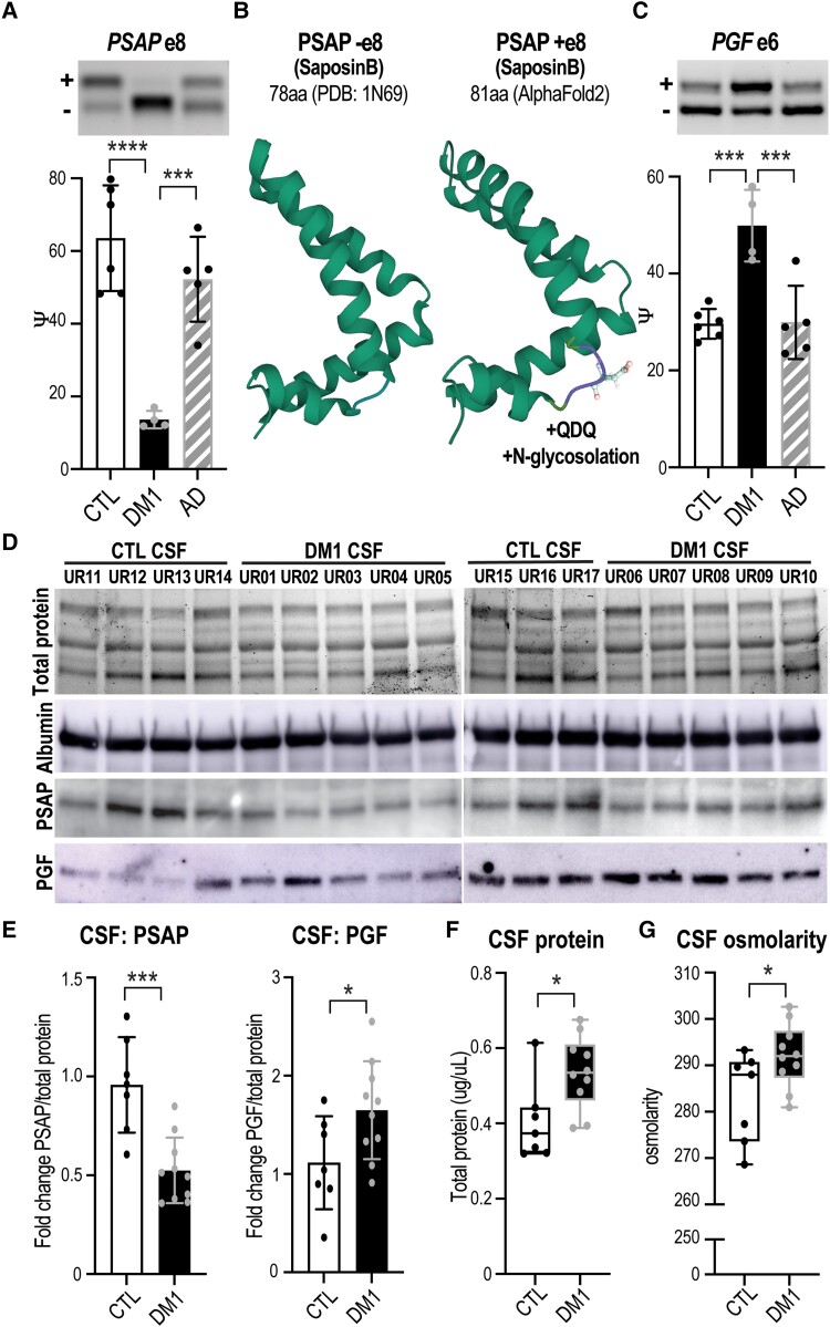
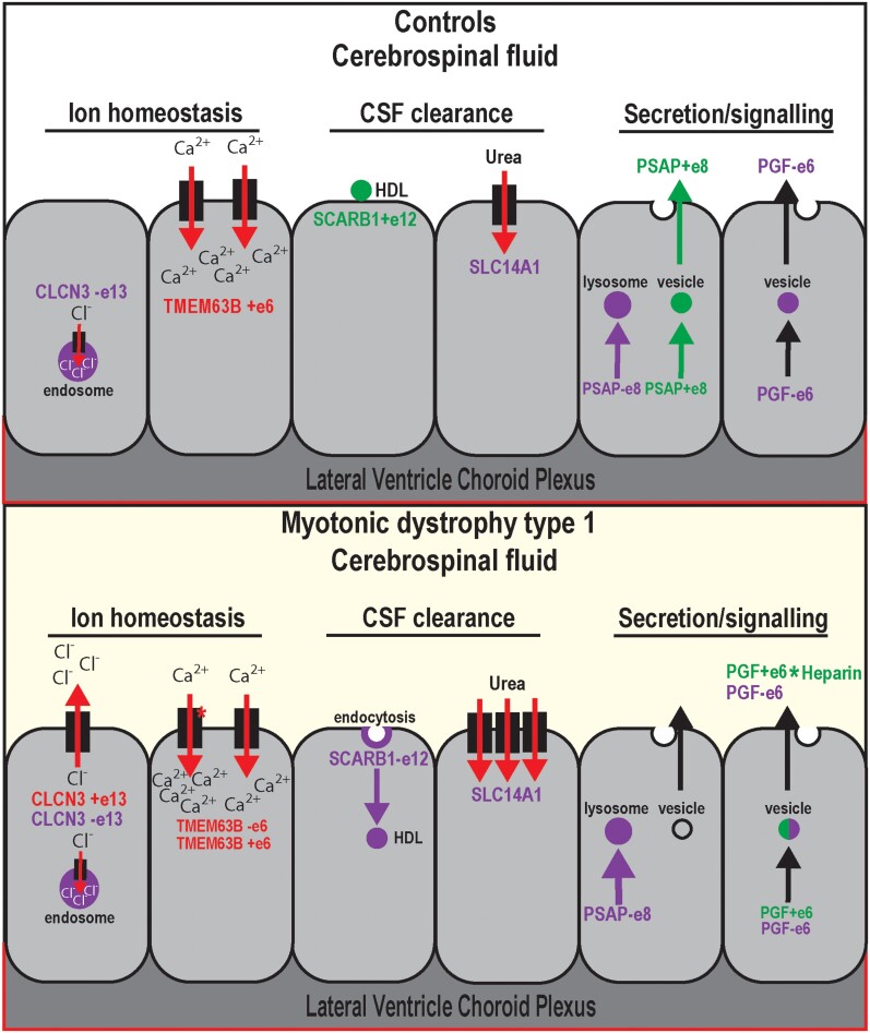
Myotonic dystrophy type 1 is a dominantly inherited multisystemic disease caused by CTG tandem repeat expansions in the DMPK 3' untranslated region. These expanded repeats are transcribed and produce toxic CUG RNAs that sequester and inhibit activities of the MBNL family of developmental RNA processing factors. Although myotonic dystrophy is classified as a muscular dystrophy, the brain is also severely affected by an unusual cohort of symptoms, including hypersomnia, executive dysfunction, as well as early onsets of tau/MAPT pathology and cerebral atrophy. To address the molecular and cellular events that lead to these pathological outcomes, we recently generated a mouse Dmpk CTG expansion knock-in model and identified choroid plexus epithelial cells as particularly affected by the expression of toxic CUG expansion RNAs. To determine if toxic CUG RNAs perturb choroid plexus functions, alternative splicing analysis was performed on lateral and hindbrain choroid plexi from Dmpk CTG knock-in mice. Choroid plexus transcriptome-wide changes were evaluated in Mbnl2 knockout mice, a developmental-onset model of myotonic dystrophy brain dysfunction. To determine if transcriptome changes also occurred in the human disease, we obtained post-mortem choroid plexus for RNA-seq from neurologically unaffected (two females, three males; ages 50-70 years) and myotonic dystrophy type 1 (one female, three males; ages 50-70 years) donors. To test that choroid plexus transcriptome alterations resulted in altered CSF composition, we obtained CSF via lumbar puncture from patients with myotonic dystrophy type 1 (five females, five males; ages 35-55 years) and non-myotonic dystrophy patients (three females, four males; ages 26-51 years), and western blot and osmolarity analyses were used to test CSF alterations predicted by choroid plexus transcriptome analysis. We determined that CUG RNA induced toxicity was more robust in the lateral choroid plexus of Dmpk CTG knock-in mice due to comparatively higher Dmpk and lower Mbnl RNA levels. Impaired transitions to adult splicing patterns during choroid plexus development were identified in Mbnl2 knockout mice, including mis-splicing previously found in Dmpk CTG knock-in mice. Whole transcriptome analysis of myotonic dystrophy type 1 choroid plexus revealed disease-associated RNA expression and mis-splicing events. Based on these RNA changes, predicted alterations in ion homeostasis, secretory output and CSF composition were confirmed by analysis of myotonic dystrophy type 1 CSF. Our results implicate choroid plexus spliceopathy and concomitant alterations in CSF homeostasis as an unappreciated contributor to myotonic dystrophy type 1 CNS pathogenesis.

Keywords: RNA disease; alternative splicing; development; neurodegeneration; short tandem repeat expansion.


Conflict of interest statement

M.S.S. is a Scientific Advisory Board member for Kate Therapeutics, Skyhawk Therapeutics and Tacit Therapeutics.

Figures

Figure 1
Figure 1
LVChP spliceopathy in Dmpk CTGexp KI mice. (A) In situ hybridization (dark stain) showing localization of LVChP and 4VChP in a sagittal brain section from an adult C57BL6 mouse using Ttr expression as the ChP marker (Allen Brain Atlas Mouse database). Scale = 839 μm. (B) Morphological comparison of isolated LVChP and 4VChP. Scale = 1 mm. (C) RNA-FISH of CUG repeat expansion (CUGexp) showing RNA foci (red) in the nucleus (blue, DAPI) of adult Dmpk+/+ FVB control versus Dmpk480/480 LVChP and 4VChP. Scale = 8 μm. WT = wild-type. (D) Mbnl1, Mbnl2, Mbnl3, and Dmpk RNA expression by RT-qPCR in LVChP (white) and 4VChP (blue) isolated from wild-type FVB littermate mice and Dmpk480/480 KI (thick crosshatch). Normalized RNA expression is based on the geometric mean of three housekeeping genes (Gorasp1, Psmb4, Rpl38) while the ratio of Dmpk/Mbnl expression was calculated as the normalized expression of Dmpk/(Mbnl1 + Mbnl2 + Mbnl3). (E) RT-PCR gels (representative top) and analysis (graph bottom) of myotonic dystrophy associated AS events (Mbnl2 exon 5, Mbnl1 exon 5, Inf2 exon 22, Kif13a exon 26) from LVChP (white) and 4VChP (blue) isolated from adult littermate Dmpk+/+ wild-type FVB (outlined), Dmpk+/480 KI (thin crosshatch), and Dmpk480/480 KI (thick crosshatch) mice *P < 0.05, **P < 0.01, ***P < 0.001, ****P < 0.0001, one-way ANOVA or paired t-test. 4V ChP = fourth ventricle choroid plexus; DE = differential expression; LV ChP = lateral ventricle choroid plexus.
Figure 2
Figure 2
Alternative splicing regulation during ChP development. (A) Developmental stage- (E13.5, P1, P14, adult) specific variations in size and morphology of LVChP. Scale = 1 mm. (B) Plot of AS events (Ψ) in wild-type FVB mice ChP comparing E13.5 to adult per cent spliced in (PSI) with the significant events in blue. (C) Numa1 exon 15 AS RNA-seq reads during development (E13.5, P0, P14, adult, light grey to black gradient). (D) RT-PCR gels (representative) and analysis (bar graph) validations for E13.5, P0, P14 and adult stages of Numa1 exon 15, Picalm exon 18, Postn exon 18 and Ndrg2 exon 3. (E) In contrast to other splicing factors, Mbnl2 expression increases during postnatal development. Relative expression (RNA-seq) is plotted for significant differentially expressed genes during LVChP development. (F) The ratio of Dmpk/Mbnl expression was calculated as the normalized expression of Dmpk/(Mbnl1 + Mbnl2 + Mbnl3) for each developmental stage. #Significance of at least P < 0.05 for all the compared stages, *P < 0.05, **P < 0.01, ***P < 0.001, ****P < 0.0001, one-way ANOVA. LVChP = lateral ventricle choroid plexus.
Figure 3
Figure 3
MBNL2 promotes adult splicing events during ChP development. (A) MBNL2 is the major MBNL paralogue in ChP. Immunofluorescence of nuclear MBNL2 (green) and the choroid plexus (ChP) apical membrane marker AQP1 (magenta) in the LVChP of wild-type (WT) (left) versus Mbnl2 KO (right) mice. Localization of AQP1 and DAPI (blue, nucleus). Scale = 8 μm. (B) LVChP RNA splicing events (Ψ) in Mbnl2−/− (Mbnl2 KO) compared to wild-type FVB (WT) littermate mice at E13.5, P1, P14 and adult (P63), with significant events in blue. (C) LVChP AS changes by RNA-seq in Mbnl2 KO compared to FVB WT littermate mice at developmental stages E13.5, P1, P14 and P63 (gradient of light to dark grey with crosshatch for Mbnl2KO) for Mbnl2 exon 5, Mbnl1 exon 5, Inf2 exon 22 and Kif13a exon 26. Right: Mbnl1 exon 5 AS RNA-seq reads during development (E13.5, P0, P14, adult) for FVB WT littermates (light grey) compared to Mbnl2 KO (black). *P < 0.05, **P < 0.01, ****P < 0.0001, unpaired t-test.
Figure 4
Figure 4
ChP spliceopathy in DM1. (A) Normalized RNA expression (RNA-seq) for choroid plexus (ChP) markers TTR, AQP1 and KL for neurologically unaffected controls (CTL), myotonic dystrophy type 1 (DM1) and Alzheimer’s disease (AD), together with non-ChP brain tissues (brain). (B) Violin plots of AS events (Ψ) quantified by change in per cent spliced in (ΔPSI). A5SS = alternative 5′ splice site; A3SS = alternative 3′ splice site; MXE = mutually exclusive exon; SE = skipped exon. (C) DE of RNA transcripts showing the log2 transformed fold change plotted against significance (P-value). (D) RT-PCR validations of splicing changes in DM1 ChP compared to CTL and AD for MBNL2 exon 5, MBNL1 exon 5, INF2 exon 22, KIF13A exon 26, ITGAV exons 5 and 6, NFAT5 exons 4 and 5, HSPH1 exon 12 and MID1 alternative first exon (afe). *P < 0.05, **P < 0.01, ***P < 0.001, one-way ANOVA.
Figure 5
Figure 5
ChP mis-splicing alters transporters and ion channels. Neurologically unaffected (CTL) versus myotonic dystrophy type 1 (DM1) LVChP transcriptomic analysis of alternative splicing (AS) and differential expression (DE) was screened for either ‘ion channel’ or ‘transporter’ candidates. (A) CLCN3 was identified as a top ion channel candidate by RNA-seq (left) and validated (right) by RT-PCR gel (representative) and analysis (bar graph) of CLCN3 exon 13 (e13) AS in DM1 versus CTL and Alzheimer’s disease (AD) LVChP. (B) TMEM63B was also identified from RNA-seq and validated by RT-PCR for AS of exon 6. (C) SCARB1 was identified as a top transporter candidate and RNA-seq revealed SCARB1 exon 12 AS for DM1 versus CTL and AD LVChP. (D) Another transporter, SLC14A1, exhibited increased DE in DM1 versus CTL patients’ ChP. ***P < 0.001, ****P < 0.0001, one-way ANOVA or unpaired t-test. LVChP = lateral ventricle choroid plexus.
Figure 6
Figure 6
Mis-splicing alters the ChP secretome and CSF composition. Neurologically unaffected (CTL) versus DM1 LVChP transcriptomic analysis of alternative splicing (AS) and differential expression (DE) was screened for ‘secreted’ GO annotation candidates. (A) PSAP was selected as a top secreted candidate and validated by RT-PCR gel (representative, top) and analysis (bar graph, bottom), which showed exclusion of exon 8 in DM1 ChP. (B) Predicted (AlphaFold2) protein structural changes due to PSAP exon 8 AS with the inclusion of a +QDQ (glutamine-aspartate-glutamine) in PSAP region SaposinB (PDB: 1N69). aa = amino acids. (C) Another top secreted candidate was PGF, which was validated by RT-PCR for increased exon 6 inclusion in DM1. (D) To test if ChP changes correlate with CSF composition, we obtained DM1 and non-DM1 (CTL) patient CSF for protein lysates and assayed top candidates, PSAP and PGF. Loading was normalized based on protein quantification, total protein signal and albumin western blot. (E) Western blot of PSAP and PGF proteins in DM1 patients’ CSF compared to CTLs was quantified as fold-change normalized over total protein signal. (F and G) To test if ChP changes also correlate with cumulative measures of CSF composition, CTL versus DM1 patients’ CSF was assayed for (F) total protein as well as (G) total osmolarity. *P < 0.05, ***P < 0.001, ****P < 0.0001, one-way ANOVA or unpaired t-test.
Figure 7
Figure 7
Choroid plexus mis-splicing correlates with CSF alterations in DM1. Proposed model of non-DM1 (unaffected and disease controls) and DM1 ChP AS of CLCN3 exon 13, TMEM63B exon 6, SCARB1 exon 12, SLC14A1 DE, PSAP exon 8 and PGF exon 6, as well as potential influences on CSF content. Both CLCN3 and TMEM63B are involved in ChP regulation of ion homeostasis. CLCN3 exon 13 is associated with a localization switch from endosomal to plasma membrane, leading to a gain in outward rectifying chloride ion flow. TMEM63B exon 6 loss is associated with a switch to a more osmosensitive isoform of the calcium permeable ion channel. Both SCARB1 and SLC14A1 are involved in clearance of the CSF. SCARB1 exon 12 loss is associated with a switch to isoform SR-BII, which has been shown to promote endocytosis of high-density lipoprotein (HDL) particles. SLC14A1 encodes urea transporter B, the primary transport protein for urea in the CNS, whose increased expression has been reported in neurodegenerative diseases. Both PSAP and PGF are highly expressed secretory proteins produced by the ChP to modulate signalling throughout the CSF. PSAP exon 8 skipping is associated with a switch from trafficking of the protein to vesicles for secretion to primarily lysosomal for non-signalling functions. PGF exon 6 splicing is associated with a change in heparin binding activity and thus altered distribution gradients.

References

    1. Vuong CK, Black DL, Zheng S. The neurogenetics of alternative splicing. Nat Rev Neurosci. 2016;17:265–281. - PMC - PubMed
    1. Manning KS, Cooper TA. The roles of RNA processing in translating genotype to phenotype. Nat Rev Mol Cell Biol. 2017;18:102–114. - PMC - PubMed
    1. Scotti MM, Swanson MS. RNA mis-splicing in disease. Nat Rev Genet. 2016;17:19–32. - PMC - PubMed
    1. Weskamp K, Olwin BB, Parker R. Post-transcriptional regulation in skeletal muscle development, repair, and disease. Trends Mol Med. 2021;27:469–481. - PubMed
    1. Brook JD, McCurrach ME, Harley HG, et al. Molecular basis of myotonic dystrophy: Expansion of a trinucleotide (CTG) repeat at the 3’ end of a transcript encoding a protein kinase family member. Cell. 1992;68:799–808. - PubMed

Publication types