

Case Series - Osteochondromas at Rare Locations
- PMID: 37143552
- PMCID: PMC10152955
- DOI: 10.13107/jocr.2023.v13.i01.3522
Case Series - Osteochondromas at Rare Locations
Abstract
Introduction: Osteochondromas are very common. They are typically seen in long bones and rarely seen in smaller bones. Some of the rare presentations include flat bones, the body of pelvis, scapula, skull, and small bones of the hand and foot. Their presentation also varies according to the site of presentation.
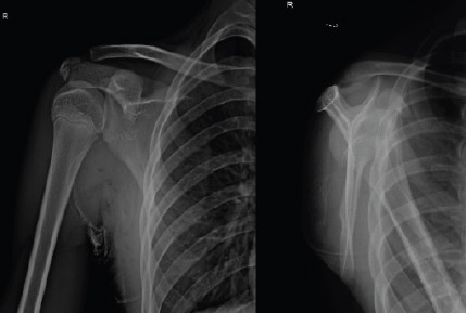
Case report: We have included five cases of osteochondromas occurring at rare locations with variable presentations and their management. We have included one case of metacarpal, one case of skull exostosis, two cases of scapula exostosis, and one case of fibula exostosis.
Conclusion: Osteochondromas can rarely occur at unusual locations. It is important to thoroughly evaluate all patients presenting with swelling and pain over bony regions to accurately diagnose osteochondromas and manage accordingly.
Keywords: Scapula; excision; fibula; metacarpal; skull.
Copyright: © Indian Orthopaedic Research Group.
Conflict of interest statement
Conflict of Interest: Nil
Figures


References
-
- Dahlin DC. General Aspects and Data on ⅞Cases. 4th ed. United States: Charles C Thomas Pub Ltd; 1986.
-
- Greenspan A, Jundt G, Remagen W, editors. Differential Diagnosis in Orthopaedic Oncology. 2nd ed. Philadelphia, PA: Lippincot and Williams; 2006.
-
- Calafiore G, Calafiore G, Bertone C, Urgelli S, Rivera F, Maniscalco P. Osteochondroma:Report of a case with atypical localization and symptomatology. Acta Biomed Ateneo Parmense. 2001;72:91–6. - PubMed
-
- Gökkuş K, Atmaca H, Sağtaş E, Saylık M, Aydın AT. Osteochondromas originating from unusual locations complicating orthopedic discipline:Case series. Eklem Hastalik Cerrahisi. 2015;26:100–9. - PubMed
-
- Gökkuş K, Aydın AT, Sağtaş E. Solitary osteochondroma of ischial ramus causing sciatic nerve compression. Eklem Hastalik Cerrahisi. 2013;24:49–52. - PubMed
Publication types
LinkOut - more resources
Full Text Sources