Review
. 2023 May 2;4(3):e261.
doi: 10.1002/mco2.261. eCollection 2023 Jun.

Protein posttranslational modifications in health and diseases: Functions, regulatory mechanisms, and therapeutic implications

Affiliations
Review

Protein posttranslational modifications in health and diseases: Functions, regulatory mechanisms, and therapeutic implications

Qian Zhong et al. MedComm (2020). .

Abstract

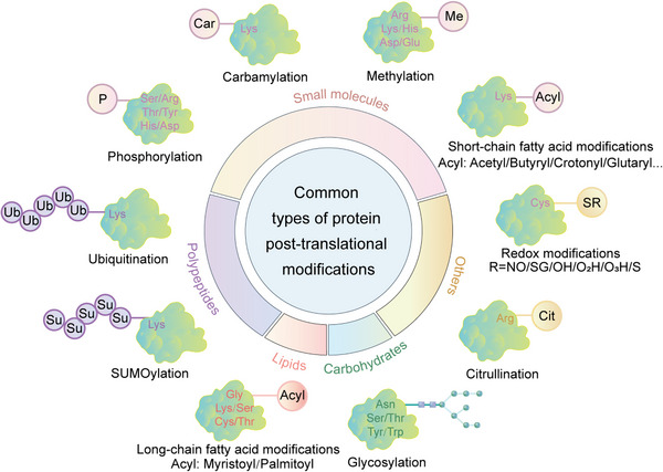
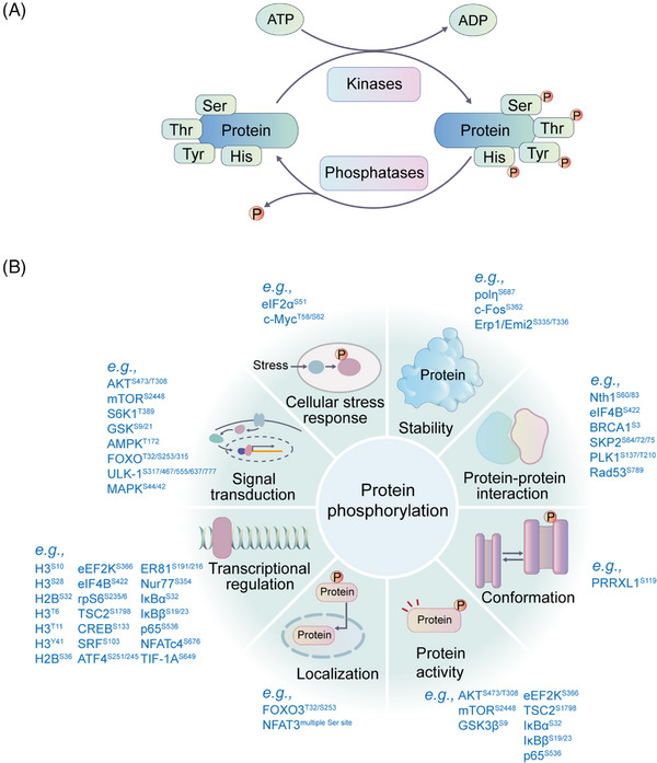
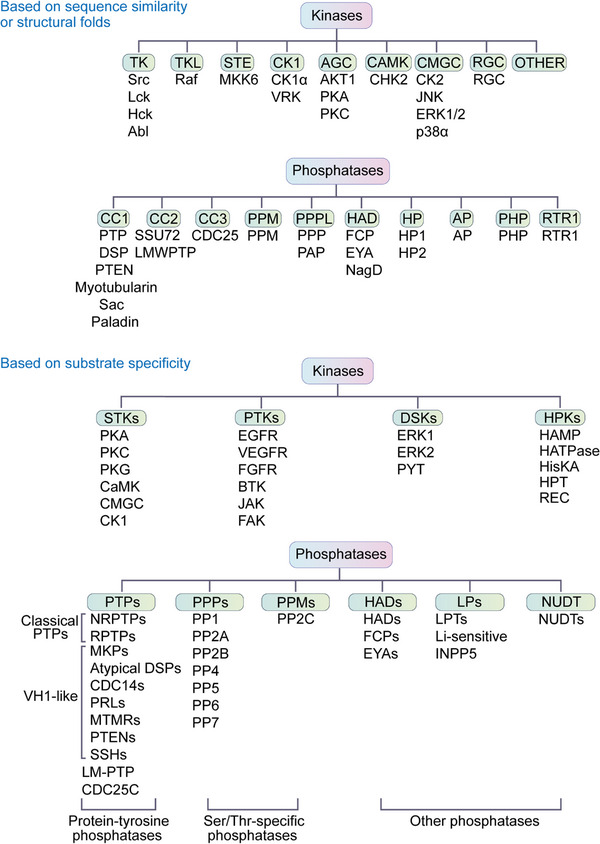
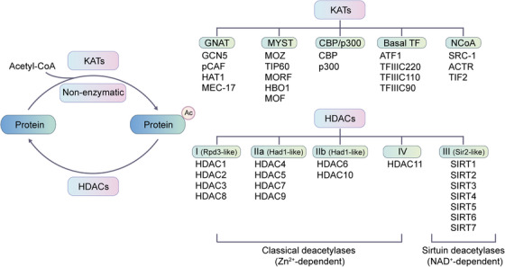
Protein posttranslational modifications (PTMs) refer to the breaking or generation of covalent bonds on the backbones or amino acid side chains of proteins and expand the diversity of proteins, which provides the basis for the emergence of organismal complexity. To date, more than 650 types of protein modifications, such as the most well-known phosphorylation, ubiquitination, glycosylation, methylation, SUMOylation, short-chain and long-chain acylation modifications, redox modifications, and irreversible modifications, have been described, and the inventory is still increasing. By changing the protein conformation, localization, activity, stability, charges, and interactions with other biomolecules, PTMs ultimately alter the phenotypes and biological processes of cells. The homeostasis of protein modifications is important to human health. Abnormal PTMs may cause changes in protein properties and loss of protein functions, which are closely related to the occurrence and development of various diseases. In this review, we systematically introduce the characteristics, regulatory mechanisms, and functions of various PTMs in health and diseases. In addition, the therapeutic prospects in various diseases by targeting PTMs and associated regulatory enzymes are also summarized. This work will deepen the understanding of protein modifications in health and diseases and promote the discovery of diagnostic and prognostic markers and drug targets for diseases.

Keywords: aging; cancers; metabolic diseases; neurodegenerative diseases; protein posttranslational modifications; targeted therapy.


Conflict of interest statement

The authors declare no competing interests.

Figures

FIGURE 1
FIGURE 1
Common types of protein posttranslational modifications. Small molecules, lipids, carbohydrates, and polypeptides can be added to amino acid side chains to form modifications. In addition, changes in the chemical properties of amino acids are also common modifications, such as citrullination.
FIGURE 2
FIGURE 2
The phosphorylation process and the potential functions of phosphorylation. (A) The phosphorylation and dephosphorylation process. Phosphorylation is catalyzed by kinases, and dephosphorylation is mediated by phosphatases. Most phosphorylation events occur on serine, threonine, and tyrosine residues. (B) Representative functions of protein phosphorylation are shown. Protein phosphorylation extensively affects cellular signal transduction, protein stability, activity, localization, conformation, protein–protein interactions, gene transcription, and so on. Representative phosphosites with related functions are shown.
FIGURE 3
FIGURE 3
The kinases and phosphatases can be classified into different groups based on their phosphorylation substrates or based on their sequence similarity.
FIGURE 4
FIGURE 4
Representative scheme of reversible acetylation regulated by HATs and HDACs is shown. The classification of well‐known HATs and HDACs are organized. KATs are classified into three major families: GCN5, p300 and MYST. The remaining KATs belong to basal TF family and NCoA family. HDACs are divided into two categories: the classical Zn2+‐dependent HDACs and NAD+‐dependent sirtuin deacetylases. HDACs can be further grouped into class I, class Iia, class Iib, class III, and class IV.
FIGURE 5
FIGURE 5
Representative functions of histone and nonhistone acetylation are shown. Protein acetylation is mainly involved in the regulation of gene transcription, metabolism, DNA damage repair, signal transduction, stress response, signal transduction, protein stability, protein activity, protein–protein interaction, and autophagy.
FIGURE 6
FIGURE 6
Chemical structures and representative biological functions of SCFA‐derived lysine acylation modifications, including propionylation (Kpr), butyrylation (Kbu), succinylation (Ksucc), 2‐hydroxyisobutyrylation (Khib), isobutyrylation (Kisobu), malonylation (Kmal), glutarylation (Kglu), crotonylation (Kcr), β‐hydroxybutyrylation (Kbhb), and lactylation (Kla).
FIGURE 7
FIGURE 7
Distribution of reported lysine acylation modifications on histones.
FIGURE 8
FIGURE 8
Origin and regulation of SCFA. SCFA‐derived lysine acylation modifications. The SCFA is converted into the corresponding acyl‐CoA in the presence of enzymes such as ACCS2. The acyl groups can be transferred onto proteins to modify the side chain of lysine residues by HATs. In addition, the acylated proteins can be deacylated by HDACs and sirtuins.
FIGURE 9
FIGURE 9
The functions of SCFA‐derived acylation modifications in various human diseases. Representative examples are shown.
FIGURE 10
FIGURE 10
The process of S‐palmitoylation and N‐myristoylation. (A) The S‐palmitoylation and depalmitoylation process. Step1, palmitoyl acyltransferases (PATs) undergo auto‐palmitoylation, and the palmitoyl group is transferred to PAT; Step2, the palmitoyl group is transferred from PAT to protein substrates. (B) The process and functions of N‐myristoylation. Myristic acid and coenzyme A are converted to myristic acid coenzyme A by acetyl‐CoA synthetase. If the starting amino acid of the protein is methionine, it needs to be removed by methionyl aminopeptidase (MetAP2) before N‐myristoylation. N‐myristoyltransferase (NMT) is responsible for the addition of the myristoyl group to the glycine residue at the N‐terminal of the protein.
FIGURE 11
FIGURE 11
The functions of LCFA‐derived acylation modifications in various diseases, including palmitoylation and myristoylation. Representative examples are shown.
FIGURE 12
FIGURE 12
The methylation and demethylation process. (A) Lysine residues undergo mono‐, di‐ or trimethylation through the addition of a methyl group to its side chain. (B) There are three types of methylation occurring at the side chain of arginine, including monomethylated arginine (MMA), asymmetric dimethylated arginine (ADMA), symmetric dimethylated arginine (SDMA). (C) The common lysine methyltransferases and demethylases are listed.
FIGURE 13
FIGURE 13
Protein methylation on histone H3 and H4 and their regulatory enzymes. The same residues can be regulated by multiple methylases and demethylases.
FIGURE 14
FIGURE 14
Functions of protein methylation. Representative methylation substrates are presented. Protein methylation is extensively involved in signal transduction, protein stability, protein activity, protein–protein interaction, mRNA splicing, transcriptional regulation, DNA damage repair, nuclear, and cytoplasmic shuttling.
FIGURE 15
FIGURE 15
The protein ubiquitination pathway. The ubiquitin (Ub) moiety is activated by E1 through the cysteine (Cys) residue of E1. Ub at E1 is transferred to the Cys residue of E2. Ub conjugated with E2 is transferred to the lysine residue of a substrate protein by E3. Ubiquitinated proteins with ubiquitination are subjected to 26S proteasome‐dependent degradation or execute other functions and activities. Deubiquitination is the opposite mechanism of ubiquitination mediated by DUBs. The process includes reversing ubiquitin conjugation and recycling ubiquitin molecules through the UPS. Based on the enzymatic cleavage mechanism, the DUB family is divided into two subfamilies. The cysteine protease family consists of USP, UCH, OTH, MJD, MINDY, MCPIP, and ZUFSP. Metalloprotease family includes JAMM.
FIGURE 16
FIGURE 16
Ubiquitin linkage types and their roles. Ubiquitination can occur as single or multiple monoubiquitin or as homotypic/heterotypic/branched chains linked through K6, K11, K27, K29, K33, K48, or K63, as well as M1. The functional consequences of ubiquitin signals are determined by cellular and substrate‐context, ubiquitin chain position, linkage type and conformation, ranging from proteasomal degradation to nonproteolytic functions.
FIGURE 17
FIGURE 17
SUMOylation process and its functions. (A) The catalytic cycle of SUMOylation. SENP have endopeptidase activity to cleave SUMO precursors by exposing the carboxy‐terminal diglycine motifs essential for the conjugation to lysine residues in target proteins. SUMOylation is catalyzed by SAE1‐SAE2 (E1) and UBC9 (E2). The E3 ligases can facilitate the last step of SUMO conjugation. SENPs isopeptidase activity allows for the release of SUMO from target proteins. (B) Functions of SUMOylation. SUMOylation is extensively involved in signal transduction, and the regulation of protein activity, protein stability, protein–protein interaction, cell apoptosis, translocation, and DNA damage repair. Representative SUMOylation proteins and substrates are shown.
FIGURE 18
FIGURE 18
Overview of human N‐ and O‐glycosylation in the ER and Golgi apparatus. On the right side, the biosynthesis of complex‐type N‐glycans is shown. On the left side, the biosynthesis O‐glycosylation is shown.
FIGURE 19
FIGURE 19
Functions of glycosylation in health and diseases, such as congenital diseases, immune regulation, neurodegenerative diseases, IBD, cancers, aging, and infectious diseases.
FIGURE 20
FIGURE 20
The schematic reaction for citrullination regulated by PADs. (A) Cartoon depicting PAD‐mediated conversion of arginine to citrulline. (B) Table listing the tissue distribution of PADs and their representative citrullination substrates.
FIGURE 21
FIGURE 21
Representative citrullination substrates and their functions in various diseases, such as RA, cancer, MS, and SLE.
FIGURE 22
FIGURE 22
The different pathways of carbamylation and its roles in several diseases. (A) There are two main pathways for the production of isocyanic acid for carbamylation. (B) Representative protein substrates and roles of carbamylation in various diseases.
FIGURE 23
FIGURE 23
Redox modifications of cysteine. (A) Different‐types‐of‐redox modifications of cysteine. (B–E) The mechanisms of different types of redox modifications and associated physiological functions.
FIGURE 24
FIGURE 24
Representative redox modification events in aging, metabolic disorders, neurological disorders, cancers, and CVDs are shown.

References

    1. Millan‐Zambrano G, Burton A, Bannister AJ, Schneider R. Histone post‐translational modifications: cause and consequence of genome function. Nat Rev Genet. 2022;23(9):563‐580. - PubMed
    1. Li W, Li F, Zhang X, Lin H‐K, Xu C. Insights into the post‐translational modification and its emerging role in shaping the tumor microenvironment. Signal Transduct Target Ther. 2021;6(1):422. - PMC - PubMed
    1. Müller MM. Post‐translational modifications of protein backbones: unique functions, mechanisms, and challenges. Biochemistry. 2018;57(2):177‐185. - PMC - PubMed
    1. Keenan EK, Zachman DK, Hirschey MD. Discovering the landscape of protein modifications. Mol Cell. 2021;81(9):1868‐1878. - PMC - PubMed
    1. Prabakaran S, Lippens G, Steen H, Gunawardena J. Post‐translational modification: nature's escape from genetic imprisonment and the basis for dynamic information encoding. Wiley Interdiscip Rev Syst Biol Med. 2012;4(6):565‐583. - PMC - PubMed