. 2023 Apr 18:14:1163066.
doi: 10.3389/fimmu.2023.1163066. eCollection 2023.

Dsg3 epitope-specific signalling in pemphigus

Affiliations

Dsg3 epitope-specific signalling in pemphigus

Thomas Schmitt et al. Front Immunol. .

Abstract

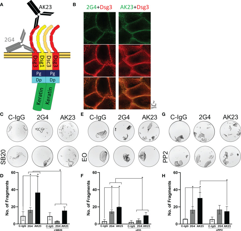
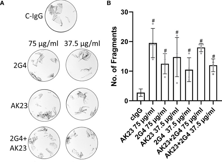
Introduction: Pemphigus is an autoantibody driven disease that impairs the barrier function of the skin and mucosa by disrupting desmosomes and thereby impeding cellular cohesion. It is known that the different clinical phenotypes of pemphigus vulgaris (PV) and pemphigus foliaceus (PF) are dependent on the autoantibody profile and target antigens that, amongst others, are primarily desmoglein (Dsg)1 and/or Dsg3 for PV and Dsg1 for PF. However, it was reported that autoantibodiesagainst different epitopes of Dsg1 and Dsg3 can be pathogenic or not. The underlying mechanisms are very complex and involve both direct inhibition of Dsg interactions and downstream signalling. The aim of this study was to find out whether there is target-epitope-specific Dsg3 signalling by comparing the effects of the two pathogenic murine IgGs, 2G4 and AK23.

Methods: Dispase-based dissociation assay, Western Blot analysis, Stimulated emission depletion microscopy, Fura-based Ca2+ flux measurements, Rho/Rac G-Protein-linked immunosorbent assay, Enzyme-linked immunosorbent assay.

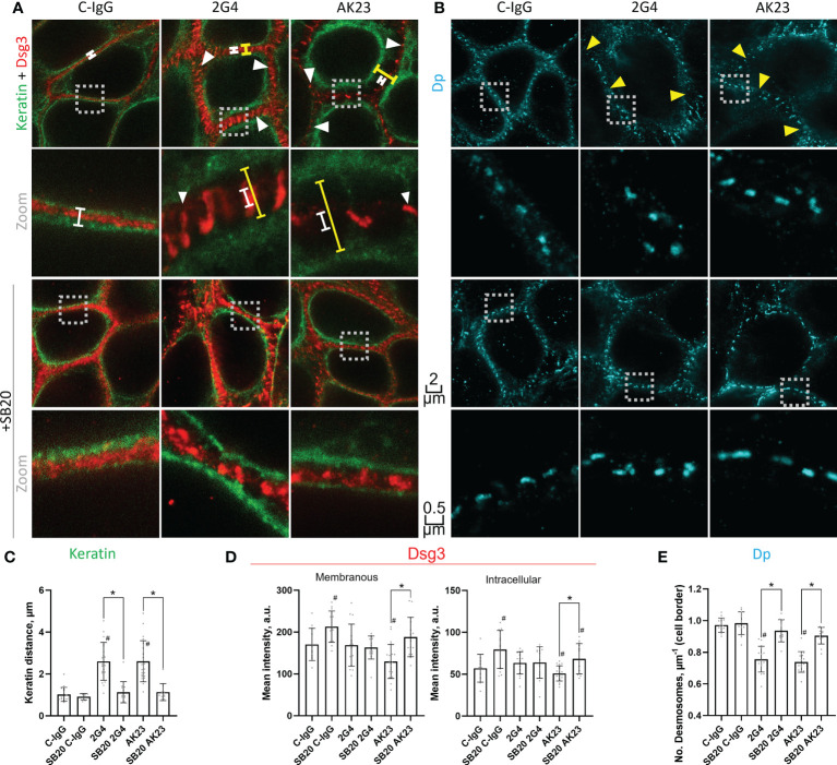
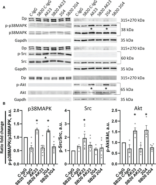
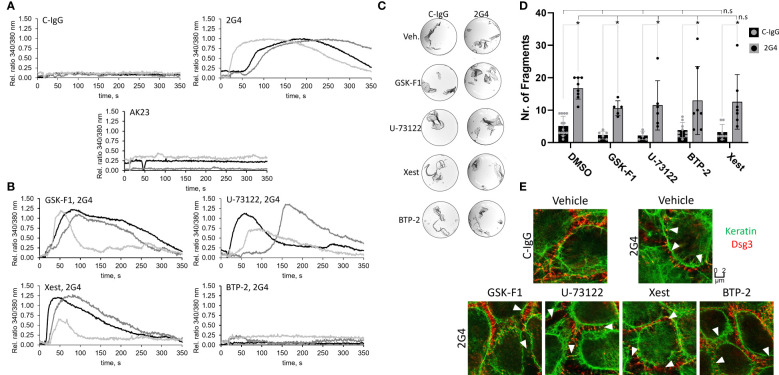
Results: The IgGs are directed against the EC5 and EC1 domain of Dsg3, respectively. The data show that 2G4 was less effective in causing loss of cell adhesion, compared to AK23. STED imaging revealed that both autoantibodies had similar effects on keratin retraction and reduction of desmosome number whereas only AK23 induced Dsg3 depletion. Moreover, both antibodies induced phosphorylation of p38MAPK and Akt whereas Src was phosphorylated upon treatment with AK23 only. Interestingly, Src and Akt activation were p38MAPK-dependent. All pathogenic effects were rescued by p38MAPK inhibition and AK23-mediated effects were also ameliorated by Src inhibition.

Discussion: The results give first insights into pemphigus autoantibody-induced Dsg3 epitope-specific signalling which is involved in pathogenic events such as Dsg3 depletion.

Keywords: adhesion; autoimmune disease; desmoglein (dsg); desmosomes; epidermis; keratin; pemphigus; skin.


Conflict of interest statement

The authors declare that the research was conducted in the absence of any commercial or financial relationships that could be construed as a potential conflict of interest.

Figures

Figure 1
Figure 1
2G4 is a pathogenic Dsg3 specific IgG. (A) Schematic depiction of binding positions of AK23 (Black), directed against the EC1 domain and 2G4 (Grey) directed against the EC5 domain of Dsg3, on a desmosome-bound Dsg3. (B) Co-staining of either 2G4 or AK23 (green) with Dsg3 (intracellular domain, red) and an overlay of both channels in HaCaT cells after 24 hours incubation with the IgGs (1:50), imaged with the STED microscope (N=4). (C) Representative images of dispase-based dissociation assay results in HaCaT cells. Vehicle control (left) pre-treated with SB20 (60 µM, inhibiting p38MAPK, right). (D) Quantification of dispase-based dissociation assay results using SB20 (N=4). (E) Representative images of dispase-based dissociation assay results in HaCaT cells. Vehicle control (left) pre-treated with EO (40 nM, inhibiting p38MAPK, right). (F) Quantification of dispase-based dissociation assay results using EO (N=4). (G) Representative images of dispase-based dissociation assay results in HaCaT cells. Vehicle control (left) pre-treated with PP2 (10 µM, inhibiting Src, right). (H) Quantification of dispase-based dissociation assay results using PP2 (N=4). * indicates statistically significant differences in two-way-ANOVA with Sidaks correction for multiple comparisons p<0.05.
Figure 2
Figure 2
(A) Representative images of dispase-based dissociation assay results in HaCaT cells. Control, treated with C-IgG only (top), treated with either single 2G4, AK23 or a mixture of both at a concentration of 75 µg/ml eacht (left) and at half concentration of 37.5 µg/ml right. (B) Quantification of dispase-based dissociation assay (N=4). # indicates statistically significant differences towards control conditions, one-way-ANOVA with Dunett correction for multiple comparisons, p<0.05.
Figure 3
Figure 3
STED microscopy images reveal specific pathogenic effects of 2G4 and AK23. (A) Co-staining of Dsg3 (red) and pan-cytokeratin (green) in HaCaT cells treated with either C-IgG, AK23 or 2G4 for 24 h after 1 h preincubation with vehicle or SB20 (60 µM, inhibiting p38MAPK). Spans illustrate the degree of keratin filament retraction (yellow) compared to control (white). White arrowheads showcase the fragmented staining of Dsg3, which is mostly associated with keratin filaments. (B) Staining of Dp (with anti-Dp-pAb, A7169) after 24 h treatment with IgGs and 1 h preincubation with vehicle or SB20. Yellow arrowheads indicate reduced numbers of desmosomes per µm of cell border. Two different magnifications are shown for each condition (Scalebars as indicated, boxes with dotted outline indicate the area which was zoomed in on); Quantification of the STED images: (C) Keratin filament retraction form the cell borders towards the nucleus. (D) Dsg3 staining intensity at the cell borders/membranes (left) or in the cytoplasm (right). (E) Number of desmosomes per µm membrane (Experiments (N)=4-6, Evaluated images (n)=12-22). * marks statistically significant differences between the two conditions indicated, # indicates statistically significant differences towards control conditions, both in two-way-ANOVA with Sidaks correction for multiple comparisons, p<0.05.
Figure 4
Figure 4
Western blot analysis reveals anti-Dsg3 epitope-specific kinase phosphorylation. (A) Representative Western blot images, with respective controls for Triton-soluble (membranous+cytosolic fraction, right) Gapdh and Triton-insoluble (cytoskeleton/desmosome-bound fraction, left) Dp. The cells were treated with either C-IgG, AK23 or 2G4 with or without pre-treatment with SB20 for 1 h, to inhibit p38 MAPK. (B) Quantification of differences in phosphorylation of the investigated proteins from the WB results. Significant changes were observed and quantified in the soluble fraction for p38MAPK and Akt and the insoluble fraction for Src (N=4-7). * marks statistically significant differences between the two conditions indicated in two-way-ANOVA with Sidaks correction for multiple comparisons, p<0.05.
Figure 5
Figure 5
Alterations of intracellular Ca2+ signalling induced by 2G4. (A) Changes in intracellular Ca2+-concentration determined in HaCaT cells, after addition of 2G4, AK23 or C-IgG, by a measured change in ratio of 340/380 nm fluorescence of intracellular FURA-2AM upon Ca2+ binding (One curve represents one independent experiment, N=3, obtained from the mean of 8 cells, n=8). (B) Changes in intracellular Ca2+-concentration after addition of 2G4 to HaCaT cells previously treated with inhibitors of the anti-Dsg1-IgG-dependent Ca-influx pathway (see Table S2 ). (C) Representative images of dispase-based dissociation assay results after pre-treatment of HaCaT cells with Ca2+-influx pathway mediators for 1 h. (D) quantification of the dispase-based dissociation assay results (N=5-13, * makrs statistically significant differences between the two conditions indicated, n.s. (not significant) indicates, that there are no significant differences to the corresponding C-IgG (negative) or 2G4 (positive) control condition, both in two-way-ANOVA with Sidaks correction for multiple comparisons, p<0.05). (E) STED images of cells pre-treated with vehicle or Ca2+-influx pathway mediators for 1 h and C-IgG or 2G4 for 24 h. White arrowheads indicate fragmented Dsg3 staining.

References

    1. Spindler V, Gerull B, Green KJ, Kowalczyk AP, Leube R, Marian AJ, et al. Meeting report - desmosome dysfunction and disease: Alpine desmosome disease meeting. J Cell Sci (2023) 136(1):jcs260832. doi: 10.1242/jcs.260832 - DOI - PubMed
    1. Carson PJ, Hameed A, Ahmed AR. Influence of treatment on the clinical course of pemphigus vulgaris. J Am Acad Dermatol (1996) 34:645–52. doi: 10.1016/s0190-9622(96)80066-1 - DOI - PubMed
    1. Kasperkiewicz M, Ellebrecht CT, Takahashi H, Yamagami J, Zillikens D, Payne AS, et al. Pemphigus. Nat Rev Dis Primers (2017) 3:17026. doi: 10.1038/nrdp.2017.26 - DOI - PMC - PubMed
    1. Schmidt E, Kasperkiewicz M, Joly P. Pemphigus. Lancet (2019) 394:882–94. doi: 10.1016/S0140-6736(19)31778-7 - DOI - PubMed
    1. Amagai M, Stanley JR. Desmoglein as a target in skin disease and beyond. J Invest Dermatol (2012) 132:776–84. doi: 10.1038/jid.2011.390 - DOI - PMC - PubMed

Publication types