

Detailed analysis of the association between urate deposition and bone erosion in gout: a dual-energy computed tomography study
- PMID: 37143721
- PMCID: PMC10153093
- DOI: 10.3389/fendo.2023.1167756
Detailed analysis of the association between urate deposition and bone erosion in gout: a dual-energy computed tomography study
Abstract
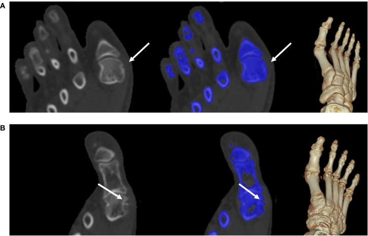
Objective: This study aimed to analyze the effect of urate deposition (UD) on bone erosion and examine the association between the volume of monosodium urate (MSU) crystals and an improved bone erosion score method, as measured in the metatarsophalangeal (MTP) joints of patients with gout.
Materials and methods: Fifty-six patients diagnosed with gout using the 2015 European League Against Rheumatism and American College of Rheumatology criteria were enrolled. MSU crystals volume at each MTP joint was measured using dual-energy computed tomography (DECT) images. The degree of bone erosion was evaluated with the modified Sharp/van der Heijde (SvdH) erosion scoring system based on CT images. Differences in clinical features between patients with (UD group) and without (non-UD group) UD were assessed, and the correlation between erosion scores and urate crystal volume was analyzed.
Results: The UD and non-UD groups comprised 30 and 26 patients, respectively. Among the 560 MTP joints assessed, 80 showed MSU crystal deposition, and 108 showed bone erosion. Bone erosion occurred in both groups but was significantly less severe in the non-UD group (p <0.001). Both groups had equivalent levels of serum uric acid (p=0.200). Symptom duration was significantly longer in the UD group (p=0.009). The UD group also had a higher rate of kidney stones (p=0.023). The volume of MSU crystals was strongly and positively associated with the degree of bone erosion (r=0.714, p <0.001).
Conclusion: This study found that patients with UD show significant increased bone erosion than those without UD. The volume of MSU crystals is associated with the improved SvdH erosion score based on CT images, regardless of serum uric acid level, demonstrating the potential of combining DECT and serum uric acid measurements in helping optimize the management of patients with gout.
Keywords: bone erosion; dual-energy computed tomography; gout; serum uric acid; urate deposition.
Copyright © 2023 Zheng, Zhan, Wang, Deng, Hung, Wang and Jiang.
Conflict of interest statement
Author J-YW is employed by United Imaging Healthcare. The remaining authors declare that the research was conducted in the absence of any commercial or financial relationships that could be construed as a potential conflict of interest.
Figures


Similar articles
-
Relationship between structural joint damage and urate deposition in gout: a plain radiography and dual-energy CT study.Ann Rheum Dis. 2015 Jun;74(6):1030-6. doi: 10.1136/annrheumdis-2013-204273. Epub 2014 Feb 12. Ann Rheum Dis. 2015. PMID: 24521739
-
Investigation on monosodium urate deposition in the first metatarsophalangeal joint and ankle of primary gout patients using dual-energy computed tomography.Med J Malaysia. 2022 May;77(3):279-283. Med J Malaysia. 2022. PMID: 35638482
-
Urate crystal deposition and bone erosion in gout: 'inside-out' or 'outside-in'? A dual-energy computed tomography study.Arthritis Res Ther. 2016 Sep 15;18(1):208. doi: 10.1186/s13075-016-1105-z. Arthritis Res Ther. 2016. PMID: 27629724 Free PMC article.
-
What Has Dual Energy CT Taught Us About Gout?Curr Rheumatol Rep. 2021 Jul 14;23(9):71. doi: 10.1007/s11926-021-01035-5. Curr Rheumatol Rep. 2021. PMID: 34259946 Review.
-
Dual-Energy Computed Tomography of the Knee, Ankle, and Foot: Noninvasive Diagnosis of Gout and Quantification of Monosodium Urate in Tendons and Ligaments.Semin Musculoskelet Radiol. 2016 Feb;20(1):130-6. doi: 10.1055/s-0036-1579709. Epub 2016 Apr 14. Semin Musculoskelet Radiol. 2016. PMID: 27077593 Review.
Cited by
-
Serum ionized magnesium acts as an independent protective factor against bone erosion in patients with gouty arthritis: a cross-sectional study.Front Endocrinol (Lausanne). 2024 Sep 30;15:1375871. doi: 10.3389/fendo.2024.1375871. eCollection 2024. Front Endocrinol (Lausanne). 2024. PMID: 39403581 Free PMC article.
-
A Study on the Diagnostic Value of Dual-Energy CT (DECT) Imaging in Patients With Gouty Arthritis.Int J Rheum Dis. 2024 Dec;27(12):e15431. doi: 10.1111/1756-185X.15431. Int J Rheum Dis. 2024. PMID: 39618108 Free PMC article.
-
The comprehensive role of dual-energy CT in gout as an advanced diagnostic innovation.Skeletal Radiol. 2024 Dec 17. doi: 10.1007/s00256-024-04856-4. Online ahead of print. Skeletal Radiol. 2024. PMID: 39690304 Review.
-
Innovative modeling: a diet-induced quail model for progressive pathological changes in uric acid metabolism disorders.Front Nutr. 2025 Jul 23;12:1612479. doi: 10.3389/fnut.2025.1612479. eCollection 2025. Front Nutr. 2025. PMID: 40771212 Free PMC article.
References
Publication types
MeSH terms
Substances
LinkOut - more resources
Full Text Sources
Medical