. 2023 Jul 1;30(Pt 4):815-821.
doi: 10.1107/S1600577523003168. Epub 2023 May 5.

Full-field hard X-ray nano-tomography at SSRF

Affiliations

Full-field hard X-ray nano-tomography at SSRF

Fen Tao et al. J Synchrotron Radiat. .

Abstract

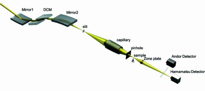
An in-house designed transmission X-ray microscopy (TXM) instrument has been developed and commissioned at beamline BL18B of the Shanghai Synchrotron Radiation Facility (SSRF). BL18B is a hard (5-14 keV) X-ray bending-magnet beamline recently built with sub-20 nm spatial resolution in TXM. There are two kinds of resolution mode: one based on using a high-resolution-based scintillator-lens-coupled camera, and the other on using a medium-resolution-based X-ray sCMOS camera. Here, a demonstration of full-field hard X-ray nano-tomography for high-Z material samples (e.g. Au particles, battery particles) and low-Z material samples (e.g. SiO2 powders) is presented for both resolution modes. Sub-50 nm to 100 nm resolution in three dimensions (3D) has been achieved. These results represent the ability of 3D non-destructive characterization with nano-scale spatial resolution for scientific applications in many research fields.

Keywords: X-ray nano-imaging; ellipsoidal capillary; spatial resolution; synchrotron radiation facility.


Figures

Figure 1
Figure 1
General schematic of the layout of beamline BL18B at SSRF.
Figure 2
Figure 2
Photograph of the TXM endstation at SSRF BL18B, with the mounting of the sample shown in the bottom-right corner.
Figure 3
Figure 3
Design drawing of the two resolution targets and the corresponding SEM images of the Siemens star pattern. (a) 30 nm finest features from ZEISS; (b 50 nm finest features from ANT.
Figure 4
Figure 4
Results of the Siemens star pattern tests for the different resolution modes. (ac) Results for high-resolution mode. (a) Siemens star pattern with 30 nm finest feature size as indicated by circle L1 by absorption contrast, with an insert of an enlarged quarter of the center in high-resolution mode at 8 keV. (b) Line profile of L1. (c) Power spectrum density analysis on the star patterns by absorption contrast (a). (df) Results for medium-resolution mode. (d) Siemens star pattern with 50 nm finest feature size as indicated by circle L2 by absorption contrast, with an insert of an enlarged quarter of the center in medium-resolution mode at 5.2 keV. (e) Line profile of L2. (f) Power spectrum density analysis on the star patterns by absorption contrast (d).
Figure 5
Figure 5
High- and low-Z reference sample nano-imaging by the two resolution modes. (ac) Image of a high-Z material – gold particles. (a) A 2D slice of the gold particles with 20 µm field of view taken at 8 keV. (b) A rending of the 3D structure of a local region. (c) The same batch of gold particles imaged with SEM. (de) Image of a low-Z material – SiO2 powder. (d) A 2D slice of the SiO2 with 20 µm field of view taken at 5.2 keV. (e) A rending of the 3D structure of a local region. (f) The same batch of SiO2 powders imaged with SEM.
Figure 6
Figure 6
Image of an NCM622 battery in high-resolution mode at 8 keV. (a) A 2D slice of a 10 µm-diameter particle of the charged NCM622 material. (b) Enlarged view of the yellow square in (a). (c) 3D reconstruction of NCM622.

References

    1. Alemu, T. & Wang, F.-M. (2018). J. Synchrotron Rad. 25, 151–165. - PubMed
    1. Chao, W., Harteneck, B. D., Liddle, J. A., Anderson, E. H. & Attwood, D. T. (2005). Nature, 435, 1210–1213. - PubMed
    1. Chen, T., Chen, Y., Wang, C., Kempson, I. M., Lee, W., Chu, Y. S., Hwu, Y. & Margaritondo, G. (2011). Opt. Express, 19, 19919–19924. - PubMed
    1. Ge, M., Coburn, D. S., Nazaretski, E., Xu, W., Gofron, K., Xu, H., Yin, Z. & Lee, W. (2018). Appl. Phys. Lett. 113, 083109.
    1. Jiang, Z., Li, J., Yang, Y., Mu, L., Wei, C., Yu, X., Pianetta, P., Zhao, K., Cloetens, P., Lin, F. & Liu, Y. (2020). Nat. Commun. 11, 2310. - PMC - PubMed

Substances