

Racial differences in quantitative optical coherence tomography angiography findings between older non-diabetics with co-morbidities
- PMID: 37146056
- PMCID: PMC10162566
- DOI: 10.1371/journal.pone.0285360
Racial differences in quantitative optical coherence tomography angiography findings between older non-diabetics with co-morbidities
Abstract
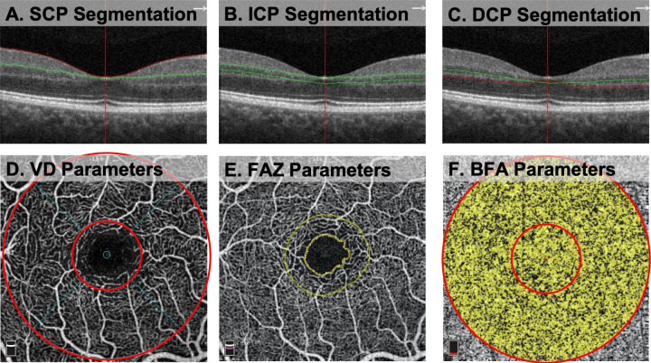
This cross-sectional study compared optical coherence tomography angiography (OCTA) parameters between older Black and White adults with systemic comorbidities in an effort to further understand racial differences in the retinal microvasculature. We analyzed vessel density at the superficial (SCP), intermediate (ICP), and deep capillary plexuses (DCP), foveal avascular zone (FAZ) parameters, and blood flow area (BFA) at the choriocapillaris. We used a mixed-effects linear regression model, controlling for hypertension and two eyes from the same subject, to compare OCTA parameters. Black subjects had lower foveal vessel density at the SCP and ICP, while no differences were observed at the parafovea or 3x3 mm macular area of any capillary layer. Black subjects had greater FAZ area, perimeter, and FD-300, a measurement of vessel density in a 300 μm wide ring around the FAZ. Black subjects also had lower BFA at the choriocapillaris. Within a cohort of subjects without hypertension, these differences remained statistically significant, with the exception of foveal vessel density at the SCP and foveal BFA of the choriocapillaris. These findings suggest that normative databases of OCTA parameters must strive to be diverse in nature to adequately capture differences across patient populations. Further study is required to understand if baseline differences in OCTA parameters contribute to epidemiological disparities in ocular diseases.
Copyright: © 2023 Moir et al. This is an open access article distributed under the terms of the Creative Commons Attribution License, which permits unrestricted use, distribution, and reproduction in any medium, provided the original author and source are credited.
Conflict of interest statement
The authors have declared that no competing interests exist.
Figures
References
Publication types
MeSH terms
LinkOut - more resources
Full Text Sources
Medical
Miscellaneous
