. 2023 May 12;8(83):eadh3455.
doi: 10.1126/sciimmunol.adh3455. Epub 2023 May 5.

Cytokinopathy with aberrant cytotoxic lymphocytes and profibrotic myeloid response in SARS-CoV-2 mRNA vaccine-associated myocarditis

Affiliations

Cytokinopathy with aberrant cytotoxic lymphocytes and profibrotic myeloid response in SARS-CoV-2 mRNA vaccine-associated myocarditis

Anis Barmada et al. Sci Immunol. .

Abstract

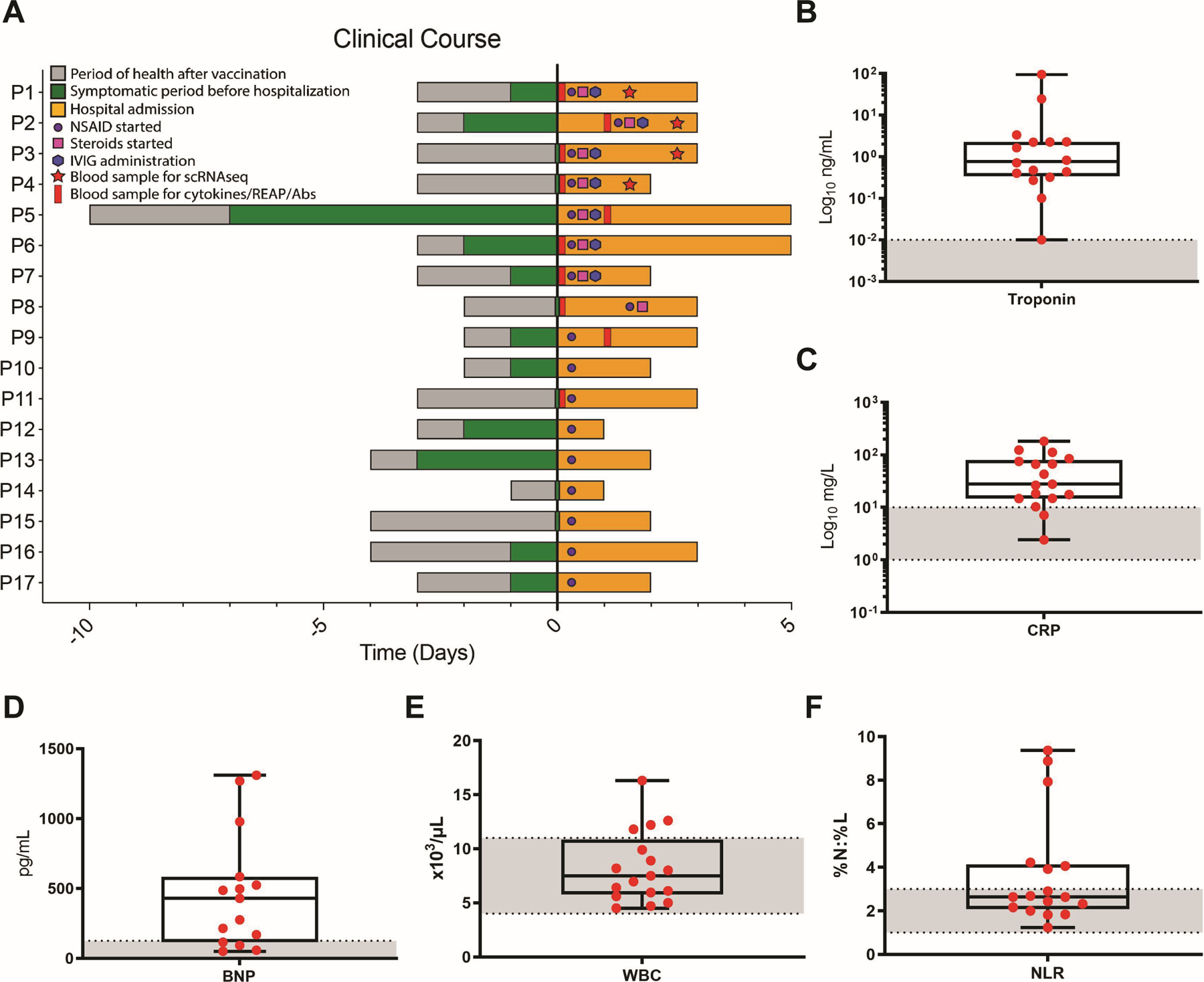
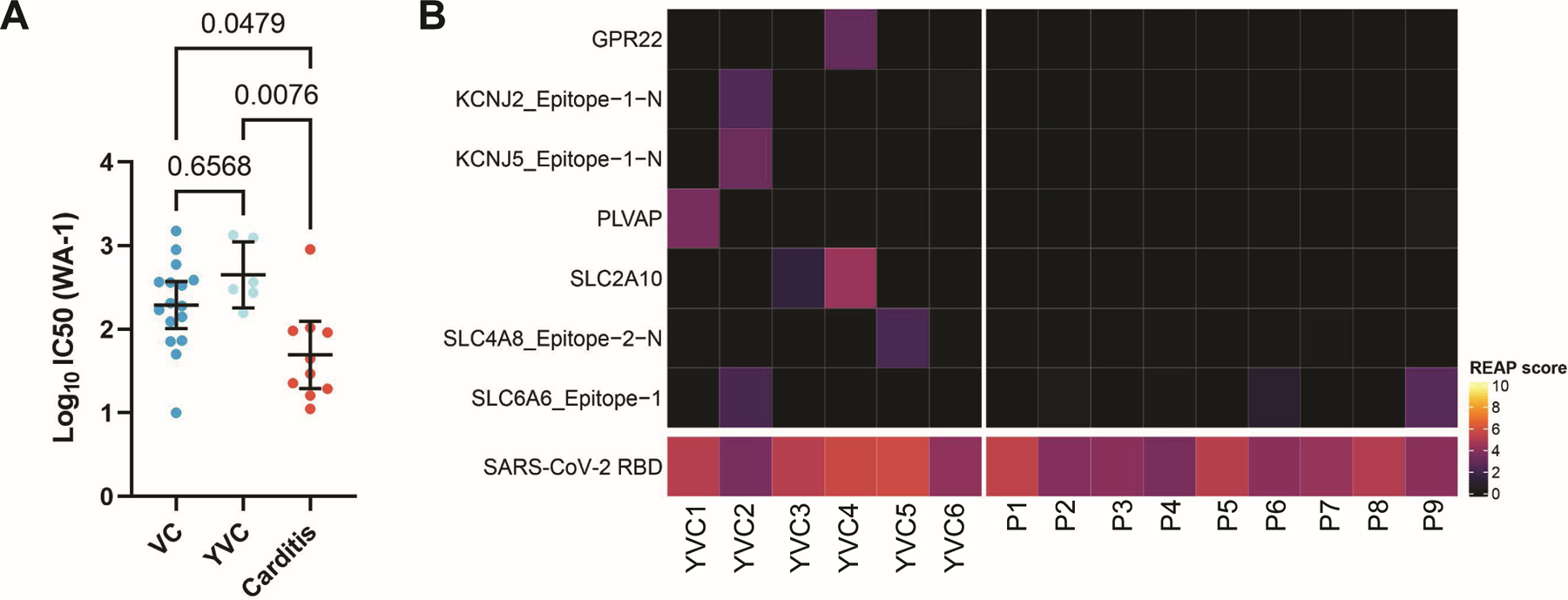
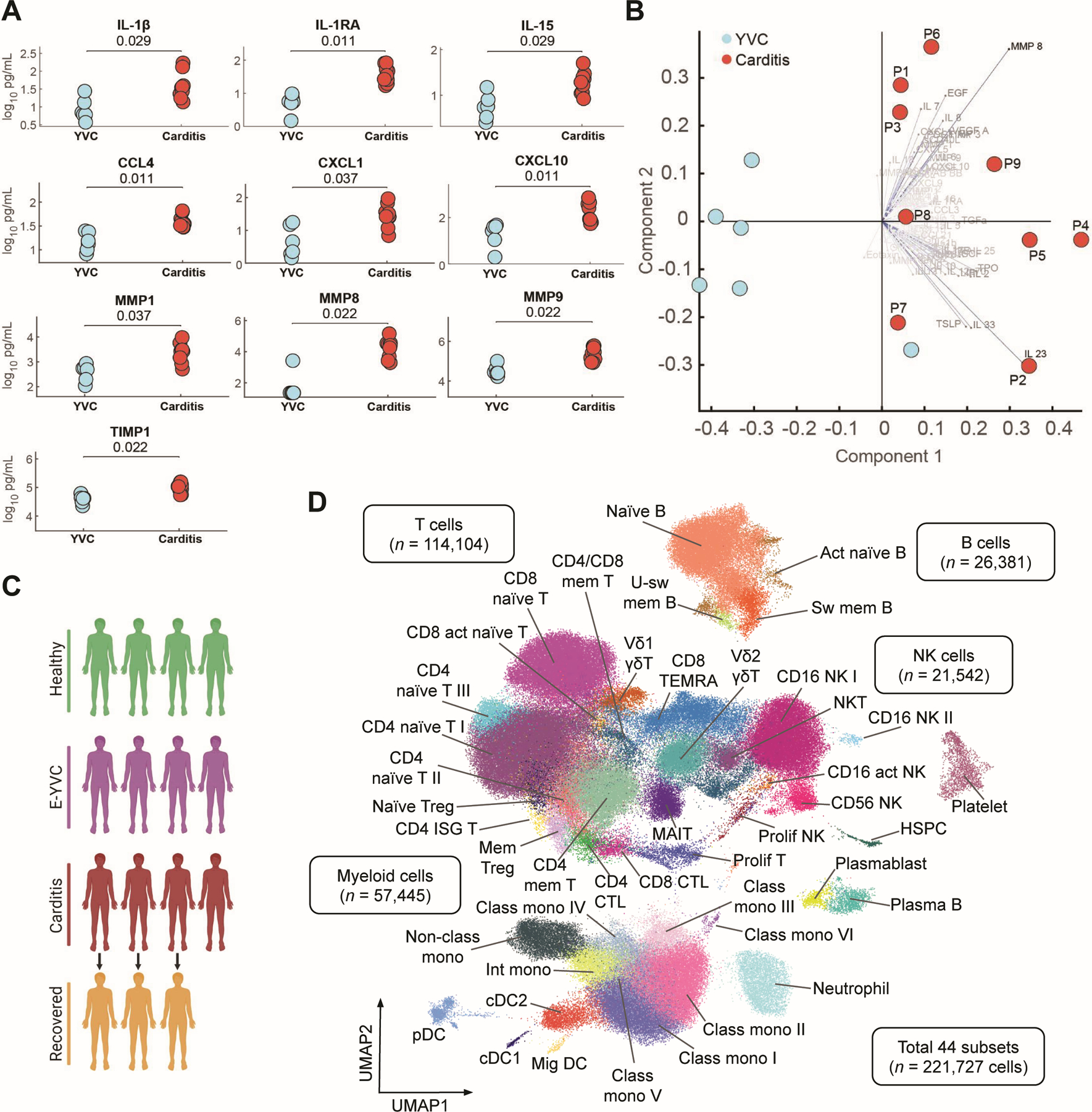
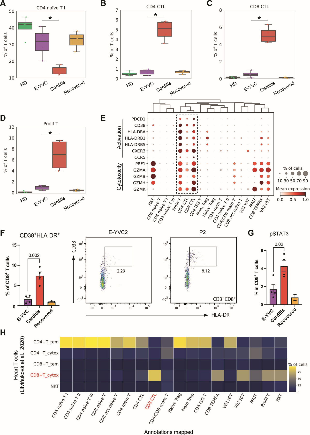
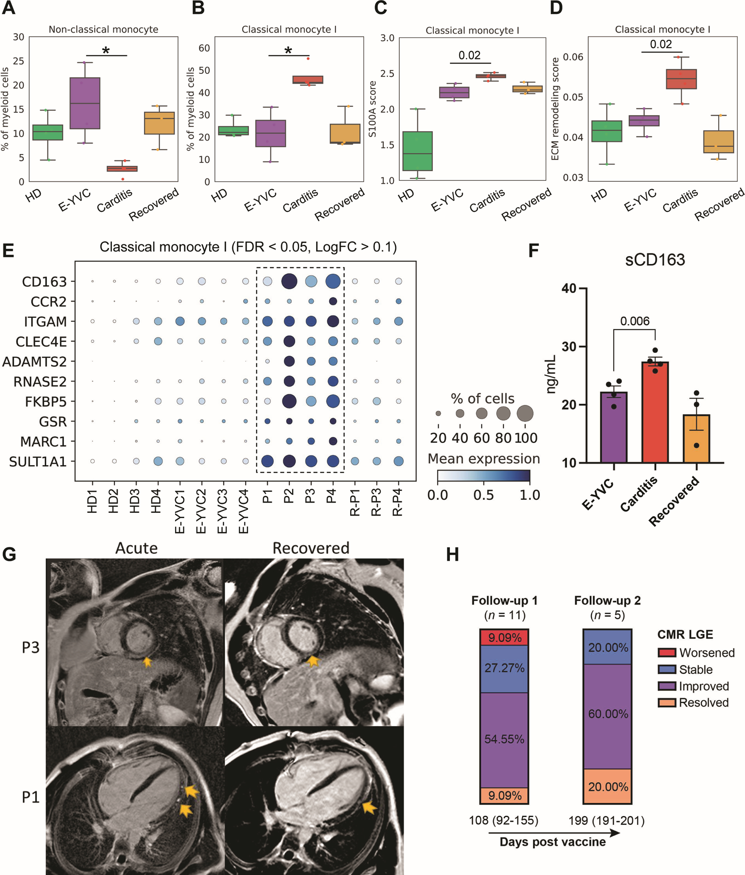
Rare immune-mediated cardiac tissue inflammation can occur after vaccination, including after SARS-CoV-2 mRNA vaccines. However, the underlying immune cellular and molecular mechanisms driving this pathology remain poorly understood. Here, we investigated a cohort of patients who developed myocarditis and/or pericarditis with elevated troponin, B-type natriuretic peptide, and C-reactive protein levels as well as cardiac imaging abnormalities shortly after SARS-CoV-2 mRNA vaccination. Contrary to early hypotheses, patients did not demonstrate features of hypersensitivity myocarditis, nor did they have exaggerated SARS-CoV-2-specific or neutralizing antibody responses consistent with a hyperimmune humoral mechanism. We additionally found no evidence of cardiac-targeted autoantibodies. Instead, unbiased systematic immune serum profiling revealed elevations in circulating interleukins (IL-1β, IL-1RA, and IL-15), chemokines (CCL4, CXCL1, and CXCL10), and matrix metalloproteases (MMP1, MMP8, MMP9, and TIMP1). Subsequent deep immune profiling using single-cell RNA and repertoire sequencing of peripheral blood mononuclear cells during acute disease revealed expansion of activated CXCR3+ cytotoxic T cells and NK cells, both phenotypically resembling cytokine-driven killer cells. In addition, patients displayed signatures of inflammatory and profibrotic CCR2+ CD163+ monocytes, coupled with elevated serum-soluble CD163, that may be linked to the late gadolinium enhancement on cardiac MRI, which can persist for months after vaccination. Together, our results demonstrate up-regulation in inflammatory cytokines and corresponding lymphocytes with tissue-damaging capabilities, suggesting a cytokine-dependent pathology, which may further be accompanied by myeloid cell-associated cardiac fibrosis. These findings likely rule out some previously proposed mechanisms of mRNA vaccine--associated myopericarditis and point to new ones with relevance to vaccine development and clinical care.


Conflict of interest statement

Competing interests: C.L.L. reported an unrelated advisory/consulting role for Pharming Healthcare, Inc. and unrelated funding to her institution from Ono Pharma. I.Y. reported being a member of the NIAID/COVPN mRNA-1273 Study Group; has received funding to her institution to conduct clinical research from Pfizer and Moderna outside the submitted work; an unrelated advisory/consulting role for Sanofi Pasteur and Merck. A. I. co-founded and consults for RIGImmune, Xanadu Bio and PanV, consults for Paratus Sciences, InvisiShield Technologies, and is a member of the Board of Directors of Roche Holding Ltd. A.M.R. is an inventor of a patent describing the REAP technology, and is the founder of and holds equity in Seranova Bio, the commercial licensee of this patent. D.B. is the founder of Lab11 Therapeutics. S.A. serves on various WHO advisory committees and on the boards of Gavi, the Vaccine Alliance, and Savin Vaccine Institute.

Figures

Fig. 1:
Fig. 1:. Clinical parameters of the SARS-CoV-2 vaccine-associated myopericarditis cohort.
(A) Time course for patients showing the day of vaccine administration, symptom onset, treatment, and sample collection relative to hospital admission (Day 0). (B to F) Maximum values of selected blood markers in patients tested during hospital admission. Boxes depict the interquartile range (IQR), horizontal bars represent the median, whiskers extend to 1.5 × IQR, and red dots show the value of each patient. Dashed lines and gray area represent normal reference ranges as provided by the CLIA-certified Yale New Haven Hospital Department of Laboratory Medicine. Abbreviations: C-reactive protein (CRP), B-type natriuretic peptide (BNP), white blood cells (WBC), neutrophil-to-lymphocyte ratio (NLR).
Fig. 2:
Fig. 2:. Myopericarditis patients mount humoral response to vaccination with absence of reactive autoantibodies.
(A) IC50 estimates for SARS-CoV-2 neutralizing antibody titers against ancestral SARS-CoV-2 (WA-1) in patients (n = 10) compared to healthy vaccinated controls (VC, n = 16), in addition to those closer in age to patients where there is an increased risk for vaccine-associated myopericarditis (younger vaccinated controls (YVC), n = 6). Black bars denote group means and error bars represent 95% confidence intervals. Statistical significance was assessed using the Kruskal-Wallis test with Dunn’s correction for multiple comparisons. (B) Heat map of Rapid Extracellular Antigen Profiling (REAP) scores showing cardiac-related autoantibodies present in at least one donor (either patients or YVC), from a total of 526 antigens/epitopes defined by Gene Ontology (circulatory system process (GO:0003013) or heart contraction (GO:0060047)) or the Human Protein Atlas (heart tissue enriched/enhanced), across individual patients (n = 9) and YVC (n = 6). SARS-CoV-2 RBD antigen is used as a positive control in vaccinated individuals. Positive autoantibody reactivity is defined as REAP score ≥ 2, protein-wise z-score ≥ 1.96, and protein-wise mean < 0.5, with higher REAP scores correlating with higher antibody titer and/or abundance as validated previously (43, 44). Negative findings for all cardiac antigens/epitopes tested from the Human Protein Atlas (81 total) are further shown in Fig. S2D.
Fig. 3:
Fig. 3:. Elevation of immune cytokines in myopericarditis and identification of immune cell subsets.
(A) Scatter plots of selected cytokines in patients (n = 9) and healthy younger vaccinated controls (YVC, n = 6). Statistical significance was determined using the unpaired two-sided Wilcoxon rank-sum test with Benjamini-Hochberg FDR correction for multiple comparisons. All additional serum proteins assayed are shown in Fig. S3 (B) Biplot of all serum proteins assayed (84 total) between patients and healthy YVC (see related principal component analysis (PCA) in Fig. S4, A and B). (C) Schematic diagram showing the cohort studied in subsequent scRNA-seq analyses, including pediatric male healthy donors (HD, n = 4), healthy early-young vaccinated controls (E-YVC, n = 4), acute myopericarditis patients (n = 4), and matched patients at follow-up/recovery (n = 3). (D) UMAP visualization of immune cell subsets identified from the cohort in (C), with additional four CITE-seq multisystem inflammatory syndrome in children (MIS-C) after SARS-CoV-2 samples included to refine cluster annotation using surface proteins. Cell subset abbreviations: activated (act), class-switched (sw), class-unswitched (u-sw), memory (mem), natural killer (NK), natural killer T (NKT; CD3+ CD161+ KLRF1+), regulatory T (Treg), interferon-stimulated gene (ISG), cytotoxic T lymphocyte (CTL), proliferating (prolif), mucosal-associated invariant T (MAIT), terminally differentiated effector memory CD45RA+ T (TEMRA), non-classical (non-class), intermediate (int), classical (class), monocyte (mono), conventional dendritic cell (cDC), plasmacytoid dendritic cell (pDC), migratory (mig), hematopoietic stem and progenitor cell (HSPC).
Fig. 4:
Fig. 4:. Expansion of activated cytotoxic T lymphocytes in myopericarditis.
(A to D) Box plots showing the average proportions of four T cell subsets (CD4+ naïve T I, CD4+ and CD8+ cytotoxic T lymphocytes (CTLs), as well as proliferating T cells) across the groups. The boxes denote the interquartile range (IQR), horizontal bars represent the median, whiskers extend to 1.5 × IQR, and dots show the values of each donor. Statistical significance was determined using the Bayesian model scCODA (49) accounting for the compositional dependencies between cell subsets in the scRNA-seq data while controlling for false discoveries (FDR < 0.05 in myopericarditis vs. E-YVC). (E) Dot plot showing the expression of activation markers (PD-1 and CD38/HLA-DR), chemokine receptors (CXCR3 and CCR5), and cytotoxicity genes (perforin and granzymes) characterizing the T cell subsets shown in (B to D). (F) Flow cytometry quantifying the percentage of the CD38+ HLA-DR+ population out of CD8+ T cells across the groups (left), with representative plots for E-YVC and patient (P2) donors (right). Statistical significance was determined using the unpaired two-tailed t-test between the E-YVC and myopericarditis groups, and error bars represent the standard error (SE). (G) Percentage phosphorylated STAT3 (pSTAT3) in CD8+ T cells across the groups by flow cytometry; statistical significance was determined as in (F), and error bars represent SE. (H) Heat map depicting the percentage of each T cell subset identified in this study that mapped, using a logistic regression model based on gene expression with mean prediction probability of 0.9, to each subset from published single-cell data of heart T cell populations (60).
Fig. 5:
Fig. 5:. Inflammatory and profibrotic signatures of monocytes in myopericarditis.
(A and B) Box plots showing the average proportions of non-classical (CD14dim CD16+) and classical (CD14+ CD16) monocyte subsets across the groups. The boxes denote the interquartile range (IQR), horizontal bars represent the median, whiskers extend to 1.5 × IQR, and dots show the values of each donor. Statistical significance was determined using the Bayesian model scCODA (49) accounting for the compositional dependencies between cell subsets in the scRNA-seq data while controlling for false discoveries (FDR < 0.05 in myopericarditis vs. E-YVC). (C and D) Average expression score of (C) inflammatory genes from the S100A family of alarmins (S100A8–12; FDR < 0.05, LogFC > 0.1 in myopericarditis vs. E-YVC) and (D) 238 genes from a published dataset of extracellular matrix (ECM) remodeling (GSEA Molecular Signatures Database M3468) in the same classical monocyte subset shown in (B) across groups. Statistical significance between scores was determined using the unpaired two-sided Wilcoxon rank-sum test comparing the E-YVC and myopericarditis groups. (E) Dot plot showing top differentially expressed and upregulated genes in the same classical monocyte subset shown in (B) across donors (FDR < 0.05, LogFC > 0.1 in myopericarditis vs. E-YVC). (F) Enzyme-linked immunosorbent assay (ELISA) measurement of soluble CD163 (sCD163) in serum across the groups. Statistical significance was determined using the unpaired two-tailed t-test between the E-YVC and myopericarditis groups, and error bars represent the standard error (SE). (G) Representative cardiac magnetic resonance (CMR) images of acute myopericarditis and follow-up/recovery (191 days for P1 and 82 days for P3 after vaccination) showing persistent late gadolinium enhancement (LGE; yellow arrows) seen in a subset of patients (from 17 patients included in our cohort, at admission, 11 were LGE +, 4 were LGE −, and 2 had no CMR). Particularly, for P1, four chamber phase sequence inversion recovery (PSIR) demonstrating patch sub-epicardial LGE along the left ventricular lateral wall from base to apex (acute), with improvement in both quantity and intensity at follow-up (recovered). For P3, mid ventricle short axis PSIR demonstrating sub-epicardial to nearly transmural LGE sparing the sub-endocardial region (acute), which is mildly improved in intensity and quantity at follow-up (recovered). (H) Stacked bar plots depicting the percentage of patients categorized by CMR LGE changes at two follow-ups after vaccination/first admission (median days (IQR)). For exact patients with LGE at admission and follow-up as well as details of imaging findings, see Table S1.

Comment in

References

    1. Baden LR, El Sahly HM, Essink B, Kotloff K, Frey S, Novak R, Diemert D, Spector SA, Rouphael N, Creech CB, McGettigan J, Khetan S, Segall N, Solis J, Brosz A, Fierro C, Schwartz H, Neuzil K, Corey L, Gilbert P, Janes H, Follmann D, Marovich M, Mascola J, Polakowski L, Ledgerwood J, Graham BS, Bennett H, Pajon R, Knightly C, Leav B, Deng W, Zhou H, Han S, Ivarsson M, Miller J, Zaks T, Group CS. 2021. Efficacy and Safety of the mRNA-1273 SARS-CoV-2 Vaccine. N Engl J Med 384: 403–16 - PMC - PubMed
    1. Barda N, Dagan N, Ben-Shlomo Y, Kepten E, Waxman J, Ohana R, Hernán MA, Lipsitch M, Kohane I, Netzer D, Reis BY, Balicer RD. 2021. Safety of the BNT162b2 mRNA Covid-19 Vaccine in a Nationwide Setting. New England Journal of Medicine 385: 1078–90 - PMC - PubMed
    1. Polack FP, Thomas SJ, Kitchin N, Absalon J, Gurtman A, Lockhart S, Perez JL, Perez Marc G, Moreira ED, Zerbini C, Bailey R, Swanson KA, Roychoudhury S, Koury K, Li P, Kalina WV, Cooper D, Frenck RW Jr., Hammitt LL, Tureci O, Nell H, Schaefer A, Unal S, Tresnan DB, Mather S, Dormitzer PR, Sahin U, Jansen KU, Gruber WC, Group CCT. 2020. Safety and Efficacy of the BNT162b2 mRNA Covid-19 Vaccine. N Engl J Med 383: 2603–15 - PMC - PubMed
    1. Shimabukuro TT, Kim SY, Myers TR, Moro PL, Oduyebo T, Panagiotakopoulos L, Marquez PL, Olson CK, Liu R, Chang KT, Ellington SR, Burkel VK, Smoots AN, Green CJ, Licata C, Zhang BC, Alimchandani M, Mba-Jonas A, Martin SW, Gee JM, Meaney-Delman DM, Team CDCv-sC-PR. 2021. Preliminary Findings of mRNA Covid-19 Vaccine Safety in Pregnant Persons. N Engl J Med 384: 2273–82 - PMC - PubMed
    1. Patone M, Mei XW, Handunnetthi L, Dixon S, Zaccardi F, Shankar-Hari M, Watkinson P, Khunti K, Harnden A, Coupland CAC, Channon KM, Mills NL, Sheikh A, Hippisley-Cox J. 2021. Risks of myocarditis, pericarditis, and cardiac arrhythmias associated with COVID-19 vaccination or SARS-CoV-2 infection. Nature Medicine - PMC - PubMed

Publication types