. 2023 May 5;9(18):eabq7866.
doi: 10.1126/sciadv.abq7866. Epub 2023 May 5.

A rationally designed ICAM1 antibody drug conjugate eradicates late-stage and refractory triple-negative breast tumors in vivo

Affiliations

A rationally designed ICAM1 antibody drug conjugate eradicates late-stage and refractory triple-negative breast tumors in vivo

Peng Guo et al. Sci Adv. .

Abstract

Triple-negative breast cancer (TNBC) remains the most lethal form of breast cancer, and effective targeted therapeutics are in urgent need to improve the poor prognosis of TNBC patients. Here, we report the development of a rationally designed antibody drug conjugate (ADC) for the treatment of late-stage and refractory TNBC. We determined that intercellular adhesion molecule-1 (ICAM1), a cell surface receptor overexpressed in TNBC, efficiently facilitates receptor-mediated antibody internalization. We next constructed a panel of four ICAM1 ADCs using different chemical linkers and warheads and compared their in vitro and in vivo efficacies against multiple human TNBC cell lines and a series of standard, late-stage, and refractory TNBC in vivo models. An ICAM1 antibody conjugated with monomethyl auristatin E (MMAE) via a protease-cleavable valine-citrulline linker was identified as the optimal ADC formulation owing to its outstanding efficacy and safety, representing an effective ADC candidate for TNBC therapy.


Figures

Fig. 1.
Fig. 1.. Differential expression of ICAM1 in human TNBC cells versus normal cells.
(A) ICAM1 mRNA levels were quantitatively compared in different breast cancer subtypes, molecular subtypes of TNBC, cancer grades of TNBC, and breast tumors with BRCA1/2 or TP53 mutation. WT, wild type; BLIS, basal-like immunosuppressed; BLIA, basal-like immunoactivated; L-AR, luminal androgen receptor; MES, mesenchymal. (B) Human TNBC cell surface expression of ICAM1 and TROP2 was compared versus normal MCF10A cells by flow cytometry using FL2-H channel (PE-labeled antibody). Nontargeting IgG was used as a control. (C) Immunofluorescent staining of ICAM1 and TROP2 on human TNBC cells and normal MCF10A cells. Scale bars, 50μm. (D) Representative imaging flow cytometry images showing cellular internalization of ICAM1 antibodies in human TNBC and normal MCF10A cells. (E) Signal intensity analysis for ICAM1 antibody-mediated cell internalization (n = 5000 cells). ***P < 0.001. PE, phycoerythrin; IgG, immunoglobulin G. BF, bright field.
Fig. 2.
Fig. 2.. Selectively ablating human TNBC cells by ICAM1 ADCs.
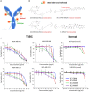
(A) Schematic illustration of an ICAM1 ADC. (B) Chemical structures of ADC linkers and warheads used in ICAM1 ADCs. (C) In vitro cytotoxicity of four ICAM1 ADCs against a panel of four human TNBC cell lines (MDA-MB-436, MDA-MB-468, MDA-MB-157, and MDA-MB-231) and two non-neoplastic cell lines (MCF10A and HEK293).
Fig. 3.
Fig. 3.. Tumor specificity and biodistribution of ICAM1 antibody in nude mice.
(A) Schematic design of TNBC biodistribution in an immunocompromised nude mouse model. (B) In vivo NIR fluorescent images of nude mice at 48 hours after the administration of IgG-Cy5.5, IC1-Cy5.5, or IC1-MMAE-Cy5.5 (n = 5 per group). (C) Quantified MDA-MB-436 tumor accumulation of IgG-Cy5.5, IC1-Cy5.5, or IC1-MMAE-Cy5.5. (D) Ex vivo NIR fluorescent images of MDA-MB-436 tumors treated by IgG-Cy5.5, IC1-Cy5.5, or IC1-MMAE-Cy5.5. (E) Representative ex vivo NIR fluorescent images of six major organs including the brain (B), lung (LU), heart (H), liver (L), spleen (S), and kidney (K). (F) Quantified normal organ distribution of IgG-Cy5.5, IC1-Cy5.5, or IC1-MMAE-Cy5.5 (n = 5). *P < 0.05 and **P < 0.01. NS, not significant.
Fig. 4.
Fig. 4.. Tumor specificity and biodistribution of ICAM1 antibody in BALB/c mice.
(A) Schematic design of tumor biodistribution in an immunocompetent BALB/c mouse model. (B) Ex vivo NIR fluorescent images of 4T1 tumors and six normal organs treated by IgG-Cy5.5 and IC1-Cy5.5 (anti-mouse) (n = 7 per group). (C) Quantified 4T1 tumor and normal organ accumulation of IgG-Cy5.5 and IC1-Cy5.5 (anti-mouse). (D) Circulating leukocyte uptake of IgG-Cy5.5 and IC1-Cy5.5 (anti-mouse) quantified by flow cytometry. **P < 0.01 and ***P < 0.001.
Fig. 5.
Fig. 5.. ICAM1 ADCs eradicate standard and late-stage TNBC tumors in vivo.
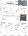
(A) Schematic design of in vivo efficacy of ICAM1 ADC in standard and late-stage settings of an orthotopic TNBC model. (B) Image of excised orthotopic MDA-MB-436 tumors from mice treated with PBS (sham), free Dox, IC1 Ab, IC1-MMAF, or IC1-MMAE in standard setting. (n = 7 to 10 per group). (C) Tumor progression in the standard setting was monitored by tumor volume measurement using a caliper. (D) Tumor mass at the end point (day 24) of the standard setting was quantified by weight. (E) Mouse body weights in standard setting receiving PBS (sham), free Dox, IC1 Ab, IC1-MMAF, or IC1-MMAE. (F) Tumor progression receiving PBS (sham), IC1-MMAF, or IC1-MMAE in a late-stage setting was monitored by tumor volume. (G) Tumor mass at the end point (day 34) of the late-stage setting was quantified by weight. (H) Mouse body weights in a late-stage setting receiving PBS (sham), IC1-MMAF, or IC1-MMAE. **P < 0.01 and ***P < 0.001.
Fig. 6.
Fig. 6.. ICAM1 ADCs eradicate refractory TNBC tumors in vivo.
(A) Schematic design of in vivo efficacy of ICAM1 ADCs in a refractory TNBC mouse model. (B) Image of excised orthotopic MDA-MB-231 tumors from mice treated with PBS (sham), IC1-MMAF, or IC1-MMAE. (C) Refractory tumor progression receiving PBS (sham), IC1-MMAF, or IC1-MMAE was monitored by tumor volume measurement using a caliper. (D) Tumor mass at the end point (day 24) was quantified by weight. (E) Quantified mouse body weights during receiving PBS (sham), free Dox, IC1 Ab, IC1-MMAF, or IC1-MMAE. (F) Schematic design of dosage-dependent efficacy of IC1-MMAE in an orthotopic TNBC tumor model. (G) Image of excised orthotopic MDA-MB-436 tumors from mice treated with PBS (sham) or IC1-MMAE at three different dosages. (H) Tumor progression receiving PBS (sham) or IC1-MMAE at different dosages was monitored by tumor volume. (I) Tumor mass at the end point (day 24) was quantified by weight. (J) Quantified mouse body weights during receiving IC1-MMAE at different dosages. (K) Chronic liver and renal toxicities of IC1-MMAE were analyzed by blood chemistry. ***P < 0.001.

References

    1. Siegel R. L., Miller K. D., Fuchs H. E., Jemal A., Cancer statistics, 2021. CA Cancer J. Clin. 71, 7–33 (2021). - PubMed
    1. Lebert J. M., Lester R., Powell E., Seal M., McCarthy J., Advances in the systemic treatment of triple-negative breast cancer. Curr. Oncol. 25, S142–S150 (2018). - PMC - PubMed
    1. Lee A., Djamgoz M. B. A., Triple negative breast cancer: Emerging therapeutic modalities and novel combination therapies. Cancer Treat. Rev. 62, 110–122 (2018). - PubMed
    1. Foulkes W. D., Smith I. E., Reis-Filho J. S., Triple-negative breast cancer. N. Engl. J. Med. 363, 1938–1948 (2010). - PubMed
    1. Dent R., Trudeau M., Pritchard K. I., Hanna W. M., Kahn H. K., Sawka C. A., Lickley L. A., Rawlinson E., Sun P., Narod S. A., Triple-negative breast cancer: Clinical features and patterns of recurrence. Clin. Cancer Res. 13, 4429–4434 (2007). - PubMed

MeSH terms

Substances