. 2023 May 4;30(5):611-616.e7.
doi: 10.1016/j.stem.2023.04.003.

Bovine blastocyst-like structures derived from stem cell cultures

Affiliations

Bovine blastocyst-like structures derived from stem cell cultures

Carlos A Pinzón-Arteaga et al. Cell Stem Cell. .

Abstract

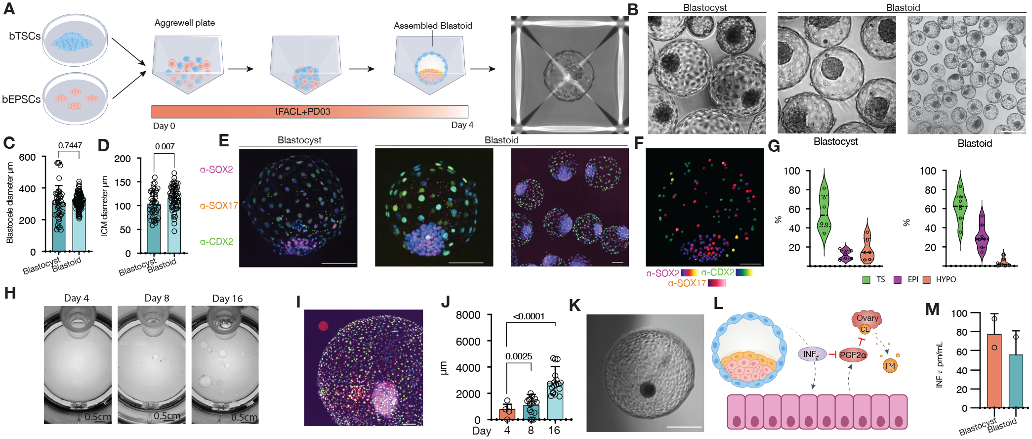
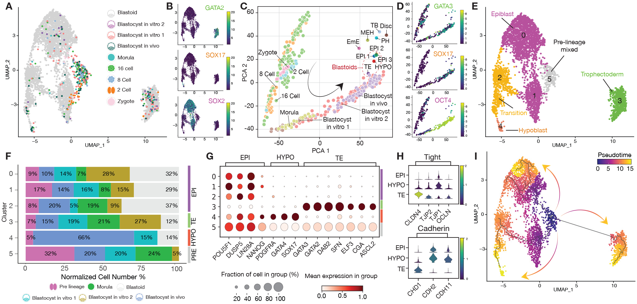
Understanding the mechanisms of blastocyst formation and implantation is critical for improving farm animal reproduction but is hampered by a limited supply of embryos. Here, we developed an efficient method to generate bovine blastocyst-like structures (termed blastoids) via assembling bovine trophoblast stem cells and expanded potential stem cells. Bovine blastoids resemble blastocysts in morphology, cell composition, single-cell transcriptomes, in vitro growth, and the ability to elicit maternal recognition of pregnancy following transfer to recipient cows. Bovine blastoids represent an accessible in vitro model for studying embryogenesis and improving reproductive efficiency in livestock species.

Keywords: Bovine-blastocyst-like structures; blastoids; bovine blastoids; bovine embryonic stem cells; bovine expanded potential stem cells; bovine trophoblast stem cells.


Conflict of interest statement

Declaration of interests C.A.P.-A., Y. Wang., Y. Wei., Z.J., and J.W. are co-inventors on US provisional patent application 63/370,068 relating to bovine-blastocyst-like structures and uses thereof.

Figures

Figure 1.
Figure 1.. Assembly of bovine blastoids from EPSCs and TSCs cultures.
(A) Illustration of the assembly process via bovine EPSCs and TSCs aggregation. (B) Phase-contrast image comparing blastoids vs blastocysts. (C) Blastocele diameter measurement. (D) Inner cell mass (ICM) diameter measurement. (E) Immunostaining for epiblast marker SOX2 (magenta, EPI), hypoblast marker SOX17(red, HYPO) and trophectoderm marker CDX2(green, TS), individual markers in Figure S1. (F) Blastocyst and Blastoid lineage composition quantified via confocal microscopy 3D reconstruction and spots colocalization quantification using IMARIS. (G) Snapshots of in vitro growth of blastoids in a rotating culture system (Clinostar Incubator, Celvivo). (H) Representative image via immunostaining of all three lineages as in e, individual markers in Figure S4. (I) Blastoid diameter quantification. (J) representative micrographs of in vitro grown blastoid. (K) A schematic of the maternal recognition of the action of pregnancy signal interferon TAU (INFt). (L) Enzyme-linked immunosorbent assay (ELISA) measurement of (INFt) in surrogate recipients following embryo transfers. PGF2α: Prostaglandin F2α. CL: Corpus luteum. P4: Progesterone.
Figure 2.
Figure 2.. Single-cell characterization of bovine assembled blastoids.
(A) Joint uniform manifold approximation and projection (UMAP) embedding of 10x Genomics single-cell transcriptomes of bovine blastoids (grey) and bovine zygote (pink), 2 cell (orange), 8 cell (blue), 16 cell (green), Morula (cyan) and in vivo and in vitro Blastocyst stage embryos (purple, dark green, light red). (B) UMAP Heatmap showing expression of Trophectoderm (TE), Hypoblast (HYPO), and epiblast (EPI) markers, GATA2, SOX17 and SOX2, respectively. (C) Principal component analysis (PCA) of pseudo bulk conversion of blastoid data. Gastrulation markers: Disc: Embryonic disc (Day 14 Stage 4). EmE: Embryonic ectoderm (Day 14 Stage 5). MEH: Mesoderm, endoderm, and visceral hypoblast. (Day 14 Stage 5). PH: Parietal hypoblast. (Day 14 Stage 5). TB: Trophoblast. (Day 14 Stage 5). (D) PCA heatmaps showing expression of Trophectoderm (TE), Hypoblast (HYPO), and epiblast (EPI) markers, GATA3, SOX17 and OCT4 (also known as POU5F1), respectively. (E) Major cluster classification based on marker expression. (F) Normalized percentage of cells in each cluster. (G) Dot plot indicating the expression of markers of epiblast (EPI), trophectoderm (TE) and hypoblast (HYPO). (H) Violin plot of lineage-specific cell junction markers. (I) RNA velocity pseudotime analysis depicting the cell trajectories.

References

    1. Rivron NC, Frias-Aldeguer J, Vrij EJ, Boisset J-C, Korving J, Vivié J, Truckenmüller RK, van Oudenaarden A, van Blitterswijk CA, and Geijsen N (2018). Blastocyst-like structures generated solely from stem cells. Nature 557, 106–111. 10.1038/s41586-018-0051-0. - DOI - PubMed
    1. Sozen B, Cox AL, De Jonghe J, Bao M, Hollfelder F, Glover DM, and Zernicka-Goetz M (2019). Self-Organization of Mouse Stem Cells into an Extended Potential Blastoid. Developmental cell 51, 698–712.e698. - PMC - PubMed
    1. Li R, Zhong C, Yu Y, Liu H, Sakurai M, Yu L, Min Z, Shi L, Wei Y, Takahashi Y, et al. (2019). Generation of Blastocyst-like Structures from Mouse Embryonic and Adult Cell Cultures. Cell 179, 687–702.e618. 10.1016/j.cell.2019.09.029. - DOI - PMC - PubMed
    1. Yu L, Wei Y, Duan J, Schmitz DA, Sakurai M, Wang L, Wang K, Zhao S, Hon GC, and Wu J (2021). Blastocyst-like structures generated from human pluripotent stem cells. Nature 591, 620–626. 10.1038/s41586-021-03356-y. - DOI - PubMed
    1. Yanagida A, Spindlow D, Nichols J, Dattani A, Smith A, and Guo G (2021). Naive stem cell blastocyst model captures human embryo lineage segregation. Cell Stem Cell 28, 1016–1022.e1014. 10.1016/j.stem.2021.04.031. - DOI - PMC - PubMed

Publication types