. 2023 May 4;30(5):632-647.e10.
doi: 10.1016/j.stem.2023.04.006.

Deriving Schwann cells from hPSCs enables disease modeling and drug discovery for diabetic peripheral neuropathy

Affiliations

Deriving Schwann cells from hPSCs enables disease modeling and drug discovery for diabetic peripheral neuropathy

Homa Majd et al. Cell Stem Cell. .

Abstract

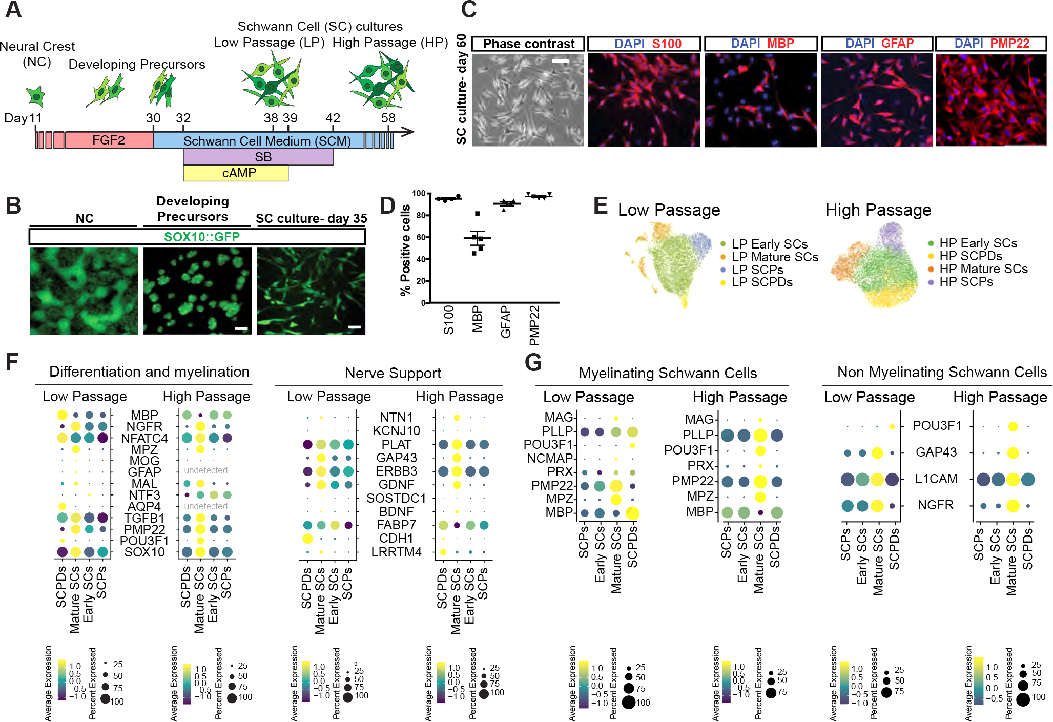
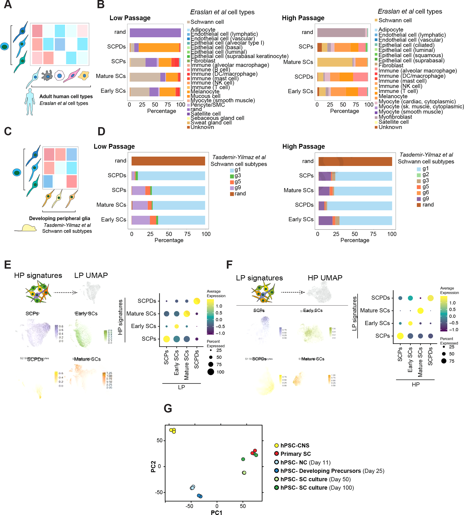
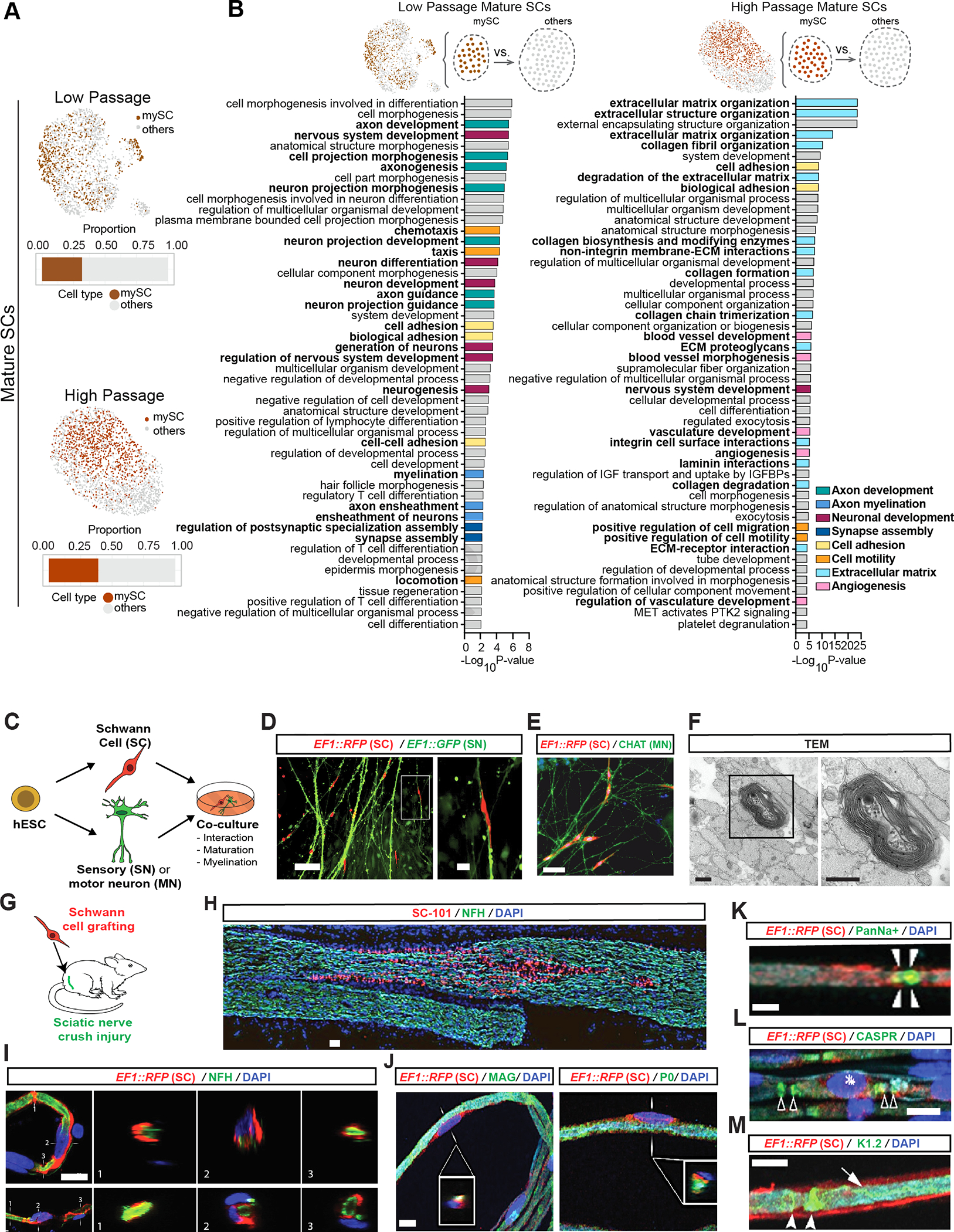
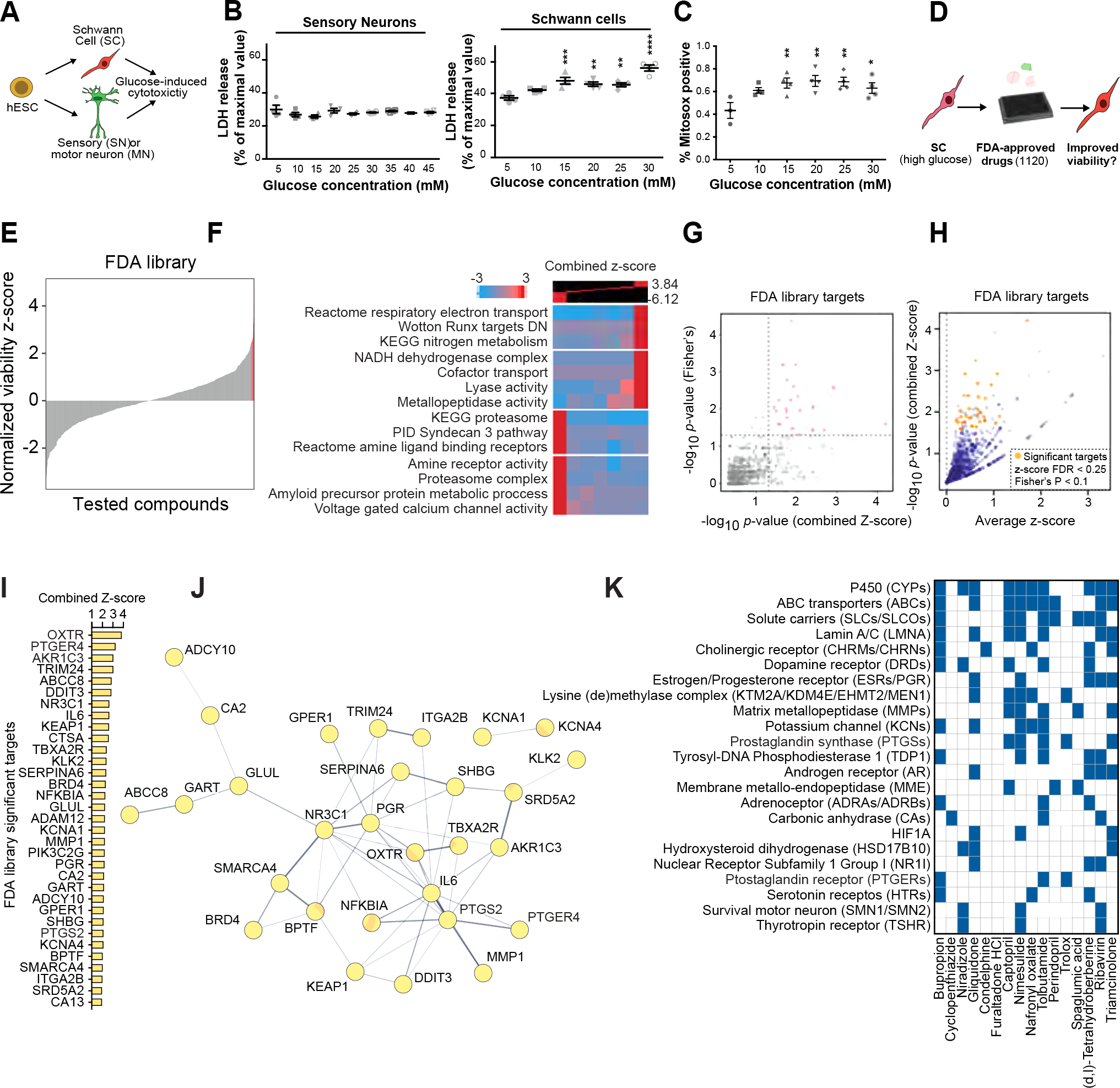
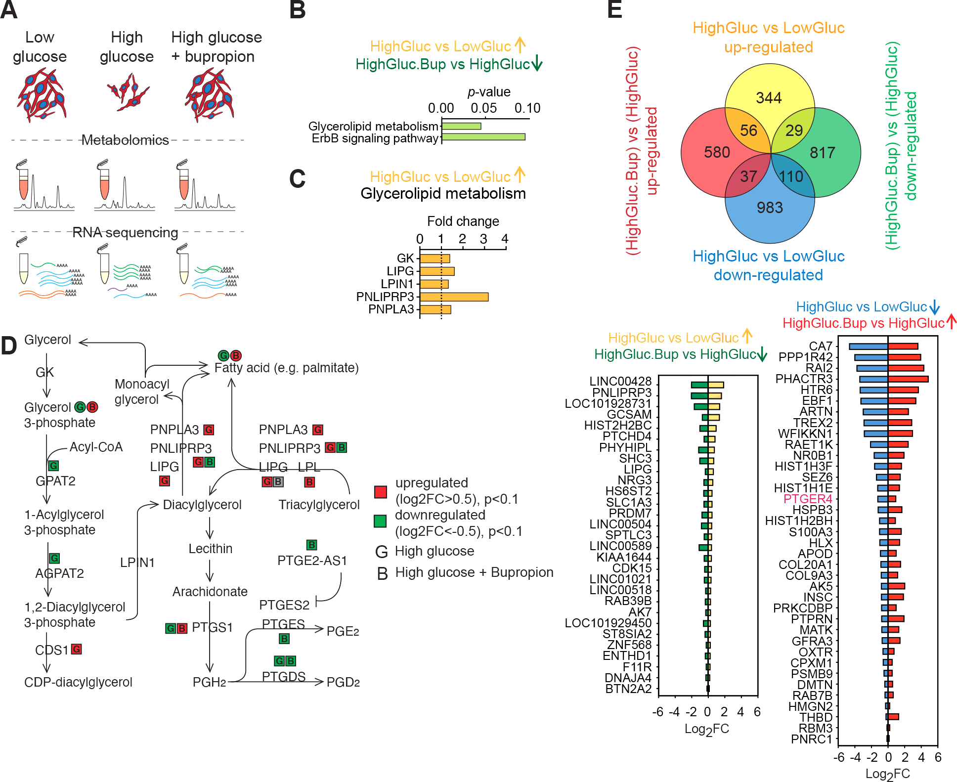
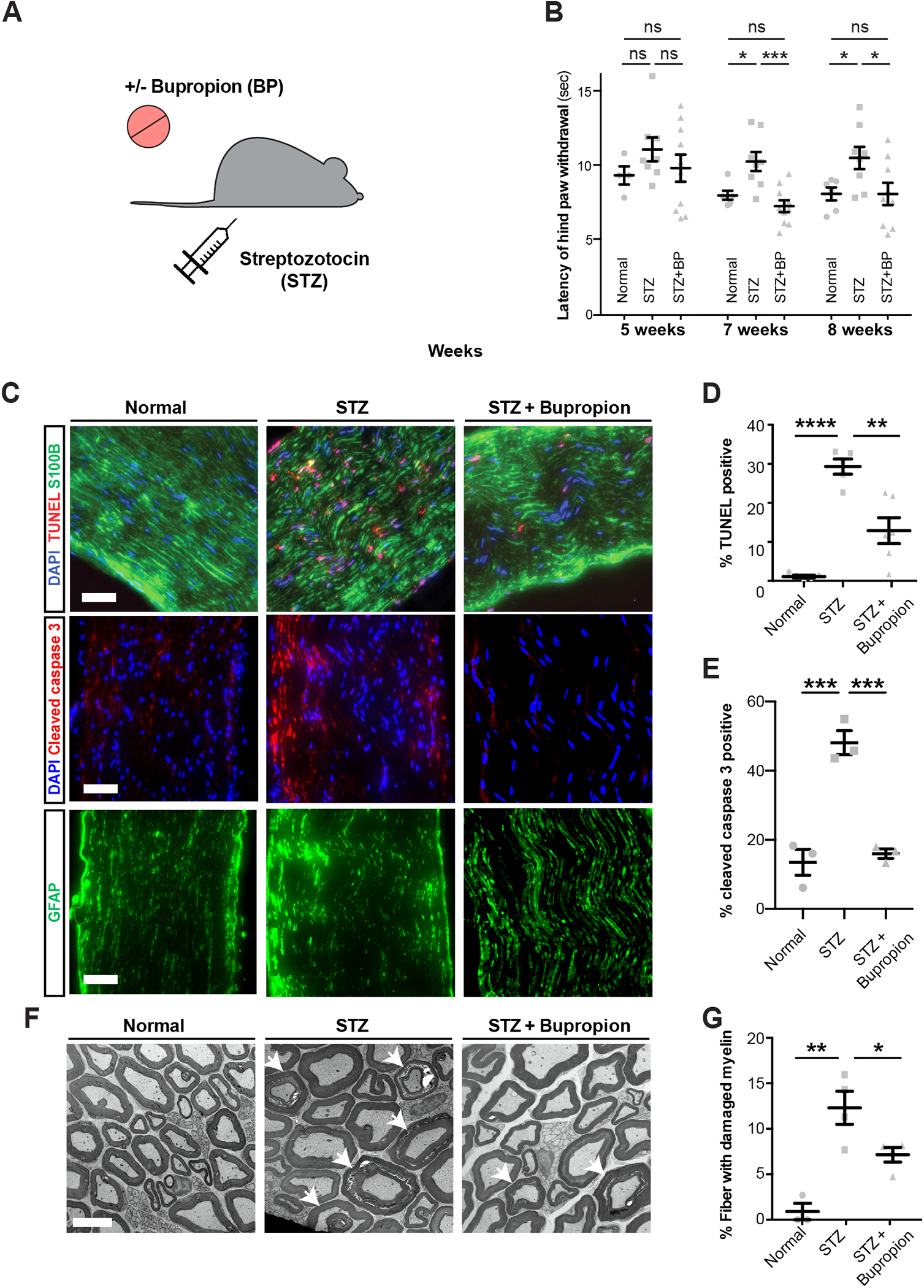
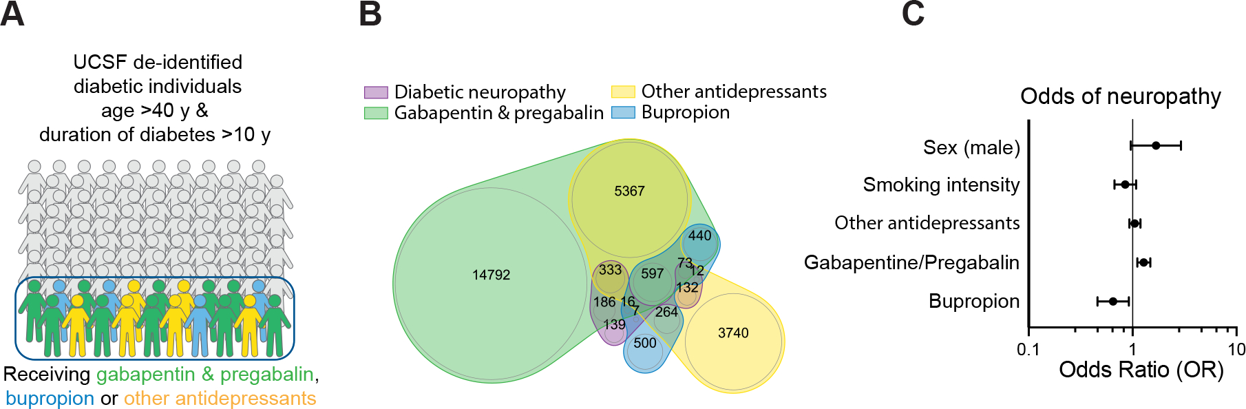
Schwann cells (SCs) are the primary glia of the peripheral nervous system. SCs are involved in many debilitating disorders, including diabetic peripheral neuropathy (DPN). Here, we present a strategy for deriving SCs from human pluripotent stem cells (hPSCs) that enables comprehensive studies of SC development, physiology, and disease. hPSC-derived SCs recapitulate the molecular features of primary SCs and are capable of in vitro and in vivo myelination. We established a model of DPN that revealed the selective vulnerability of SCs to high glucose. We performed a high-throughput screen and found that an antidepressant drug, bupropion, counteracts glucotoxicity in SCs. Treatment of hyperglycemic mice with bupropion prevents their sensory dysfunction, SC death, and myelin damage. Further, our retrospective analysis of health records revealed that bupropion treatment is associated with a lower incidence of neuropathy among diabetic patients. These results highlight the power of this approach for identifying therapeutic candidates for DPN.

Keywords: demyelination; drug repurposing; metabolic stress; myelination disorders; nerve repair.


Conflict of interest statement

Declaration of interests F.F., L.S., S.C., and Z.G. are inventors on several patent applications owned by UCSF, MSKCC, and Weill Cornell Medicine related to hPSC differentiation technologies including the technologies reported in this manuscript. L.S. is a scientific co-founder and paid consultant of BlueRock Therapeutics and DaCapo BrainScience.

Figures

Figure 1:
Figure 1:. Deriving Schwann cells from hPSCs
A) Schematic of the protocol for deriving developing precursors and SC cultures from hPSCs-derived neural crests (NCs). B) SOX10::GFP expression at days 11, 25 and 35 of differentiation. Scale bars = 100 μm (left and middle) and 25 μm (right). C, D) Representative (C) and quantification (D) of immunofluorescence images of hPSC-derived SCs for lineage markers (D60). Scale bar= 25 μm. E) UMAP visualization of scRNA-seq data for low passage (LP, Day 38) and high passage (HP, Day 58) SC cultures. SCPD: SC precursor derived; SCP: SC precursor. F, G) Dot plot of the scaled average expression of SC differentiation and myelination (left) and nerve support (right), myelinating (mySC) and non-myelinating (nmSC) SC markers in scRNA-seq data of LP and HP SCs.
Figure 2:
Figure 2:. hPSC-derived Schwann cells are transcriptionally similar to primary Schwann cells
A) Schematic of comparing SCs with primary human adult cell types. B) SingleCellNet classification of LP (left) and HP (right) SC clusters using the human cell atlas dataset as a reference. C) Schematic of comparing SCs with primary mouse developing peripheral glia. D) SingleCellNet classification of HP (left) and HP (right) SC clusters using a single cell transcriptomics mouse peripheral glia dataset as reference. E, F) Module scoring of top 100 HP SC type specific differentially expressed (DE) marker genes in LP SC types (left). Feature (left) and dot plot (right) visualizations are depicted. Panel E shows the HP dataset module scored in LP dataset and panel F shows the other way comparison. G) Principal component analysis (PCA) of NC cells, developing precursors, human primary SCs, and hPSC-derived SC cultures at D50 and D100 of differentiation in comparison with central nervous system (CNS) precursors.
Figure 3:
Figure 3:. hPSC-derived Schwann cells myelinate hPSC-derived sensory neurons and engraft in injured rat sciatic nerves
A) Feature plots of mature SC clusters isolated from LP (top) and HP (bottom) scRNA-seq data. dark colors: myelinating (mySC), bar plots: relative population of mySCs. B) Pathway enrichment analysis of top 250 DEGs of myelinating mature SCs in LP (left) and HP (right). Top 50 pathways from combined gene ontology biological process (GOBP), Reactome and KEGG analysis. C) Schematic of the SC co-cultures with sensory or motor neurons. D, E) Immunofluorescence imaging of SCs (EF1::RFP) co-cultured with sensory (EF1::GFP,D) or motor neurons (stained for CHAT,E). F) TEM of myelin in sensory neuron and SC co-cultures. Images were taken at 80 kV. Scale bars= 500 nm. G) Schematic of SC transplantation in adult rat sciatic nerves. RFP+ SCs were injected following nerve crush at the site of injury. H) Immunofluorescence staining of grafted sciatic nerves for human specific nuclear marker SC101 at 8 weeks post transplantation. I and J) Confocal analysis of teased sciatic nerve fibers for RFP (grafted human cells), axonal marker (NFH, I), myelin markers (MAG and P0, J), node markers Pan-Na+ (sodium channel, arrow heads, K), CASPR (arrow heads, L) and Kv1.2 (K+ channel, arrow heads, M) and DAPI. Scale bars= 100 μm in D left, E and B 20 μm in D right, I and J, 10 μm in K-M.
Figure 4:
Figure 4:. Schwann cells are selectively vulnerable to high glucose exposure
A) Schematic of the experimental paradigm for modeling diabetic nerve damage. B) Cytotoxicity analysis of SCs and sensory neurons at different glucose concentrations. C) Oxidative stress measurement of SCs exposed to increasing concentration of glucose. Statistical analysis (one-way ANOVA) comparing to low glucose (5 mM) condition. ns, not significant. * p<0.05; ** p<0.01. D) Schematic of high-throughput drug screening for identification of compounds that enhance the viability of high glucose-treated hPSC-SCs. E) Presentation of the distribution of library compounds by their corresponding normalized viability z-score. F) Gene-set enrichment analysis using iPAGE for the library compounds’ targets identifies GO terms associated with hits improving and worsening SC viability. G) p-value correlation plot to identify significant drug targets. See methods for details. H) One-sided volcano plot for all genes with positive z-scores. Gold: thresholds of combined z-score FDR<0.25 and Fisher’s p-value <0.1. I, J) Identified target genes (gold in H) and their protein-protein interaction network (J). K) Predicted targets of hits compiled from the following databases: BindingDB, Carlsbad, Dinies, PubChem bioassays, SEA, Superdrug 2 and SwisTargetPrediction.
Figure 5:
Figure 5:. Bupropion treatment counteracts high-glucose induced molecular changes in Schwann cells
A) Schematic of the unbiased metabolite and transcriptional profiling of differentially treated SCs. B) Pathway enrichment analysis of genes upregulated in high glucose and downregulated upon BP treatment. C) Glycerolipid metabolism enzymes upregulated in high glucose condition. D) Glycerolipid metabolism schematic adopted from KEGG shows changes in the enzymes and metabolites in response to high glucose and BP treatments in SCs. E) Venn diagram showing DEGs in SCs treated with high glucose, with or without BP (top). DEGs in hyperglycemia that show a reversed pattern of expression when treated with BP (bottom).
Figure 6:
Figure 6:. Bupropion treatment prevents diabetic nerve damage in vivo
A) Schematic of modeling diabetes and bupropion treatment in mice. B) Thermal sensitivity test measuring the latency of hind paw withdrawal in untreated mice or treated with STZ and bupropion. C-E) TUNEL and cleaved caspase-3 staining (C) and quantification (D, E) in sciatic nerves of untreated mice or treated with STZ and bupropion. Scale bar= 100 μm. F, G) Transmission electron microscopy and quantification of damaged myelin structures in sciatic nerves of untreated mice or treated with STZ and bupropion. Error bars: standard errors. Scale bar= 5 μm. * p<0.05; ** p<0.01; *** p<0.001; **** p < 0.0001.
Figure 7:
Figure 7:. Bupropion treatment is associated with lower odds of neuropathy in diabetic patients
A, B) Schematic (A) and Venn diagram (B) of the cohort of diabetes individuals derived from health records. C) Association of Bupropion with neuropathy in diabetic patients in multivariate logistic models adjusted for age, duration of diabetes, sex, smoking, and antidepressant drug treatment.

Comment in

References

    1. Jessen KR, and Mirsky R (2005). The origin and development of glial cells in peripheral nerves. Nat. Rev. Neurosci. 6, 671–682. 10.1038/nrn1746. - DOI - PubMed
    1. Lavdas AA, Papastefanaki F, Thomaidou D, and Matsas R (2008). Schwann cell transplantation for CNS repair. Curr. Med. Chem. 15, 151–160. - PubMed
    1. Adameyko I, Lallemend F, Aquino JB, Pereira JA, Topilko P, Müller T, Fritz N, Beljajeva A, Mochii M, Liste I, et al. (2009). Schwann Cell Precursors from Nerve Innervation Are a Cellular Origin of Melanocytes in Skin. Cell 139, 366–379. 10.1016/j.cell.2009.07.049. - DOI - PubMed
    1. Bonnamour G, Soret R, and Pilon N (2021). Dhh-expressing Schwann cell precursors contribute to skin and cochlear melanocytes, but not to vestibular melanocytes. Pigment Cell Melanoma Res. 34, 648–654. 10.1111/pcmr.12938. - DOI - PubMed
    1. Nitzan E, Pfaltzgraff ER, Labosky PA, and Kalcheim C (2013). Neural crest and Schwann cell progenitor-derived melanocytes are two spatially segregated populations similarly regulated by Foxd3. Proc. Natl. Acad. Sci. U. S. A. 110, 12709–12714. 10.1073/pnas.1306287110. - DOI - PMC - PubMed

Publication types