. 2023 May 11;186(10):2238-2255.e20.
doi: 10.1016/j.cell.2023.04.018. Epub 2023 May 4.

Plasma membrane preassociation drives β-arrestin coupling to receptors and activation

Affiliations

Plasma membrane preassociation drives β-arrestin coupling to receptors and activation

Jak Grimes et al. Cell. .

Abstract

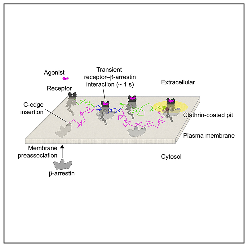
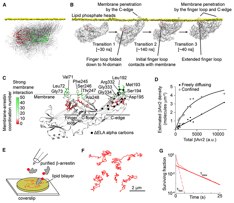
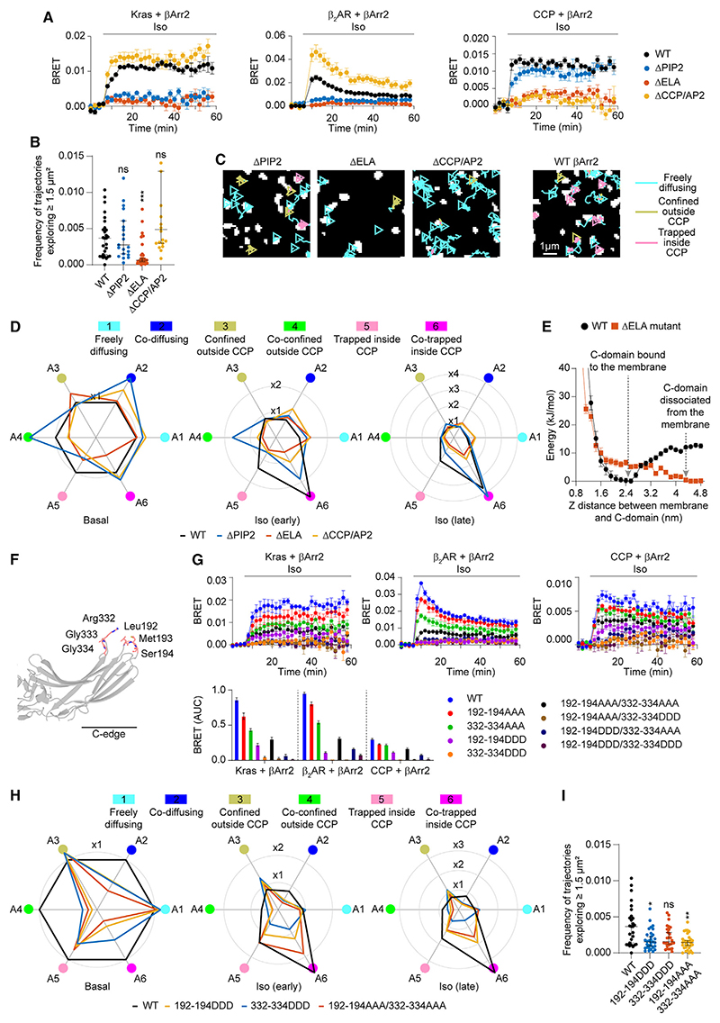
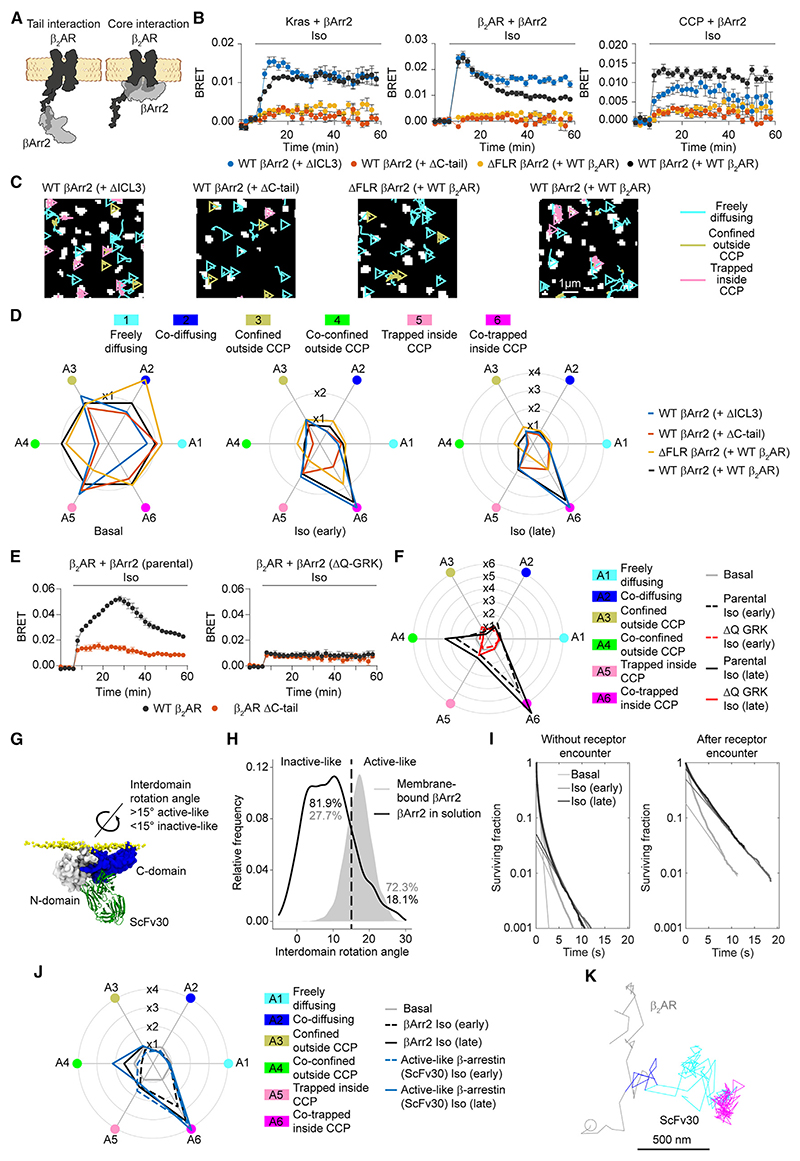
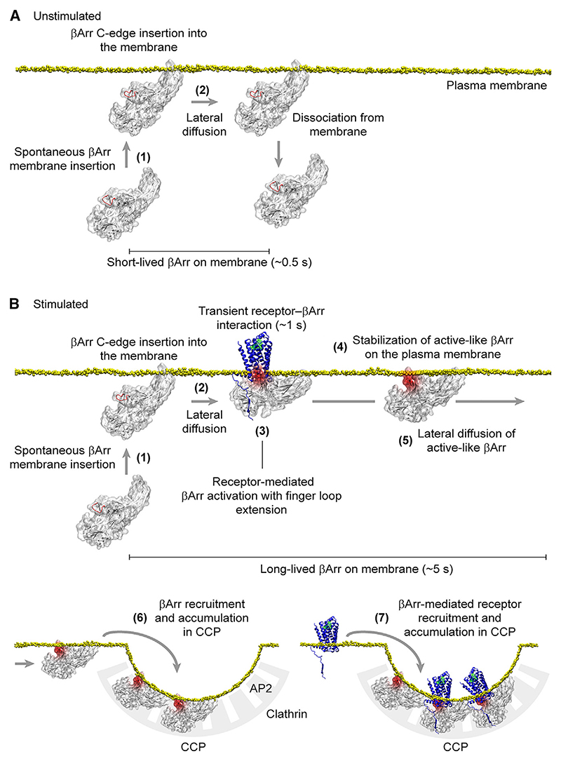
β-arrestin plays a key role in G protein-coupled receptor (GPCR) signaling and desensitization. Despite recent structural advances, the mechanisms that govern receptor-β-arrestin interactions at the plasma membrane of living cells remain elusive. Here, we combine single-molecule microscopy with molecular dynamics simulations to dissect the complex sequence of events involved in β-arrestin interactions with both receptors and the lipid bilayer. Unexpectedly, our results reveal that β-arrestin spontaneously inserts into the lipid bilayer and transiently interacts with receptors via lateral diffusion on the plasma membrane. Moreover, they indicate that, following receptor interaction, the plasma membrane stabilizes β-arrestin in a longer-lived, membrane-bound state, allowing it to diffuse to clathrin-coated pits separately from the activating receptor. These results expand our current understanding of β-arrestin function at the plasma membrane, revealing a critical role for β-arrestin preassociation with the lipid bilayer in facilitating its interactions with receptors and subsequent activation.

Keywords: G protein-coupled receptors; GPCR; TIRF; arrestin; plasma membrane; protein-protein interactions; single-molecule microscopy.


Conflict of interest statement

Declaration of interests The authors declare no competing interests.

Figures

Graphic abstract
Graphic abstract
Figure 1
Figure 1. Single-molecule imaging of β2AR and βArr2
(A) Labeling strategy. (B) Representative single-molecule results. (C) Representative trajectory of a βArr2 molecule appearing and transiently diffusing on the plasma membrane. (D) Sites of βArr2 appearance on the plasma membrane. (E) Rates of βArr2 appearance on the plasma membrane. (F) Survival curves of βArr2 molecules at the plasma membrane. (G) Diffusivity states of β2AR and βArr2 molecules. (H) Transient single-molecule co-localization event between β2AR and βArr2 on the plasma membrane. (I) Survival curves of β2AR-βArr2 interactions, based on deconvolution of apparent co-localization times. CD86 was used as a non-interacting control. (J) Estimated kon and koff of β2AR-βArr2 interactions. (K) Representative spatial map (left) and overall distributions (right) of β2AR-βArr2 co-localization events in cells stimulated with isoproterenol (10 μM; late), color-coded based on the diffusivity states of the involved molecules. (L) β2AR-βArr2 single-molecule co-localizations over super-resolved (SRRF) image of actin filaments. Data are median ± 95% confidence interval. Differences in (G), (J) (kon), and (K) are statistically significant by Kruskal-Wallis test. *p < 0.05, **p < 0.01, ***p < 0.001, ****p < 0.0001 versus basal by t test with Bonferroni correction. ns, statistically not significant. See also Figure S1 and Table S5.
Figure 2
Figure 2. Affinity for receptor C-tail governs βArr2 interaction with receptors and plasma membrane behavior
(A) Schematic of the investigated receptors. (B) Kinetics of βArr2 recruitment to the plasma membrane (Kras) and receptor upon isoproterenol (10 μM) stimulation monitored by BRET. (C) Diffusivity states of receptor and βArr2 molecules. Trajectories (left) are after isoproterenol stimulation (10 μM; late). (D and E) Estimated kon (D) and koff (E) values for β1AR, β2AR, and β2V2R-βArr2 interactions. Data are mean ± SEM in (B) and median ± 95% confidence interval in (C)–(E). Results in (C) and (D) are statistically significant by Kruskal-Wallis test. **p < 0.01, ***p < 0.001, ****p < 0.0001 versus corresponding basal condition, and ++p<0.01, +++p < 0.001, ++++p < 0.0001 versus corresponding β1AR condition by t test with Bonferroni correction. ns, statistically not significant. See also Figure S2 and Table S5.
Figure 3
Figure 3. Sequence of events in β2AR-βArr2 interactions
(A) Simplified representation of the results of the Markov chain analysis. Dashed circles, corresponding basal occupancies. See Table S3 for full transition probabilities. (B) Changes in single-molecule state occupancies induced by isoproterenol (10 μM) stimulation. Data are normalized to the corresponding basal levels. (C) Example of a βArr2 molecule undergoing a transient interaction with a β2AR to then reach a CCP without an accompanying receptor. (D) Examples of βArr2 molecules visiting multiple CCPs via lateral diffusion. See also Figures S3 and S4 and Table S5.
Figure 4
Figure 4. Spontaneous β-arrestin insertion into the lipid bilayer
(A) Superposition of βArr2 conformations sampled in solution in MD simulations. A selected conformation is highlighted to show the positions of the N- (red) and C- (green) domains. (B) Linked molecular dynamics (MD) simulations showing spontaneous insertion of the βArr2 C-edge into the lipid bilayer followed by a conformational rearrangement of the finger loop and its penetration into the bilayer. (C) Extended MD simulations (3 μs accumulated time) of membrane-bound βArr2. The results are shown on a representative structure of fully membrane-inserted βArr2 obtained in the simulations. Color indicates the lipid coordination numbers of βArr2 residues. Main interacting residues (lipid coordination number > 20) are labeled. (D) Densities of freely diffusing βArr2 molecules on the plasma membrane of cells in which βArr2 expression was varied ~25-fold. (E) Schematic of the in vitro reconstitution experiments with purified β-arrestin and supported lipid bilayers. (F) Representative single-molecule trajectories showing lateral diffusion of purified β-arrestin in supported lipid bilayers. (G) Survival curve of β-arrestin molecules on supported lipid bilayers. See also Table S5.
Figure 5
Figure 5. Functional consequences of βArr2 membrane preassociation
(A) Kinetics of βArr2 mutant(ΔPIP2, ΔELA, ΔCCP/AP2) recruitment to the plasma membrane (Kras; left), β2AR(middle), or CCPs(right) upon isoproterenol (10 μM) stimulation monitored by BRET. (B) Propensity of βArr2 mutants to explore the plasma membrane. (C) Diffusivity states of βArr2 mutants. Shown are representative trajectories after stimulation with isoproterenol (10 μM; late). (D) Changes in single-molecule state occupancies induced by isoproterenol (1 0 μM) stimulation. Data are normalized to Halo-tagged WT βArr2 with SNAP-tagged WT β2AR basal. (E) Well-tempered metadynamics simulations comparing the ΔELA mutant and WT βArr2. Shown are the free-energy landscapes as the proteins are pulled out of the membrane, using as collective variable the distance between the C-domain and the plasma membrane. (F) Positions of the targeted mutations introduced in the C-edge of βArr2. (G) Kinetics of targeted C-edge mutant recruitment to the plasma membrane (Kras; left), β2AR (middle), or CCPs (right) upon isoproterenol (10 μM) stimulation monitored by BRET. (H) Changes in single-molecule state occupancies of targeted C-edge mutants induced by isoproterenol (10 μM) stimulation. Data are normalized to Halo-tagged WT βArr2 with SNAP-tagged WT β2AR basal. (I) Propensity of targeted C-edge mutants to explore the plasma membrane. Shown are the relative frequencies of molecules exploring ≥1.5 μm in unstimulated cells. Halo-tagged WT βArr2 is included in (A)-(D) and (G)-(I) for comparison. Data are mean ± SEM in (A) and (G) and median ± 95% confidence interval in (B) and (I). Differences in (B) and (I) are statistically significant by Kruskal-Wallis test. *p < 0.05, **p < 0.01, ***p < 0.001 versus Halo-tagged WT by t test with Bonferroni correction. See also Figure S1 and Table S5.
Figure 6
Figure 6. Mechanisms of βArr2 activation and stabilization at the plasma membrane
(A) Schematic of C-tail and core receptor-arrestin interactions. (B) Kinetics of βArr2 recruitment to the plasma membrane (Kras; left), β2AR (middle), or CCPs (right) upon isoproterenol (10 μM) stimulation for the indicated construct combinations monitored by BRET. (C) βArr2 diffusivity states in cells expressing the same construct combinations. Shown are representative trajectories after stimulation with isoproterenol (10 μM; late). The results with SNAP-tagged WT β2AR and Halo-tagged WT βArr2 are included for comparison. (D) Corresponding changes in single-molecule state occupancies induced by isoproterenol (10 μM) stimulation. Data are normalized to Halo-tagged WT βArr2 with SNAP-tagged WT β2AR basal, included for comparison. (E) Kinetics of βArr2 recruitment to β2AR and β2AR ΔC-tail upon isoproterenol (10 μM) stimulation in parental (left) and ΔQ-GRK KO cells (right) monitored by BRET. (F) Corresponding changes in single-molecule state occupancies induced by isoproterenol (10 μM) stimulation. (G) Recognition of active-like membrane-bound βArr2 by Fab30/ScFv30. The structural model was obtained by aligning the membrane-bound βArr2 conformation obtained in MD simulations to the crystal structure of the active βArr1-Fab30 complex (PDB: 4JQI). (H) MD simulations comparing βArr2 conformations in solution and bound to the lipid bilayer. (I) Survival curves at the plasma membrane of βArr2 molecules without receptor encounter and after receptor encounter. (J) Radar plot obtained from single-molecule experiments comparing the behavior of ScFv30, recognizing active-like β-arrestin, and total βArr2. (K) Example of an active-like β-arrestin molecule, visualized with ScFv30, undergoing transient interaction with a β2AR molecule (blue) to then diffuse away alone (cyan) and meet another receptor in a CCP (magenta). Data are mean ± SEM in (B) and (E). See also Figures S5, S6, and S7 and Table S5.
Figure 7
Figure 7. Proposed model of receptor-β-arrestin interactions
(A) Behavior of β-arrestin at the plasma membrane under unstimulated conditions. Inactive β-arrestin in the cytosol spontaneously binds to the plasma membrane via insertion of the C-edge into the lipid bilayer (1), allowing it to explore space via lateral diffusion (2). Most β-arrestin molecules remain on the plasma membrane for a short time before dissociating and returning to the cytosol. (B) Behavior of β-arrestin at the plasma membrane in the presence of a stimulated receptor. After spontaneous insertion into the plasma membrane (1), β-arrestin reaches the receptor via lateral diffusion (2). Transient interaction with the receptor catalyzes β-arrestin activation, including β-arrestin inter-domain rotation and extension of the finger loop (3). Following dissociation from the receptor, the interaction of the extended finger loop with the lipid bilayer likely contributes to stabilizing β-arrestin in a membrane-bound, active-like conformation (4). This causes β-arrestin molecules to stay longer and accumulate on the plasma membrane, allowing them to reach CCPs vial lateral diffusion separately from the activating receptors (5). The increase in the number of active β-arrestin molecules and time they spend diffusing on the plasma membrane leads their recruitment and accumulation in CCPs via interaction with AP2 and clathrin (6). β-arrestin molecules tethered to CCPs bind receptors diffusing on the plasma membrane, also causing their recruitment and accumulation in CCPs (7).

Comment in

References

    1. Pierce KL, Premont RT, Lefkowitz RJ. Seven-transmembrane receptors. Nat Rev Mol Cell Biol. 2002;3:639–650. doi: 10.1038/nrm908. - DOI - PubMed
    1. Hauser AS, Attwood MM, Rask-Andersen M, Schioth HB, Glo-riam DE. Trends in GPCR drug discovery: new agents, targets and indications. Nat Rev Drug Discov. 2017;16:829–842. doi: 10.1038/nrd.2017.178. - DOI - PMC - PubMed
    1. Lohse MJ, Benovic JL, Codina J, Caron MG, Lefkowitz RJ. β-arrestin: a protein that regulates β-adrenergic receptor function. Science. 1990;248:1547–1550. doi: 10.1126/science.2163110. - DOI - PubMed
    1. Pierce KL, Lefkowitz RJ. Classical and new roles of β-ar-restins in the regulation of G-protein-coupled receptors. Nat Rev Neuro-sci. 2001;2:727–733. doi: 10.1038/35094577. - DOI - PubMed
    1. Daaka Y, Luttrell LM, Ahn S, Della Rocca GJ, Ferguson SS, Caron MG, Lefkowitz RJ. Essential role for G protein-coupled receptor endocytosis in the activation of mitogen-activated protein kinase. J Biol Chem. 1998;273:685–688. doi: 10.1074/jbc.273.2.685. - DOI - PubMed

Publication types

LinkOut - more resources