. 2023 May 5;13(1):7322.
doi: 10.1038/s41598-023-34353-y.

An inducible model for genetic manipulation and fate-tracing of PDGFRβ-expressing fibrogenic cells in the liver

Affiliations

An inducible model for genetic manipulation and fate-tracing of PDGFRβ-expressing fibrogenic cells in the liver

Florian Hamberger et al. Sci Rep. .

Abstract

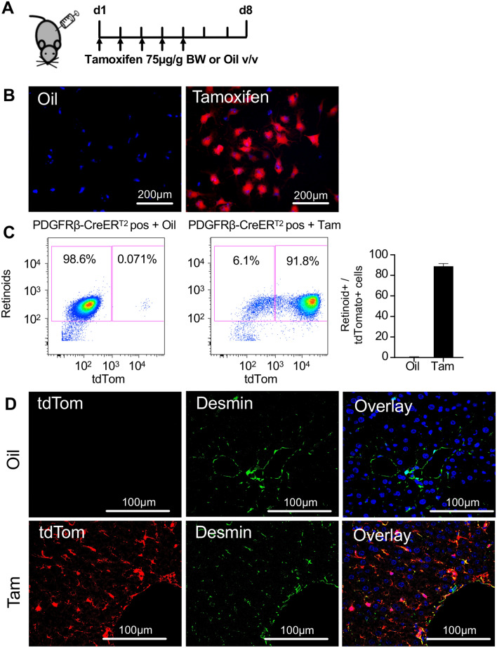
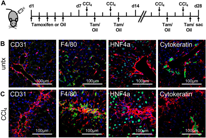
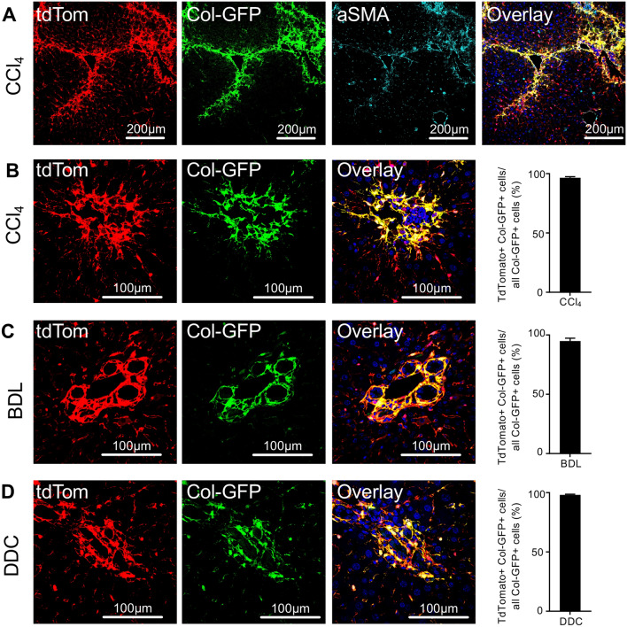
Myofibroblasts are the source of extracellular matrix protein during liver fibrogenesis. Fibroblasts, hepatic stellate cells (HSCs) and vascular smooth muscle cells are mesenchymal subpopulations in the liver that are characterized by the expression of PDGFRβ and contribute to the pool of these myofibroblasts. Conditional knockout models are important to better understand the function of specific liver cell populations including mesenchymal cells. While there is a limited number of constitutive mouse models for liver mesenchymal cell specific transgene expression, there is no established model for inducible gene targeting in HSCs or PDGFRβ-expressing mesenchymal cell populations in the liver. To address this, we investigated whether the tamoxifen inducible PDGFRβ-P2A-CreERT2 mouse can be used as a reliable tool to specifically express transgens in liver mesenchymal cells. Our data demonstrate, that PDGFRβ-P2A-CreERT2 specifically and efficiently marks over 90% of retinoid positive HSCs in healthy and fibrotic liver in mice upon tamoxifen injection, and that those cells give rise to Col1a1-expressing myofibroblasts in different models of liver fibrosis. Together with a negligible background recombination of only about 0.33%, this confirms that the PDGFRβ-P2A-CreERT2 mouse is nearly as efficient as established constitutive LratCre and PDGFRβ-Cre mouse models for recombination in HSCs, and that it is a powerful model for mesenchymal liver cell studies that require an inducible Cre approach.


Conflict of interest statement

The authors declare no competing interests.

Figures

Figure 1
Figure 1
PDGFRβ-P2A-CreERT2 efficiently labels retinoid positive HSCs in the liver upon tamoxifen induced Cre activation. (A) PDGFRβ-P2A-CreERT2 mice were crossed with mice containing a tdTomato (tdTom) Cre reporter. Cre activity was induced by tamoxifen injection (i.p.) at 75 µg/g BW for five consecutive days. Animals were sacrificed three days after the last tamoxifen or oil injection, respectively. (B,C) Fluorescent images (B) and pseudocolor plots (C) of freshly isolated HSCs from PDGFRβ-P2A-CreERT2 x tdTomato mice that received tamoxifen or oil show Cre-mediated tdTomato expression only in tamoxifen treated animals (n = 4). (D) Fluorescent staining shows co-localization of desmin with PDGFRβ-P2A-CreERT2 induced tdTomato expression.
Figure 2
Figure 2
Tamoxifen induced tdTomato expression in PDGFRβ-P2A-CreERT2 mice is restricted to mesenchymal cell populations. (A) PDGFRβ-P2A-CreERT2 was induced by tamoxifen injection (i.p.) at 75 µg/g BW for five consecutive days. Subsequently, liver fibrosis was induced by biweekly injections of CCl4 for a total of three weeks. Tamoxifen was continued during liver fibrosis induction. (B,C) Staining for markers of endothelial cells (CD31), macrophages (F4/80), hepatocytes (HNF4a), and cytokeratin show that PDGFRβ-P2A-CreERT2 driven tdTomato expression is exclusive to mesenchymal cell populations both in untreated and fibrotic mice.
Figure 3
Figure 3
PDGFRβ-P2A-CreERT2 labelled mesenchymal cell populations give rise to myofibroblasts in different models of liver injury. (A) Staining for myofibroblast marker aSMA combined with Col1a1GFP (ColGFP) overlaps with PDGFRβ-P2A-CreERT2 driven tdTomato expression in fibrotic liver. (B–D) Co-expression of ColGFP and PDGFRβ-P2A-CreERT2 driven tdTomato in different liver fibrosis models (CCl4 (n = 4), bile duct ligation BDL (n = 4), 3,5-diethoxycarbonyl-1,4-dihydrocollidine DDC (n = 4)). Means ± SEM.

References

    1. Roth GA, et al. Global, regional, and national age-sex-specific mortality for 282 causes of death in 195 countries and territories, 1980–2017: A systematic analysis for the Global Burden of Disease Study 2017. Lancet. 2018;392(10159):1736–1788. doi: 10.1016/S0140-6736(18)32203-7. - DOI - PMC - PubMed
    1. Fallowfield JA, Jimenez-Ramos M, Robertson A. Emerging synthetic drugs for the treatment of liver cirrhosis. Expert. Opin. Emerg. Drugs. 2021;26(2):149–163. doi: 10.1080/14728214.2021.1918099. - DOI - PubMed
    1. Mederacke I, et al. Fate tracing reveals hepatic stellate cells as dominant contributors to liver fibrosis independent of its aetiology. Nat. Commun. 2013;4:2823. doi: 10.1038/ncomms3823. - DOI - PMC - PubMed
    1. Filliol A, et al. Opposing roles of hepatic stellate cell subpopulations in hepatocarcinogenesis. Nature. 2022;610(7931):356–365. doi: 10.1038/s41586-022-05289-6. - DOI - PMC - PubMed
    1. Dobie R, et al. Single-cell transcriptomics uncovers zonation of function in the mesenchyme during liver fibrosis. Cell Rep. 2019;29(7):1832–18478. doi: 10.1016/j.celrep.2019.10.024. - DOI - PMC - PubMed

Publication types