. 2023 May 5;13(1):7339.
doi: 10.1038/s41598-023-34087-x.

The expression pattern of pyruvate dehydrogenase kinases predicts prognosis and correlates with immune exhaustion in clear cell renal cell carcinoma

Affiliations

The expression pattern of pyruvate dehydrogenase kinases predicts prognosis and correlates with immune exhaustion in clear cell renal cell carcinoma

Caroline E Nunes-Xavier et al. Sci Rep. .

Erratum in

Abstract

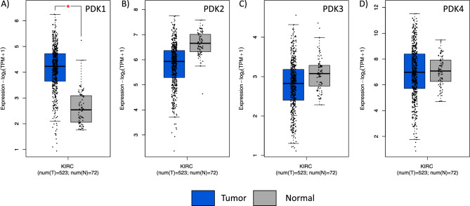
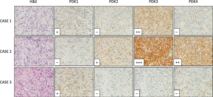
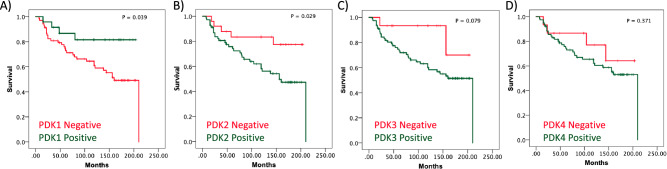
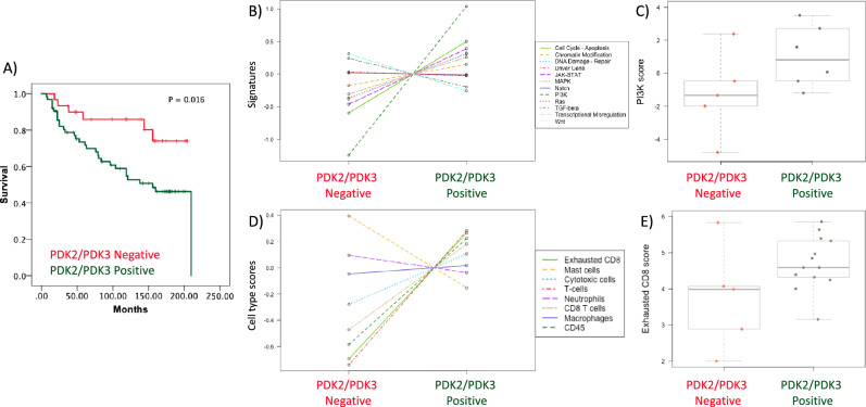
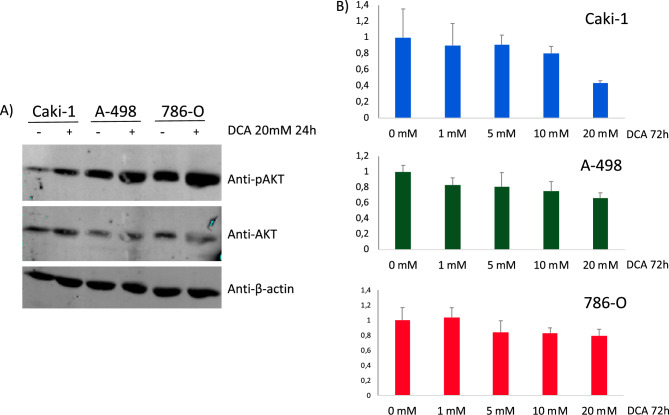
Renal cancer cells constitute a paradigm of tumor cells with a glycolytic reprogramming which drives metabolic alterations favouring cell survival and transformation. We studied the expression and activity of pyruvate dehydrogenase kinases (PDK1-4), key enzymes of the energy metabolism, in renal cancer cells. We analysed the expression, subcellular distribution and clinicopathological correlations of PDK1-4 by immunohistochemistry of tumor tissue microarray samples from a cohort of 96 clear cell renal cell carcinoma (ccRCC) patients. Gene expression analysis was performed on whole tumor tissue sections of a subset of ccRCC samples. PDK2 and PDK3 protein expression in tumor cells correlated with lower patient overall survival, whereas PDK1 protein expression correlated with higher patient survival. Gene expression analysis revealed molecular association of PDK2 and PDK3 expression with PI3K signalling pathway, as well as with T cell infiltration and exhausted CD8 T cells. Inhibition of PDK by dichloroacetate in human renal cancer cell lines resulted in lower cell viability, which was accompanied by an increase in pAKT. Together, our findings suggest a differential role for PDK enzymes in ccRCC progression, and highlight PDK as actionable metabolic proteins in relation with PI3K signalling and exhausted CD8 T cells in ccRCC.


Conflict of interest statement

The authors declare no competing interests.

Figures

Figure 1
Figure 1
mRNA expression of PDK1-4 in normal kidney and in ccRCC. Box plots for PDK1 (A), PDK2 (B), PDK3 (C) and PDK4 (D) showing mRNA expression in ccRCC tumor tissue (shown in blue) in comparison to normal kidney tissue (in grey). Data is represented in a logarithmic scale (Log2) and obtained from 523 tumor samples and 72 normal tissue samples (KIRC, TCGA). Statistically significant results (p < 0.01) are marked with an asterisk.
Figure 2
Figure 2
Protein expression of PDK1-4 in ccRCC tumors. Representative immunohistochemical staining of PDK1-4 protein expression in three representative ccRCC patient samples. H&E, Hematoxylin and Eosin staining. Case 1, positive staining of PDK1 and PDK3, and negative staining of PDK2 and PDK4. Case 2, moderate and high positive staining of PDK2, PDK3 and PDK4, and negative staining of PDK1. Case 3, positive staining of PDK1, and negative staining of PDK2, PDK3 and PDK4. Magnification: × 250.
Figure 3
Figure 3
Kaplan–Meier survival curves of ccRCC patients according to PDK1-4 tumor expression. Kaplan–Meier curves of overall survival based on PDK1 (A), PDK2 (B), PDK3 (C), and PDK4 (D) protein expression in the ccRCC cohort.
Figure 4
Figure 4
Molecular analysis of ccRCC patients according to PDK2 and PDK3 tumor expression. (A) Kaplan–Meier curves of overall survival based on PDK2/PDK3 expression in the ccRCC cohort. Patients with high PDK2/PDK3 protein expression and patients with low PDK2/PDK3 protein expression had significantly different time to recurrence (p = 0.016). (B) PanCancer Pathway signature in ccRCC tumor samples. Note PI3K pathway as most differentially expressed signalling pathway in high PDK2/PDK3 protein expression vs low PDK2 and PDK3 protein expression tumors. (C) Box plot of PI3K pathway score in high PDK2 and PDK3 protein expression vs low PDK2 and PDK3 protein expression tumors. (B, C) Data is represented in a logarithmic scale (Log2) and obtained from 11 ccRCC tumor samples. (D) Cell type score in ccRCC tumor samples using Immune Exhaustion panel. Note CD8 T cell exhaustion cell type as one of the higher enrichments in high PDK2/PDK3 protein expression vs low PDK2/PDK3 protein expression tumors. (E) Box plot of exhausted CD8 score in high PDK2/PDK3 protein expression vs low PDK2/PDK3 protein expression tumors. (D, E) Data is represented in a logarithmic scale (Log2) and obtained from 18 ccRCC tumor samples. Statistically significant results (p < 0.05) are marked with an asterisk.
Figure 5
Figure 5
(A) Immunoblot of pAKT, AKT and β-actin in ccRCC cells treated with PDK inhibitor DCA (20 mM for 24 h). Cropped blots are displayed. (B) Proliferation of Caki-1, A-498, and 786-O ccRCC cells, treated with dichloroacetate (DCA). Cell viability is shown, as determined by MTS analysis, after 72 h in the presence of DCA (1–20 mM for 72 h).

References

    1. Hay N. Reprogramming glucose metabolism in cancer: Can it be exploited for cancer therapy? Nat. Rev. Cancer. 2016;16:635–649. doi: 10.1038/nrc.2016.77. - DOI - PMC - PubMed
    1. Soga T. Cancer metabolism: Key players in metabolic reprogramming. Cancer Sci. 2013;104:275–281. doi: 10.1111/cas.12085. - DOI - PMC - PubMed
    1. Stine ZE, Schug ZT, Salvino JM, Dang CV. Targeting cancer metabolism in the era of precision oncology. Nat. Rev. Drug Discov. 2022;21:141–162. doi: 10.1038/s41573-021-00339-6. - DOI - PMC - PubMed
    1. Lu Z, Hunter T. Metabolic kinases moonlighting as protein kinases. Trends Biochem. Sci. 2018;43:301–310. doi: 10.1016/j.tibs.2018.01.006. - DOI - PMC - PubMed
    1. Ngo DC, Ververis K, Tortorella SM, Karagiannis TC. Introduction to the molecular basis of cancer metabolism and the Warburg effect. Mol. Biol. Rep. 2015;42:819–823. doi: 10.1007/s11033-015-3857-y. - DOI - PubMed

Publication types

Substances