. 2023 May;7(5):647-660.
doi: 10.1038/s41551-023-01032-2. Epub 2023 Apr 25.

Peptide-mediated delivery of CRISPR enzymes for the efficient editing of primary human lymphocytes

Affiliations

Peptide-mediated delivery of CRISPR enzymes for the efficient editing of primary human lymphocytes

Dana V Foss et al. Nat Biomed Eng. 2023 May.

Abstract

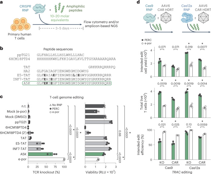
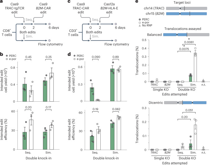
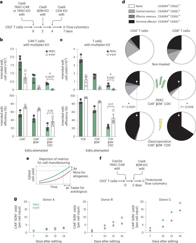
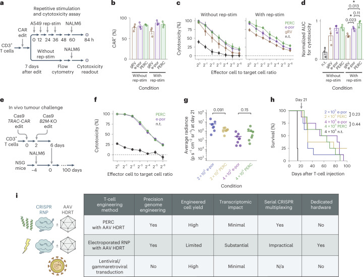
CRISPR-mediated genome editing of primary human lymphocytes is typically carried out via electroporation, which can be cytotoxic, cumbersome and costly. Here we show that the yields of edited primary human lymphocytes can be increased substantially by delivering a CRISPR ribonucleoprotein mixed with an amphiphilic peptide identified through screening. We evaluated the performance of this simple delivery method by knocking out genes in T cells, B cells and natural killer cells via the delivery of Cas9 or Cas12a ribonucleoproteins or an adenine base editor. We also show that peptide-mediated ribonucleoprotein delivery paired with an adeno-associated-virus-mediated homology-directed repair template can introduce a chimaeric antigen receptor gene at the T-cell receptor α constant locus, and that the engineered cells display antitumour potency in mice. The method is minimally perturbative, does not require dedicated hardware, and is compatible with multiplexed editing via sequential delivery, which minimizes the risk of genotoxicity. The peptide-mediated intracellular delivery of ribonucleoproteins may facilitate the manufacturing of engineered T cells.


Conflict of interest statement

D.V.F., D.N.N., D.C., S.U.S., J.M.H., A.M. and R.C.W. are named inventors on a patent application related to this work. D.N.N. is a holder of patents pertaining to but not resulting from this work. D.N.N. is a consultant for and owns stock in Navan Technologies. B.R.S. and A.M. are holders of patents pertaining to but not resulting from this work. J.E. is a compensated co-founder at Mnemo Therapeutics, a compensated scientific advisor to Cytovia Therapeutics, owns stocks in Mnemo Therapeutics and Cytovia Therapeutics, is a compensated member of the scientific advisory board at Treefrog Therapeutics, and has received a consulting fee from Casdin Capital. The J.E. laboratory has received research support from Cytovia Therapeutics, Mnemo Therapeutics and Takeda. J.E. is a holder of patents pertaining to but not resulting from this work. A.M. is a co-founder of Arsenal Biosciences, Spotlight Therapeutics and Survey Genomics, serves on the boards of directors at Spotlight Therapeutics and Survey Genomics, is a board observer (and former member of the board of directors) at Arsenal Biosciences, is a member of the scientific advisory boards of Arsenal Biosciences, Spotlight Therapeutics, Survey Genomics, NewLimit and Amgen, owns stock in Arsenal Biosciences, Spotlight Therapeutics, NewLimit, Survey Genomics and PACT Pharma, and has received fees from Arsenal Biosciences, Spotlight Therapeutics, NewLimit, 23andMe, PACT Pharma, Juno Therapeutics, Trizell, Vertex, Merck, Amgen, Genentech, AlphaSights, Rupert Case Management, Bernstein and ALDA. A.M. is an investor in and informal advisor to Offline Ventures and a client of EPIQ. The A.M. laboratory has received research support from Juno Therapeutics, Epinomics, Sanofi, GlaxoSmithKline, Gilead and Anthem. The R.C.W. laboratory has received research support from Genentech, Roche and Pfizer; the associated research is distinct from what is reported here.

Figures

Fig. 1
Fig. 1. PERC facilitates genome editing in primary human T cells.
a, Strategy for peptide-enabled RNP delivery. Peptides are mixed with pre-formed RNPs, allowing association, and the mixture is applied to primary human T cells. b, Tested peptide sequences. The influenza HA2 sequence is compared with TAT-fusion derivatives INF7-TAT and E5-TAT, as well as the A5K peptide. Differences are in bold. c, Cas9 TRAC-RNP delivery to CD4+ T cells, facilitated by peptides or electroporation (e-por). Knockout efficiency was assayed 4 days after delivery by flow cytometry for TCR surface expression. n.t. is non-treated, and mock is DMSO only or electroporated (no RNP). Total cell viability was assessed at day 2 and is reported in relative light units (RLU). P values are from an analysis of variance and Holm–Šidák multiple comparisons test. d, Editing of CD3+ T cells using Cas9 or Cas12a TRAC-RNP delivered by PERC or electroporation, paired with or without CAR AAV HDRT. n = 3 biological replicates from distinct human donors. Bars represent the mean. Error bars represent s.e.m. P values are from two-tailed Welch’s unpaired t-tests and Holm–Šidák multiple comparisons test. Source data
Fig. 2
Fig. 2. Compared with electroporation, PERC minimally perturbs T-cell state.
a, Volcano plots depict gene expression fold changes and adjusted P values (Padj) incorporating data from 6 h, 1 day and 7 days after editing. Red denotes significant upregulation, and blue denotes significant downregulation. This plot combines all timepoints, and timepoint-specific outcomes are in Supplementary Fig. 15. n.t., non-treated. b, Set of 84 genes that were significantly differentially expressed in one or more conditions in a. Left: fold changes at each timepoint. Middle: outcomes in each condition; the asterisk indicates the one gene that was significantly affected in PERC/DMSO. Right: NanoString gene category annotations. c, Fold changes across gene categories using the genes and annotations in b. The dots represent individual genes, and the shaded curves represent distributions of fold changes. Source data
Fig. 3
Fig. 3. Sequential editing with PERC minimizes translocations and maintains cell yield.
ad, Schematic (a) and data for sequential (Seq.) and simultaneous (Sim.) double knock-in editing (b) in CD8+ T cells using Cas9. Schematic (c) and data for sequential and simultaneous double knock-in editing (d) in CD3+ T cells using Cas9 and Cas12a. Plots depict cells that underwent both knock-ins. Flow cytometry was conducted 6 days after the first edit. e, Analysis of translocation frequencies by ddPCR for treatment with one or two RNPs, sequentially or simultaneously, corresponding to the experiment in a and b. In each experiment, n = 2–3 biological replicates from distinct human donors. Bars represent the mean. Error bars represent s.e.m. P values are from two-tailed Welch’s unpaired t-tests. n.t., non-treated. Source data
Fig. 4
Fig. 4. PERC supports cell viability and maintenance of phenotype in sequential editing.
a, Schematic of sequential editing of three loci in CD3+ T cells. b, Comparison of editing using PERC versus electroporation as measured by flow cytometry for TCR, CAR, B2M and CD5 surface expression. Reported CAR+ cells are also TCR. c, Editing without CAR AAV. Cell counts for each condition are scaled to an initial input of 4 × 106 T cells. n = 3 biological replicates from distinct human donors. Bars represent the mean. Error bars represent s.e.m. P values are from two-tailed Welch’s unpaired t-tests. d, Comparison of CD4+ and CD8+ cell phenotypes between delivery methods (independent of editing outcome), as measured by flow cytometry for CD62L and CD45RA surface expression. Pie segments represent the mean proportion of each phenotype. The dotted lines denote comparisons, with P values from a two-way analysis of variance and Holm–Šidák multiple comparisons test (Supplementary Figs. 24 and 25 indicate the comparisons). e, Depiction of metrics for evaluating improvement in cell manufacturing. f, Schematic of sequential editing. g, Expansion of edited cells over time, relative to the number of cells used for editing; n = 3 biological replicates from distinct human donors, plotted separately. Source data
Fig. 5
Fig. 5. Functional evaluation of CAR-T cells generated using PERC.
a, Schematic of the repetitive stimulation (rep-stim) and cytotoxicity assay. CD3+ T cells were edited to express a CAR using either gRV, Cas12a RNP electroporation and AAV, or Cas12a RNP PERC and AAV. b, Percentages of CAR+ cells in each condition with or without repetitive stimulation using CD19+ A549 cells. c,d, NALM6 cytotoxicity assay for T cells in each condition (c), and area under the curve analysis (d). n = 3 biological replicates from distinct human donors. Bars represent the mean. Error bars represent s.e.m. (3 biological replicates × 3 technical replicates). P values are from two-tailed Welch’s unpaired t-tests. e, Schematic of the in vivo tumour challenge experiment, using n = 8 mice per group. f, NALM6 cytotoxicity assay. Error bars represent s.e.m. from three technical replicates. g, BLI values on the last measurement day on which all CAR-T-cell-injected mice were alive. P values are from two-tailed Welch’s unpaired t-tests. h, Kaplan–Meier survival analysis. P values are from a log-rank test. P < 0.001 for each comparison of an edited condition versus non-treated (n.t.) cells. i, Table summarizing different delivery methods evaluated. Source data

Comment in

  • A gentler yield of ex vivo-edited T cells.
    Grünewald J, Schmidts A. Grünewald J, et al. Nat Biomed Eng. 2023 May;7(5):607-608. doi: 10.1038/s41551-023-01042-0. Nat Biomed Eng. 2023. PMID: 37208465 No abstract available.

References

    1. Weber EW, Maus MV, Mackall CL. The emerging landscape of immune cell therapies. Cell. 2020;181:46–62. doi: 10.1016/j.cell.2020.03.001. - DOI - PMC - PubMed
    1. Eyquem J, et al. Targeting a CAR to the TRAC locus with CRISPR/Cas9 enhances tumour rejection. Nature. 2017;543:113–117. doi: 10.1038/nature21405. - DOI - PMC - PubMed
    1. Rezalotfi A, Fritz L, Förster R, Bošnjak B. Challenges of CRISPR-based gene editing in primary T cells. Int. J. Mol. Sci. 2022;23:1689. doi: 10.3390/ijms23031689. - DOI - PMC - PubMed
    1. Aijaz A, et al. Biomanufacturing for clinically advanced cell therapies. Nat. Biomed. Eng. 2018;2:362–376. doi: 10.1038/s41551-018-0246-6. - DOI - PMC - PubMed
    1. Ruseska I, Zimmer A. Internalization mechanisms of cell-penetrating peptides. Beilstein J. Nanotechnol. 2020;11:101–123. doi: 10.3762/bjnano.11.10. - DOI - PMC - PubMed

Publication types