

Molecular imaging of arterial fibroblast activation protein: association with calcified plaque burden and cardiovascular risk factors
- PMID: 37147478
- PMCID: PMC10382401
- DOI: 10.1007/s00259-023-06245-w
Molecular imaging of arterial fibroblast activation protein: association with calcified plaque burden and cardiovascular risk factors
Abstract
Purpose: We aimed to assess prevalence, distribution, and intensity of in-vivo arterial wall fibroblast activation protein (FAP) uptake, and its association with calcified plaque burden, cardiovascular risk factors (CVRFs), and FAP-avid tumor burden.
Methods: We analyzed 69 oncologic patients who underwent [68 Ga]Ga-FAPI-04 PET/CT. Arterial wall FAP inhibitor (FAPI) uptake in major vessel segments was evaluated. We then investigated the associations of arterial wall uptake with calcified plaque burden (including number of plaques, plaque thickness, and calcification circumference), CVRFs, FAP-positive total tumor burden, and image noise (coefficient of variation, from normal liver parenchyma).
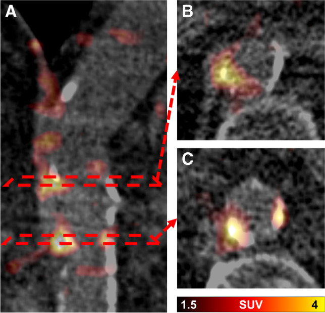
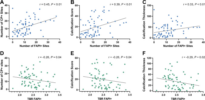
Results: High focal arterial FAPI uptake (FAPI +) was recorded in 64/69 (92.8%) scans in 800 sites, of which 377 (47.1%) exhibited concordant vessel wall calcification. The number of FAPI + sites per patient and (FAPI +)-derived target-to-background ratio (TBR) correlated significantly with the number of calcified plaques (FAPI + number: r = 0.45, P < 0.01; TBR: r = - 0.26, P = 0.04), calcified plaque thickness (FAPI + number: r = 0.33, P < 0.01; TBR: r = - 0.29, P = 0.02), and calcification circumference (FAPI + number: r = 0.34, P < 0.01; TBR: r = - 0.26, P = 0.04). In univariate analysis, only body mass index was significantly associated with the number of FAPI + sites (OR 1.06; 95% CI, 1.02 - 1.12, P < 0.01). The numbers of FAPI + sites and FAPI + TBR, however, were not associated with other investigated CVRFs in univariate and multivariate regression analyses. Image noise, however, showed significant correlations with FAPI + TBR (r = 0.30) and the number of FAPI + sites (r = 0.28; P = 0.02, respectively). In addition, there was no significant interaction between FAP-positive tumor burden and arterial wall FAPI uptake (P ≥ 0.13).
Conclusion: [68 Ga]Ga-FAPI-04 PET identifies arterial wall lesions and is linked to marked calcification and overall calcified plaque burden, but is not consistently associated with cardiovascular risk. Apparent wall uptake may be partially explained by image noise.
Keywords: Atherosclerosis; Atherosclerotic plaque; Cardiovascular risk factors; Fibroblast activation protein; Tumor burden; [68 Ga]Ga-FAPI.
© 2023. The Author(s).
Conflict of interest statement
TL is a co-inventor of a patent application for quinolone-based FAP-targeting agents for imaging and therapy in nuclear medicine. TL also has shares of a consultancy group for iTheranostics. All other authors declare no conflict of interest.
Figures


References
-
- Monslow J, Todd L, Chojnowski JE, Govindaraju PK, Assoian RK, Pure E. Fibroblast activation protein regulates lesion burden and the fibroinflammatory response in apoe-deficient mice in a sexually dimorphic manner. Am J Pathol. 2020;190:1118–1136. doi: 10.1016/j.ajpath.2020.01.004. - DOI - PMC - PubMed
MeSH terms
Substances
Grants and funding
LinkOut - more resources
Full Text Sources
Miscellaneous
