Case Reports
. 2023 May 5;19(1):69.
doi: 10.1186/s12917-023-03622-3.

First case report of pustules associated with Escherichia fergusonii in the chinese pangolin (Manis pentadactyla aurita)

Affiliations
Case Reports

First case report of pustules associated with Escherichia fergusonii in the chinese pangolin (Manis pentadactyla aurita)

Fuyu An et al. BMC Vet Res. .

Abstract

Background: Escherichia fergusonii is a common conditionally pathogenic bacterium that infects humans and animals. E. fergusonii has been reported to cause diarrhea, respiratory disease, and septicemia, but it is rarely reported to cause skin infections in animals. E. fergusonii has been isolated from the skin and muscular tissue of Chinese pangolin (Manis pentadactyla aurita). To date, there have been no reports of Chinese pangolins with clinical signs of skin diseases.

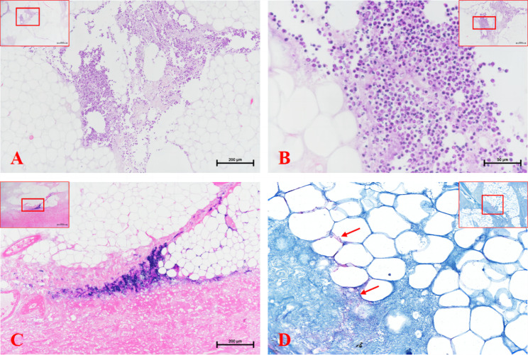
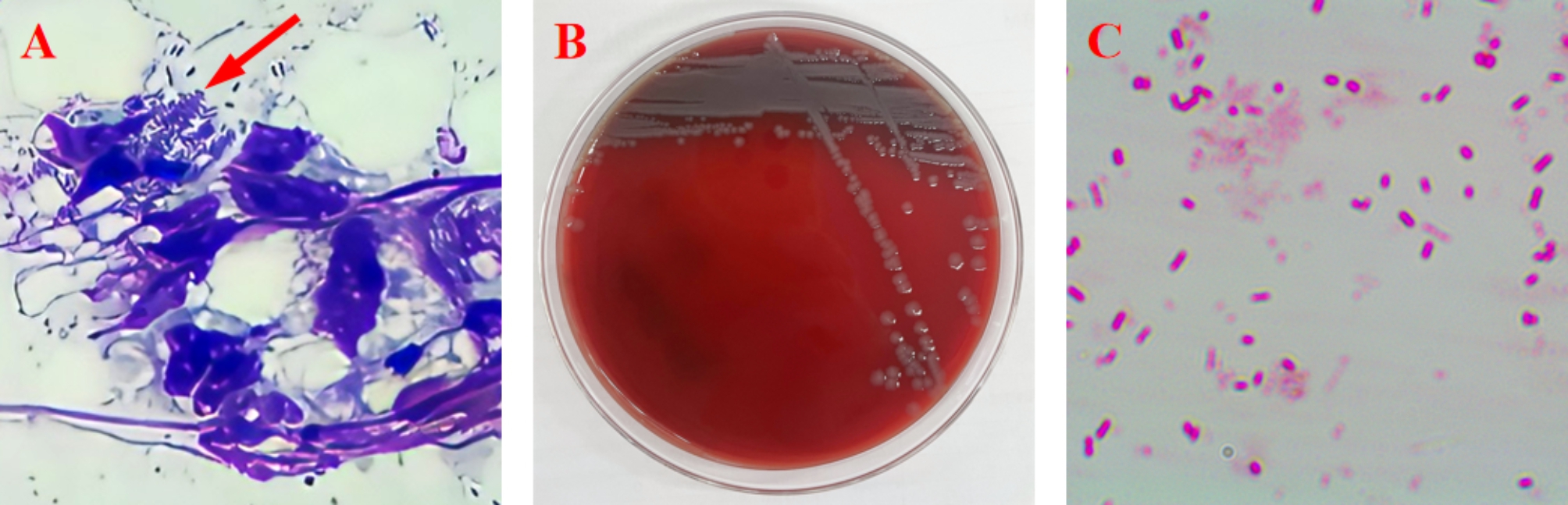
Case presentation: This case report describes the clinical case of a subadult (bodyweight: 1.1 kg) female Chinese pangolin from wild rescue with pustules and subcutaneous suppurative infection due to E. fergusonii in the abdominal skin. Bacterial culture, Biochemical analysis, PCR and histopathology were utilized to identify the bacteria in the pustule puncture fluid and infected tissue. To the best of our knowledge, this is the first report of E. fergusonii-related pustules on a Chinese pangolin.

Conclusion: This case report presents the first observed skin infection in a Chinese pangolin. E. fergusonii infection should be considered as a possible differential diagnosis of pustules and subcutaneous suppurative skin conditions in Chinese pangolins, and we also provide several recommendations for the diagnosis and treatment of this disease.

Keywords: Chinese pangolin; Escherichia fergusonii; Pustules; Skin.


Conflict of interest statement

The authors declare no conflict of interest.

Figures

Fig. 1
Fig. 1
Clinical presentation of a bacterial infection of a Chinese pangolin: (A) The abdomen is covered with pustules of the various sizes, day 1; (B) Pustules disappeared from abdominal skin, day 7.
Fig. 2
Fig. 2
Clinical presentation of Chinese pangolin with subcutaneous abscesses in the course of treatmentcellulitis during treatment: 1st day (A); the removed necrotic connective tissue of 1st day (B); the wound was debrided, 1st day (C); wounds dehiscence with infection, 18th day (D); the wound was debrided secondly, 18th day (E); healed wound site, 58th day (F).
Fig. 3
Fig. 3
Histologic section of removed wound tissue (Figue 3-A to C is hematoxilin and eosin): (A) subcutaneous fat tissue with severe inflammatory infiltration by neutrophilic granulocytes; (B) higher magnification of (A); (C) subcutaneous necrotic tissue containing numerous bacterial colonies; (D) bacterial colonies in subcutaneous necrotic tissue seen under Giemsa stain
Fig. 4
Fig. 4
(A) Diff-quik staining revealing rod-shaped bacteria in the suppurative exsudate (10×100). (B) pale, translucent bacterial colonies cultured on blood agar. (C) Gram stain: The cultured bacteria was a rod-shaped, gram-negative, and generally motile (10×100)

References

    1. Farmer J, 3rd, Fanning G, Davis B, O’hara C, Riddle C, Hickman-Brenner F, et al. Escherichia fergusonii and Enterobacter taylorae, two new species of Enterobacteriaceae isolated from clinical specimens. J Clin Microbiol. 1985;21(1):77–81. doi: 10.1128/jcm.21.1.77-81.1985. - DOI - PMC - PubMed
    1. Simmons K, Rempel H, Block G, Forgetta V, Vaillancourt R, Malouin F, et al. Duplex PCR methods for the molecular detection of Escherichia fergusonii isolates from broiler chickens. Appl Environ Microbiol. 2014;80(6):1941–8. doi: 10.1128/AEM.04169-13. - DOI - PMC - PubMed
    1. Weiss ATA, Lübke-Becker A, Krenz M, van der Grinten E. Enteritis and septicemia in a horse associated with infection by Escherichia fergusonii. J Equi Vet Sci. 2011;31(7):361–4. doi: 10.1016/j.jevs.2011.01.005. - DOI
    1. Foster G, Evans J, Tryland M, Hollamby S, MacArthur I, Gordon E, et al. Use of citrate adonitol agar as a selective medium for the isolation of Escherichia fergusonii from a captive reindeer herd. Vet Microbiol. 2010;144:484–6. doi: 10.1016/j.vetmic.2010.01.014. - DOI - PubMed
    1. Herráez P, Rodriguez F, De Espinosa A, Acosta B, et al. Fibrino-necrotic typhlitis caused by Escherichia fergusonii in ostriches (Struthio camelus) Avian Dis. 2005;49(1):167–9. doi: 10.1637/7221-061104r. - DOI - PubMed

Publication types

Supplementary concepts

LinkOut - more resources