. 2023 Jun 7;31(6):1775-1790.
doi: 10.1016/j.ymthe.2023.04.020. Epub 2023 May 5.

Targeting long non-coding RNA NUDT6 enhances smooth muscle cell survival and limits vascular disease progression

Affiliations

Targeting long non-coding RNA NUDT6 enhances smooth muscle cell survival and limits vascular disease progression

Hanna Winter et al. Mol Ther. .

Abstract

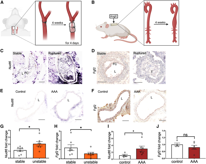
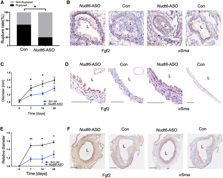
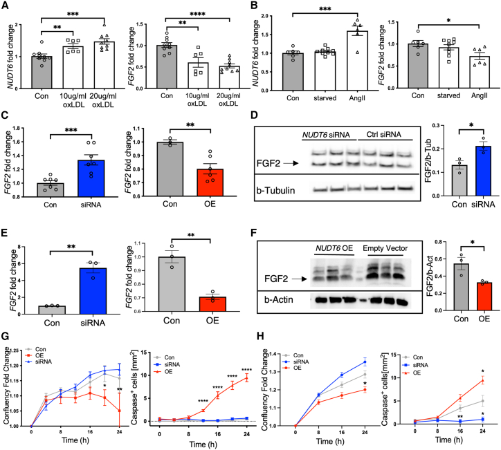
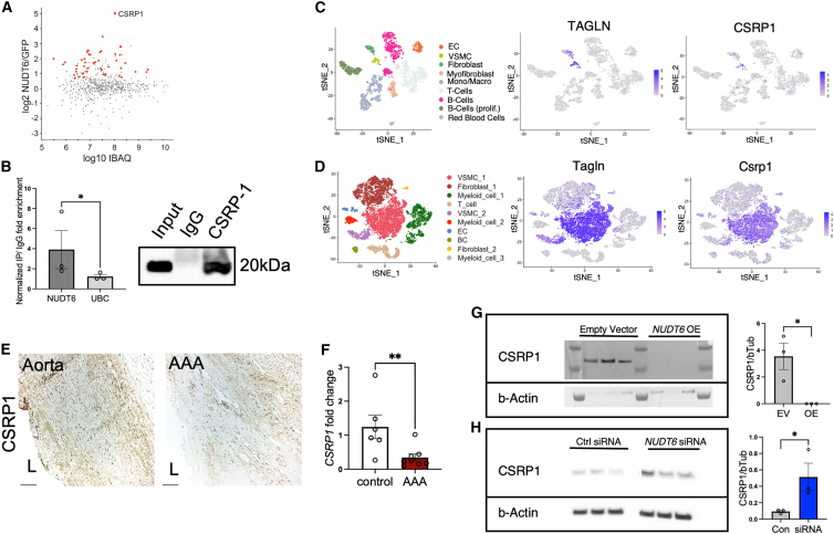
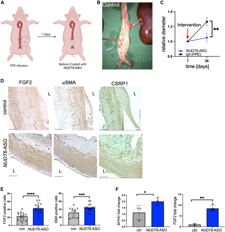
Long non-coding RNAs (lncRNAs) orchestrate various biological processes and regulate the development of cardiovascular diseases. Their potential therapeutic benefit to tackle disease progression has recently been extensively explored. Our study investigates the role of lncRNA Nudix Hydrolase 6 (NUDT6) and its antisense target fibroblast growth factor 2 (FGF2) in two vascular pathologies: abdominal aortic aneurysms (AAA) and carotid artery disease. Using tissue samples from both diseases, we detected a substantial increase of NUDT6, whereas FGF2 was downregulated. Targeting Nudt6 in vivo with antisense oligonucleotides in three murine and one porcine animal model of carotid artery disease and AAA limited disease progression. Restoration of FGF2 upon Nudt6 knockdown improved vessel wall morphology and fibrous cap stability. Overexpression of NUDT6 in vitro impaired smooth muscle cell (SMC) migration, while limiting their proliferation and augmenting apoptosis. By employing RNA pulldown followed by mass spectrometry as well as RNA immunoprecipitation, we identified Cysteine and Glycine Rich Protein 1 (CSRP1) as another direct NUDT6 interaction partner, regulating cell motility and SMC differentiation. Overall, the present study identifies NUDT6 as a well-conserved antisense transcript of FGF2. NUDT6 silencing triggers SMC survival and migration and could serve as a novel RNA-based therapeutic strategy in vascular diseases.

Keywords: aortic aneurysm; atherosclerosis; long non-coding RNAs; proliferation; smooth muscle cell; therapeutics; vascular disease.


Conflict of interest statement

Declaration of interests L.M. is a scientific consultant and adviser for Novo Nordisk (Malov, Denmark), DrugFarm (Shanghai, China), and Angiolutions (Hannover, Germany), and received research funds from Roche Diagnostics (Rotkreuz, Switzerland) and Novo Nordisk (Malov, Denmark).

Figures

None
Graphical abstract
Figure 1
Figure 1
NUDT6 expression is upregulated in human ruptured carotid plaques and human abdominal aortic aneurysm (A) Hematoxylin and eosin stained stable and unstable or ruptured atherosclerotic plaques from the Munich Vascular Biobank. The dotted lines mark the fibrous cap (FC) shielding the necrotic core (NC) from the lumen. FC > 200 μm defines stable lesions, FC < 200 μL defines unstable lesions. (B and C) NUDT6 (B) and FGF2 (C) expression in micro-dissected carotid FCs of ruptured vs. stable lesions (n = 10 per group). (D) In situ hybridization (ISH) of stable and ruptured atherosclerotic carotid lesions (n = 5 per group) indicates a stronger signal in the fibrous caps of ruptured lesions. L = Lumen. (E) Immunohistochemical analysis of FGF2 shows decreased expression in correlation with NUDT6 in FC ruptured lesions compared with stable lesions (n = 3 per group). Dashed lines mark the FC. FC = fibrous cap; L = lumen; M = media; NC = necrotic core. (F and G) qRT-PCR analysis detecting NUDT6 (F) and FGF2 (G) in whole fresh-frozen carotid arteries of controls (n = 3–5) or advanced lesions (n = 8). (H) In situ hybridization of control aorta and abdominal aortic aneurysm (AAA) (n = 3 per group) indicates higher expression of NUDT6 in the vessel’s smooth muscle cell-rich media layer (dashed lines). L = lumen. (J) Immunohistochemical staining of FGF2 shows decreased expression of both markers in AAA compared with aortic control (n = 3 per group). L = lumen. (I–K) qRT-PCR of whole fresh-frozen abdominal aortic aneurysm (n = 16) or control aorta (n = 13) detecting NUDT6 (I) and FGF2 (K). n = 13–16. Quantitative data are presented as mean +SEM. ∗p < 0.05; ∗∗p < 0.01; ∗∗∗∗p < 0.0001. Significance was determined using one-tailed Student’s t test (B), (C), (F), (G), and (I)–(K).
Figure 2
Figure 2
Increased Nudt6 mRNA and decreased Fgf2 mRNA and protein levels in two experimental animal models for vascular disease (A) Scheme describing the inducible plaque rupture mouse model. A ligation is placed distally to the carotid bifurcation. After 4 weeks, a cone-shaped cuff is placed around the vessel, leading to disturbed blood flow. (B) Scheme of the Angiotensin II mouse model. After the implantation of an AngII-filled pump, an abdominal aortic aneurysm develops. (C and D) Stable vs. ruptured carotid plaques derived from ApoE−/− mice undergoing the inducible plaque rupture (n = 7 per group) were stained for Nudt6 (C, in situ hybridization) and Fgf2 (D, immunohistochemistry), resembling the phenotype observed in human disease with increased Nudt6 mRNA expression and decreased Fgf2 protein levels in ruptured compared with stable lesions (n = 3 per group). (E and F) Saline-infused (control) vs. AngII-infused (AAA) ApoE−/− aorta from the Angiotensin II mouse model (n = 6 per group) was stained for Nudt6 (E) and Fgf2 (F). (G–J) The observed phenotype was confirmed by qRT-PCR in murine stable vs. unstable/ruptured ApoE−/− lesions (G) and (H) and murine ApoE−/− saline-infused (control) vs. AngII-infused aortas (I) and (J) (n = 4–7 per group). Quantitative statistics are presented as mean + SEM. ∗p < 0.05 in one-tailed Student’s t test. Scale bar, 100 μm.
Figure 3
Figure 3
Modulating Nudt6 in vivo leads to reduced rupture risk and reduced AAA growth (A) Nudt6-ASO treatment (n = 20) of ApoE−/− mice significantly reduced the rupture rate in the inducible plaque rupture model compared with scramble-control-treated animals (n = 20). (B) Fgf2 and αSma immunohistochemistry display restored expression after Nudt6-ASO treatment (n = 5 per group). (C) In the AngII model, local Nudt6-ASO treatment (n = 8) of ApoE−/− mice via ultrasound-targeted microbubble destruction (UTMD) led to significantly lower abdominal aortic diameter and reduced growth compared with scramble control treatment (n = 13). (D) Fgf2 and αSma protein levels are restored in Nudt6-ASO-treated animals (n = 3 per group). (E) Systemic Nudt6-ASO treatment (n = 4) in the porcine pancreatic elastase (PPE) mouse model significantly reduced abdominal aortic diameter of ApoE−/− mice and growth rate compared with control (n = 5). (F) Immunohistochemistry of Fgf2 and αSma indicates higher expression in the Nudt6-ASO group compared with the control group (n = 3 per group). Quantitative data are shown as mean ± SEM. ∗p < 0.05, ∗∗p < 0.01. Significance was calculated using χ2 test (A), and multiple t tests (C) and (D). Scale bar, 100 μm.
Figure 4
Figure 4
NUDT6 represses FGF2, thereby inducing apoptosis and inhibiting proliferation (A) Oxidized LDL treatment results in a dose-dependent increase of NUDT6 expression, whereas FGF2 decreases with increased dosage in human carotid smooth muscle cells (hCtSMCs) (n = 6 per group). (B) Angiotensin II treatment of human aortic smooth muscle cells (hAoSMCs) results in increased NUDT6 and decreased FGF2 expression (n = 6–9 per group). (C–E) By transfection of hCtSMCs (C) and hAoSMCS (E), siRNA-mediated knockdown of NUDT6 results in increased FGF2 expression, whereas vector-mediated overexpression of NUDT6 limits FGF2 mRNA levels. n = 6 per group. (D) NUDT6 silencing restores FGF2 protein, n = 3 per group. Scale bar, 100 μm. (F) NUDT6 overexpression leads to less FGF2 protein expression. (G and H) Dynamic live-cell imaging of both hCtSMCs (G) and hAoSMCs (H) show impaired proliferation capacity in NUDT6 overexpressing cells while the apoptosis rate is significantly increased. NUDT6-siRNA-treated cells behave as scramble control-treated cells. n ≥ 6. Quantitative results are presented as mean ± SEM. ∗p < 0.05; ∗∗p < 0.01; ∗∗∗p < 0.001; ∗∗∗∗p < 0.0001. Significance is determined using one-way ANOVA with Sidak (A) and (B), one-tailed Student’s t test (C)–(F), or two-way ANOVA with Tukey (G) and (H).
Figure 5
Figure 5
Identification and validation of NUDT6 interaction partner CSRP1 (A) Identified proteins in biotinylated NUDT6 vs. biotinylated GFP pulldown in hAoSMCs (n = 6 per condition), red dots represent significantly enriched proteins (Student’s t test) to NUDT6. (B) Confirmed binding of NUDT6 to CSRP1 protein in hAoSMCs. NUDT6 and UBC (unrelated target) enrichment in CSRP1 IP fraction was quantified using RT-qPCR and CSRP1 availability was verified via western blot. T-distributed stochastic neighbor embedding (t-SNE) plots of single-cell RNA sequencing data from human (C) human and (D) murine AAA (PPE-induced) with featured expression plots of Transgelin (TAGLN) and CSRP1. (E) Immunohistochemical staining of CSRP1 in human control aorta and AAA. (L indicates lumen). (F) qRT-PCR of whole fresh-frozen abdominal aortic control (n = 6) and AAA (n = 8) of CSRP1 mRNA. (G) CSRP1 protein levels after NUDT6 overexpression. (H) CSRP1 protein levels after NUDT6 siRNA-mediated knockdown (n = 3 per group). Quantitative data are shown as mean ± SEM. ∗p < 0.05; ∗∗p < 0.01. Significance was calculated using one-tailed Student’s t test. EC = endothelial cells, VSMCs = vascular smooth muscle cells; Mono/Macro = monocytes/macrophages; B-Cells (prolif.) = proliferating B cells. Scale bar, 150 μm.
Figure 6
Figure 6
Translational aspects of NUDT6 inhibition in a large animal model of AAA (A) Scheme describing the AAA induction in LDLR−/− Yucatan minipigs, in which the abdominal aorta was clamped and infused with PPE for 1 min. Seven days later, a balloon coated with NUDT6-ASO was introduced via the femoral artery and inflated for 3 min. (B) Representative image of the abdominal aorta of a control LDLR−/− minipig. (C) The relative diameter of NUDT6-ASO-treated animals (n = 4) shows a decrease compared with DEB control-treated animals (n = 3). (D) Immunohistochemical staining of the abdominal aorta of control and NUDT6-ASO-receiving pigs using antibodies against FGF2, αSMA, and CSRP1. “L” indicates lumen. (E) Quantification (four high-power fields per aorta, in total 16 counts) of immunohistochemical staining from (D) for FGF2 and αSMA. (F and G) qRT-PCR of whole porcine aortic tissue shows a significant increase in MYHC (F) and FGF2 (G) mRNA expression in NUDT6-ASO-treated animals (n = 4) compared with control (n = 3). Quantitative results are presented as mean ± SEM. ∗p < 0.05, ∗∗p < 0.01, ∗∗∗p < 0.001; ∗∗∗∗p < 0.0001. Significance was determined using two-way ANOVA with Sidak (C) and one-tailed Student’s t tests (E)–(G). Scale bar, 500 μm.

References

    1. GBD 2017 Causes of Death Collaborators Global, regional, and national age-sex-specific mortality for 282 causes of death in 195 countries and territories, 1980-2017: a systematic analysis for the Global Burden of Disease Study 2017. Lancet. 2018;392:1736–1788. doi: 10.1016/S0140-6736(18)32203-7. - DOI - PMC - PubMed
    1. Franck G., Dai J., Fifre A., Ngo S., Justine C., Michineau S., Allaire E., Gervais M. Reestablishment of the endothelial lining by endothelial cell therapy stabilizes experimental abdominal aortic aneurysms. Circulation. 2013;127:1877–1887. doi: 10.1161/CIRCULATIONAHA.113.001677. - DOI - PubMed
    1. Davignon J., Ganz P. Role of endothelial dysfunction in atherosclerosis. Circulation. 2004;109 doi: 10.1161/01.CIR.0000131515.03336.f8. III27-32. - DOI - PubMed
    1. Golledge J., Norman P.E. Atherosclerosis and abdominal aortic aneurysm: cause, response, or common risk factors? Arterioscler. Thromb. Vasc. Biol. 2010;30:1075–1077. doi: 10.1161/ATVBAHA.110.206573. - DOI - PMC - PubMed
    1. Ailawadi G., Eliason J.L., Upchurch G.R., Jr. Current concepts in the pathogenesis of abdominal aortic aneurysm. J. Vasc. Surg. 2003;38:584–588. doi: 10.1016/s0741-5214(03)00324-0. - DOI - PubMed

Publication types

MeSH terms

Substances