

A preparation of bacterial outer membrane with osmium tetroxide and uranyl acetate co-stain enables improved structural determination by transmission electron microscopy
- PMID: 37148329
- PMCID: PMC10673695
- DOI: 10.1093/jmicro/dfad027
A preparation of bacterial outer membrane with osmium tetroxide and uranyl acetate co-stain enables improved structural determination by transmission electron microscopy
Abstract
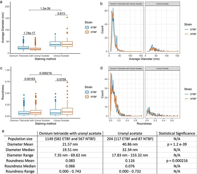
Biological nanoparticles, such as bacterial outer membrane vesicles (OMVs), are routinely characterized through transmission electron microscopy (TEM). In this study, we report a novel method to prepare OMVs for TEM imaging. To preserve vesicular shape and structure, we developed a dual fixation protocol involving osmium tetroxide incubation prior to negative staining with uranyl acetate. Combining osmium tetroxide with uranyl acetate resulted in preservation of sub-50 nm vesicles and improved morphological stability, enhancing characterization of lipid-based nanoparticles by TEM.
Keywords: bacteria; nanoparticles; osmium tetroxide; outer membrane vesicles; transmission electron microscopy; uranyl acetate.
© The Author(s) 2023. Published by Oxford University Press on behalf of The Japanese Society of Microscopy. All rights reserved. For permissions, please e-mail: journals.permissions@oup.com.
Figures


References
-
- Stanley S (2014) Biological nanoparticles and their influence on organisms. Curr. Opin. Biotechnol. 28: 69–74. - PubMed
-
- Tsatsaronis J A, Franch-Arroyo S, Resch U, and Charpentier E (2018) Extracellular vesicle RNA: a universal mediator of microbial communication? Trends Microbiol. 26: 401–410. - PubMed
MeSH terms
Substances
Grants and funding
LinkOut - more resources
Full Text Sources
