. 2023 Aug:166:133-146.
doi: 10.1016/j.actbio.2023.05.002. Epub 2023 May 5.

The cell-assembled extracellular matrix: A focus on the storage stability and terminal sterilization of this human "bio" material

Affiliations

The cell-assembled extracellular matrix: A focus on the storage stability and terminal sterilization of this human "bio" material

Diane Potart et al. Acta Biomater. 2023 Aug.

Abstract

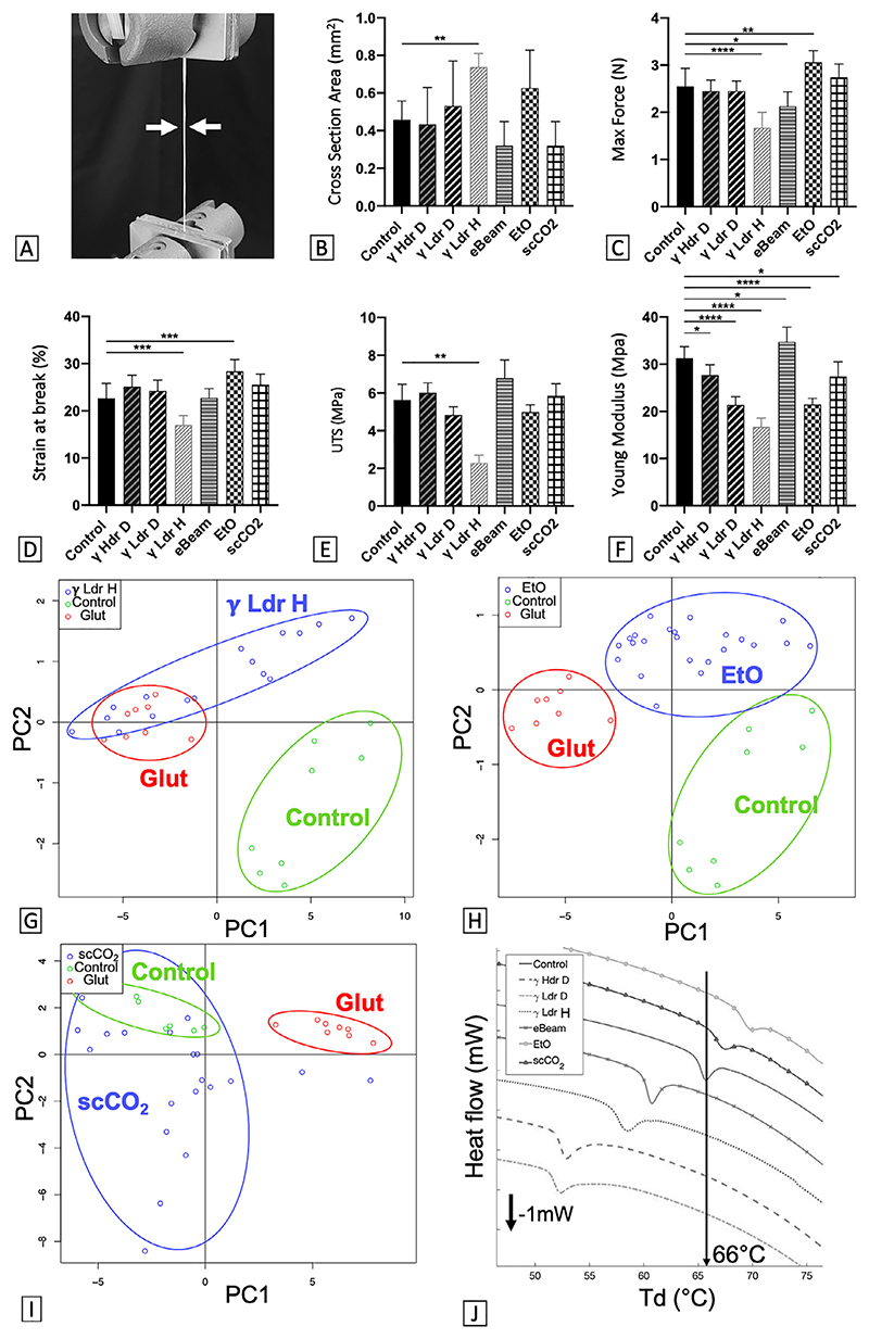
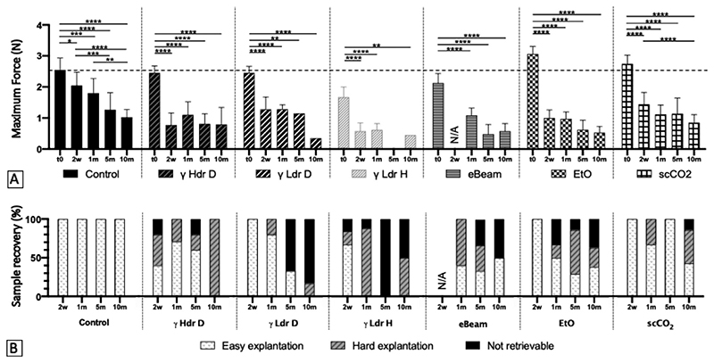
The Cell-Assembled extracellular Matrix (CAM) is an attractive biomaterial because it provided the backbone of vascular grafts that were successfully implanted in patients, and because it can now be assembled in "human textiles". For future clinical development, it is important to consider key manufacturing questions. In this study, the impact of various storage conditions and sterilization methods were evaluated. After 1 year of dry frozen storage, no change in mechanical nor physicochemical properties were detected. However, storage at 4 °C and room temperature resulted in some mechanical changes, especially for dry CAM, but physicochemical changes were minor. Sterilization modified CAM mechanical and physicochemical properties marginally except for hydrated gamma treatment. All sterilized CAM supported cell proliferation. CAM ribbons were implanted subcutaneously in immunodeficient rats to assess the impact of sterilization on the innate immune response. Sterilization accelerated strength loss but no significant difference could be shown at 10 months. Very mild and transient inflammatory responses were observed. Supercritical CO2 sterilization had the least effect. In conclusion, the CAM is a promising biomaterial since it is unaffected by long-term storage in conditions available in hospitals (hydrated at 4 °C), and can be sterilized terminally (scCO2) without compromising in vitro nor in vivo performance. STATEMENT OF SIGNIFICANCE: In the field of tissue engineering, the use of extracellular matrix (ECM) proteins as a scaffolding biomaterial has become very popular. Recently, many investigators have focused on ECM produced by cells in vitro to produce unprocessed biological scaffolds. As this new kind of "biomaterial" becomes more and more relevant, it is critical to consider key manufacturing questions to facilitate future transition to the clinic. This article presents an extensive evaluation of long-term storage stability and terminal sterilization effects on an extracellular matrix assembled by cells in vitro. We believe that this article will be of great interest to help tissue engineers involved in so-called scaffold-free approaches to better prepare the translation from benchtop to bedside.

Keywords: Cell-assembled extracellular matrix; In vivo remodeling; Mechanical properties; Physicochemical properties; Sterilization; Storage stability.


Conflict of interest statement

Declaration of Competing Interest The authors declare that they have no known competing financial interests or personal relationships that could have appeared to influence the work reported in this paper.

Figures

Fig. 1
Fig. 1. CAM can be conserved for at least 1 year with no (or minor) modification of mechanical and physicochemical properties.
(A) Stress-strain curve obtained after tensile testing of a control CAM ribbon. (B) Cross-section of devitalized CAM ribbons before (control) and after 1 year of storage in various conditions (RT: Room Temperature, HYDR = Hydrated). Tensile test results: (C) Maximum Force, (D) Ultimate Tensile Strength (UTS), (E) Strain at break and (F) Young modulus of CAM ribbons before and after 1 year storage. Results are expressed as mean of 3 experiments using 3 CAM batches (± SD, n = 5 to 8 ribbons) as % of control. *p < 0.05, ***p < 0.001, ****p < 0.0001 as compared to control. One-way ANOVA for Cross Section, Max Force, Strain at break and Kruskal-Wallis for Max Stress, Young Modulus. (G) PCA scatter plot of CAM FTIR spectra (4000 cm−1 to 400 cm−1) does not show a significant difference between control and CAM stored 1 year dry at RT (RT D). Glutaraldehyde-crosslinked CAM (Glut – positive denatured control) is easily distinguished from control. Ellipses cluster the different groups. (H) CAM denaturation temperature (Td) before and after 1 year of storage in various conditions (n = 4-11, *p < 0.05, **p < 0.01, One-way ANOVA).
Fig. 2
Fig. 2. CAM can be terminally sterilized with minor modification of its mechanical and physicochemical properties.
(A) Five-mm-wide CAM ribbon (white arrows) undergoing a tensile test. (B) Cross-section of devitalized CAM ribbons before (control) and after terminal sterilization with various methods. Tensile test results: (C) Maximum Force, (D) UTS, (E) Strain at break and (F) Young modulus of CAM ribbons before and after terminal sterilization. n = 7-8 ribbons, ± STD. *p < 0.05, **p < 0.01, ***p < 0.001, ****p < 0.0001 as compared to control. One-way ANOVA for Max Force, Strain at break and Young Modulus, Kruskal-Wallis for UTS. (G) PCA scatter plot of CAM FTIR spectra (4000 cm−1 to 400 cm−1) show a significant distinction between control and CAM sterilized with γ Hdr D, but γ Hdr D can not be distinguished from positive control (Glut). (H) PCA scatter plot of CAM FTIR spectra show a significant distinction between control, CAM sterilized with EtO and positive control. (I) PCA scatter plot of CAM FTIR spectra does not show a significant difference between control and CAM sterilized with scCO2. Ellipses cluster the different groups. (J) DSC thermogram of CAM samples shows a significant difference in Td (lowest point of endothermic peak) between control and sterilized CAM (n = 11-12, p < 0.0001, One-way ANOVA).
Fig. 3
Fig. 3. CAM can be terminally sterilized without modification of its microscopic structure and remain a favorable biomaterial for cell attachment.
Masson’s Trichrome staining on cross sections of CAM ribbons (dashed lines delimit one cross section). (A) The typical dense network of collagen (blue staining) is visible on sterilely produced control CAM ribbons. Devitalized cells are mainly visible at ribbons surface (white arrows). (B) After γ Ldr H sterilization, the microscopic structure of CAM ribbons was modified. Layers within the ribbon are delaminated, thus exposing gaps (black arrows) and increasing ribbons thickness. (C) Microscopic structure of EtO sterilized ribbons is similar to control. Dashed lines outline ribbons, scale bar = 50 μm. Microscopic images of HUVECs after VE-Cadherin (VE-Cad) and nuclear staining on sterilely produced or terminally sterilized CAM sheets. The presence of VE-Cadherin shows an organization of the cell-cell interface, suggesting a normal morphology of the endothelium regardless of the condition ((D) Control, (E) γ Ldr H, (F) scCO2). Scale bar = 50 μm. γ Ldr H: Gamma irradiation with Low dose rate Hydrated, EtO: Ethylene Oxide.
Fig. 4
Fig. 4. Terminal sterilization accelerates CAM loss of strength in early stages of in vivo remodeling, but may not affect its long-term evolution.
(A) Maximum force of 5-mm-wide CAM ribbons obtained in tensile test as a function of sterilization methods and time (γ Hdr D: Gamma irradiation with High dose rate Dry, γ Ldr D: Gamma irradiation with Low dose rate Dry, γ Ldr H: Gamma irradiation with Low dose rate Hydrated, eBeam: electron Beam irradiation, EtO: Ethylene Oxide, scCO2 : supercritical CO2). *p < 0.05, **p < 0.01, ***p < 0.001, ****p < 0.0001 as compared to control. Two-way ANOVA (n = 1-8). The dotted line indicates the maximum force of non-sterilized non-implanted CAM ribbons. (B) Retrievability of CAM ribbons (easy/hard/not retrievable) for mechanical evaluation as a function of sterilization methods and time.
Fig. 5
Fig. 5. CAM can be terminally sterilized without substantial increase of the acute immune response.
Histological analyses of sterilized CAM ribbons at 2 weeks (TM, H&E, Immunofluorescence for M1/M2 phenotype) in the immunodeficient rats. Asterisk (*) indicates the position of the ribbon delimited by the dashed lines. (A)(B) Foreign bodies (possibly rat hair, white arrows) are surrounded by numerous inflammatory cells (round dark purple). (C) Immunostaining for CD68 (red) labels macrophages and double staining with CCR7 (yellow) suggest large number of macrophages with a M1 phenotype (pro-inflammatory). (D) Immunostaining for CD68 (red) labels macrophages and double staining with CD206 (yellow) show few M2 macrophages (remodeling phenotype). Insets show high magnification views of doubly labelled cells showing membrane staining of CCR7/CD206 while CD68 stains cellular organelles. (E)(F) Very few inflammatory cells surround the control CAM ribbon, and adipocytes (yellow arrows) are present between ribbon layers. (G)(H) Double staining with CD68/CCR7 compared to double staining with CD68/CD206 suggest a majority of macrophages with a M2 phenotype. (I)(J) Inflammatory cell clusters (red arrows) are visible within the ribbon and between the folds it forms. Black arrows indicate blood vessels. (K)(L) Double staining suggests a majority of macrophages with a M1 phenotype. (M)-(P) The few macrophages present between the layers of the γ Ldr D sterilized ribbon are predominantly M1 phenotype. (Q)(R) Inflammatory cell clusters are visible within the γ Ldr H sterilized ribbon and between the folds it forms. (S)(T) CD68 labeled macrophages are either double stained with CCR7 or with CD206 showing the presence of both M1 and M2 phenotypes. (U)-(X) The few macrophages present around and between the EtO-sterilized ribbon layers are slightly more co-stained with CD206 than with CCR7, suggesting a slight majority of M2 phenotype. (Y)-(AB) The few macrophages present around and between the scCO2-sterilized ribbon layers are double-stained with either CD206 or CCR7, indicating M1 and M2 phenotypes. Scale bar = 100 μm (close up scale bar = 5 μm).
Fig. 6
Fig. 6. CAM can be terminally sterilized with no risk of chronic immune reaction.
Histological analyses of sterilized CAM ribbons at 1, 5 and 10 months (TM, H&E, M1/M2 phenotype) in the immunodeficient rats. Asterisk (*) indicates the position of the ribbon delimited by the dashed lines. (A)(B) Hardly any inflammatory cells surround the control CAM ribbon. Black arrows indicate blood vessels. (C)(D) Barely any cell is stained with CD68 (red, labelling for macrophages) and no double staining (yellow) with either CCR7 nor with CD206 shows the absence of both M1 and M2 phenotypes. (E)-(H) The few macrophages present between the layers of the γLdr H sterilized ribbon are predominantly M2 phenotype. The red arrow indicates inflammatory cells (round dark purple). (I)(J) Very few inflammatory cells are present around and between the scCO2-sterilized ribbon layers. (K)(L) Barely any cell is double stained with CD68/CCR7 nor CD68/CD206, showing the quasi-absence of both M1 and M2 phenotypes. (M)(N) Hardly any inflammatory cells surround the control CAM ribbon. (O)(P) Only one cell is double stained with CD68 and CCR7 showing the quasi-absence of both M1 and M2 phenotypes. (Q)-(T) The rare macrophages present surrounding the γ Ldr H sterilized ribbon are M2 phenotype. (U)(V) Hardly any inflammatory cells surround the control CAM ribbon. (W)(X) Only one cell is double stained with CD68 and CCR7 (M1 phenotype) showing the quasi-absence of both M1 and M2 phenotypes. (Y)(Z) Hardly any inflammatory cells surround the γ Ldr H CAM ribbon, and a few adipocytes are present between ribbons layers (yellow arrows). (AA)(AB) Only one cell is double stained with CD68 and CCR7 (M1 phenotype) showing the quasi-absence of both M1 and M2 phenotypes. Scale bar = 100 μm.

Similar articles

Cited by

References

    1. U.S. National Library of Medicine. ClinicalTrialsgov-Tissue engineering. [accessed April 9, 2022]. (n.d.). https://www.clinicaltrials.gov/ct2/results?cond=&term=tissue+engineering...
    1. Alliance for Regenerative Medicine. Alliance for regenerative medicine, Available products. 2023. (n.d.). https://alliancerm.org/available-products/
    1. Ghaemi RV, Siang LC, Yadav VG. Improving the rate of translation of tissue engineering products. Adv Healthc Mater. 2019;8:1900538. doi: 10.1002/adhm.201900538. - DOI - PubMed
    1. Wystrychowski W, Garrido SA, Marini A, Dusserre N, Radochonski S, Za-galski K, Antonelli J, Canalis M, Sammartino A, Darocha Z, Baczyn’ ski R, et al. Long-term results of autologous scaffold-free tissue-engineered vascular graft for hemodialysis access. J Vasc Access. 2022:112972982210959. doi: 10.1177/11297298221095994. - DOI - PubMed
    1. Magnan L, Labrunie G, Fénelon M, Dusserre N, Foulc M-P, Lafourcade M, Svahn I, Gontier E, Vélez JH, McAllister VTN, L’Heureux N. Human textiles: A cell-synthesized yarn as a truly “bio” material for tissue engineering applications. Acta Biomater. 2020;105:111–120. doi: 10.1016/j.actbio.2020.01.037. - DOI - PubMed

Publication types

Substances