Review
. 2023 Jun;16(2):197-211.
doi: 10.1016/j.path.2023.01.007. Epub 2023 Feb 2.

Hematopathology of Severe Acute Respiratory Syndrome Coronavirus 2 Infection and Coronavirus Disease-19

Affiliations
Review

Hematopathology of Severe Acute Respiratory Syndrome Coronavirus 2 Infection and Coronavirus Disease-19

Fabienne Lucas et al. Surg Pathol Clin. 2023 Jun.

Abstract

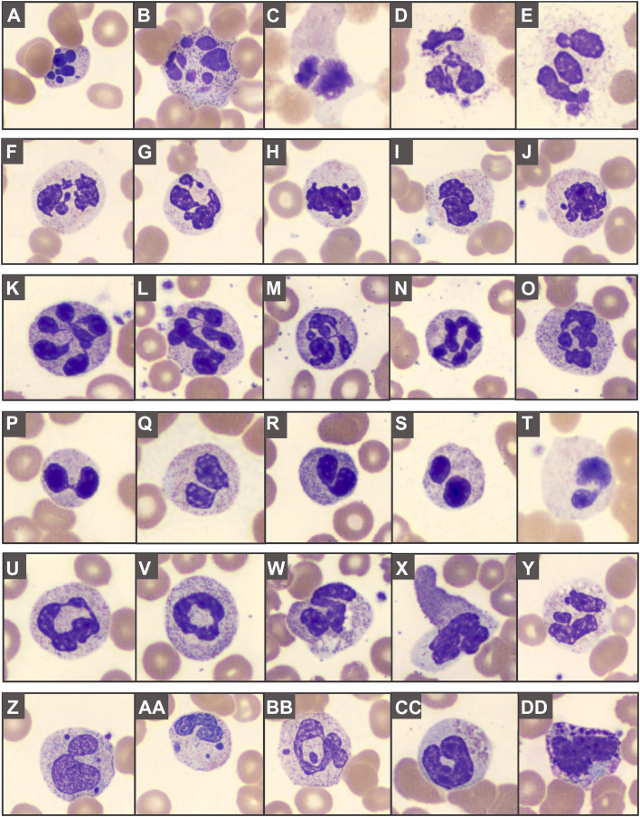
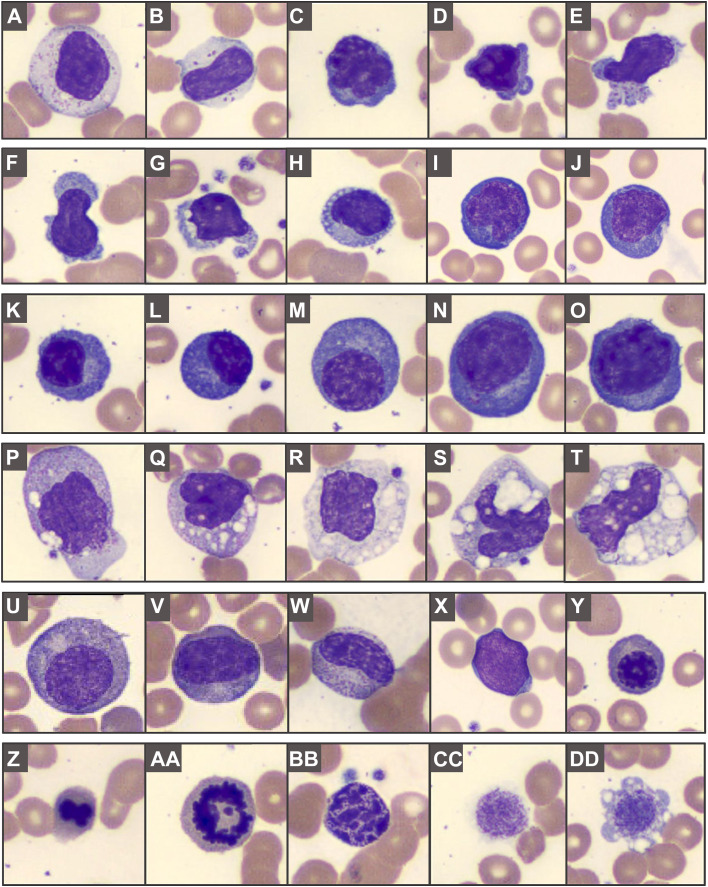
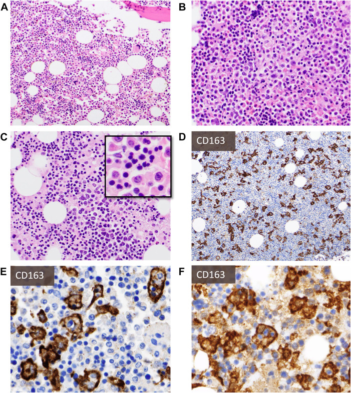
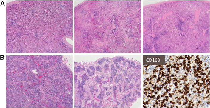
Coronavirus disease 2019 is caused by severe acute respiratory syndrome coronavirus 2 and is associated with pronounced hematopathologic findings. Peripheral blood features are heterogeneous and very often include neutrophilia, lymphopenia, myeloid left shift, abnormally segmented neutrophils, atypical lymphocytes/plasmacytoid lymphocytes, and atypical monocytes. Bone marrow biopsies and aspirates are often notable for histiocytosis and hemophagocytosis, whereas secondary lymphoid organs may exhibit lymphocyte depletion, pronounced plasmacytoid infiltrates, and hemophagocytosis. These changes are reflective of profound innate and adaptive immune dysregulation, and ongoing research efforts continue to identify clinically applicable biomarkers of disease severity and outcome.

Keywords: Atypical lymphocytes; COVID-19; Dysgranulopoiesis; Hemophagocytic lymphohistiocytosis; Lymphadenopathy; Lymphopenia; Neutrophilia; SARS-CoV-2.


Figures

Fig. 1
Fig. 1
Abnormal granulopoiesis in peripheral blood smears: (AE) apoptotic/pyknotic, disintegrating, and smudged neutrophils; (FJ) abnormally segmented nuclei, including (KO) hypersegmentation, (PT) pseudo-Pelger–Huët morphology, and (U, V) ring forms. Abnormal granulation can feature (WY) toxic granules, cytoplasmic vacuolization, patchy hypogranularity, and (Z–BB) Döhle body-like inclusions or apparent nuclear fragments. Abnormally granulated eosinophils (CC) and basophils (DD) can be seen.
Fig. 2
Fig. 2
Atypical lymphocytes and other COVID-19-related blood findings: (A, B) large granular lymphocytes; (CO) atypical lymphocytes can show a spectrum of features, with nuclear irregularities, variably coarse chromatin, deeply basophilic cytoplasm, occasionally with cytoplasmic blebs, granules or vacuolization; (KO) plasma cells or plasmacytoid forms can include irregular enlarged forms. (PT) Monocytes can show coarser granules or varying degrees of prominent coalescing vacuolization. (UY) Left-shifted myeloid cells and leukoerythroblastosis; (Z–AA) budding erythroid nucleus and mitotic figure, (BB) pyknotic cell, and (CC–DD) giant platelets.
Fig. 3
Fig. 3
Bone marrow biopsy findings: H&E sections (A–C, from four different decedents) show varying cellularity with frequent hypercellularity, maturing trilineage hematopoiesis with frequent myeloid left shift (A, B), and occasional decreased M:E ratio (C). There may be pronounced dyserythropoiesis (C-inlay) and varying degrees of histiocytosis and hemophagocytosis that may not be readily appreciated on H&E sections but are unearthed with immunohistochemistry for histiocytic markers (eg, CD163) (D, E). Autopsy cases kindly provided by Dr Olga Pozdnyakova.
Fig. 4
Fig. 4
Common findings in spleen (A) and lymph node (B): Spleens show diminished white pulp and increased red-to-white pulp ratio, with areas of hemorrhage (A). On low magnification, lymph nodes show attenuation of follicular architecture (B, left) and varying degrees of sinus histiocytosis (B, middle), with some cases also showing prominent hemophagocytosis (B, right, highlighted by CD163).
Fig. 5
Fig. 5
Lymph node showing lymphocyte depletion and prominent polyclonal plasmacytosis with increased proliferation in acute infection: (A, B) show an architecturally intact lymph node with patent subcapsular and medullary sinuses, many with reactive histiocytosis. There are almost no apparent follicles and lymphocytes are diminished, with the parenchyma (C, D) mostly replaced by a population of small to medium-sized plasma cells and plasmacytoid forms with occasional markedly enlarged, hyperchromatic nuclei or binucleated forms, as well as ones with more dispersed chromatin and prominent nucleoli. (E) CD3+ T cells and CD20+ B cells are markedly reduced, with only rare CD21+ follicular dendritic cell meshworks. The plasma cell population shows dim-to-negative CD138, uniform MUM1 expression, and considerably increased Ki-67 proliferation (approximately 50%) but polytypic kappa and lambda light chain expression.
Fig. 6
Fig. 6
Kappa-predominant plasmacytoid proliferation in a patient with a history of lymphoma: A patient with stage IV diffuse large B-cell lymphoma developed new FDG-avid lymphadenopathy concerning recurrent lymphoma (note: case has previously been published, new images were taken for this publication with permission). H&E sections (A, B) show an interfollicular expansion by numerous small lymphocytes, epithelioid histiocytes, and focal sheets of plasmacytoid cells variably positive for PAX5 (C), positive for CD79a (D), negative for CD138 (E), and positive for MUM1 (F) with marked excess kappa (G) light chain expression compared with lambda (H). However, molecular studies showed polyclonal immunoglobulin heavy chain gene (IGH) rearrangements, absence of clonal aberrancies by cytogenetics, and lymphadenopathy resolved over time without evidence of lymphoma recurrence.
Fig. 7
Fig. 7
Histologic and cytologic findings of COVID-19 vaccine-related lymphadenopathy: An individual in their twenties suddenly developed axillary, cervical, and supraclavicular lymphadenopathy (up to 2 cm in greatest dimension) 1 week after receiving the second mRNA COVID-19 vaccine in the deltoid muscle. H&E sections (A, B) and IHC for CD3 (C), CD20 (D), CD10 (E), and Bcl-2 (F) of lymph nodes (ipsilateral to vaccine site) show marked reactive lymphoid hyperplasia. Aspirate smears (G, H) show a polymorphous mixture of small and large lymphocytes, occasional admixed inflammatory cells, tingible-body macrophages, and lymphoglandular bodies (cytoplasmic fragments). Case and images kindly provided by Dr Amy Duffield.

Similar articles

Cited by

References

    1. Hooper J.E., Padera R.F., Dolhnikoff M., et al. A Postmortem Portrait of the Coronavirus Disease 2019 (COVID-19) Pandemic: A Large Multi-institutional Autopsy Survey Study. Arch Pathol Lab Med. 2021;145(5):529–535. - PubMed
    1. von Stillfried S., Bülow R.D., Röhrig R., et al. German Registry of COVID-19 Autopsies (DeRegCOVID) DeRegCOVID Collaborators First report from the German COVID-19 autopsy registry. Lancet Reg Health Eur. 2022;15:100330. - PMC - PubMed
    1. Delorey T.M., Ziegler C.G.K., Heimberg G., et al. COVID-19 tissue atlases reveal SARS-CoV-2 pathology and cellular targets. Nature. 2021;595(7865):107–113. - PMC - PubMed
    1. Ahern D.J., Ai Z., Ainsworth M., et al. A blood atlas of COVID-19 defines hallmarks of disease severity and specificity. Cell. 2022;185(5):916–938.e58. - PMC - PubMed
    1. Terpos E., Ntanasis-Stathopoulos I., Elalamy I., et al. Hematological findings and complications of COVID-19. Am J Hematol. 2020;95(7):834–847. - PMC - PubMed