. 2023 May 6;23(1):323.
doi: 10.1186/s12884-023-05562-0.

Differential immunophenotype of circulating monocytes from pregnant women in response to viral ligands

Affiliations

Differential immunophenotype of circulating monocytes from pregnant women in response to viral ligands

Marcelo Farias-Jofre et al. BMC Pregnancy Childbirth. .

Abstract

Background: Viral infections during pregnancy can have deleterious effects on mothers and their offspring. Monocytes participate in the maternal host defense against invading viruses; however, whether pregnancy alters monocyte responses is still under investigation. Herein, we undertook a comprehensive in vitro study of peripheral monocytes to characterize the differences in phenotype and interferon release driven by viral ligands between pregnant and non-pregnant women.

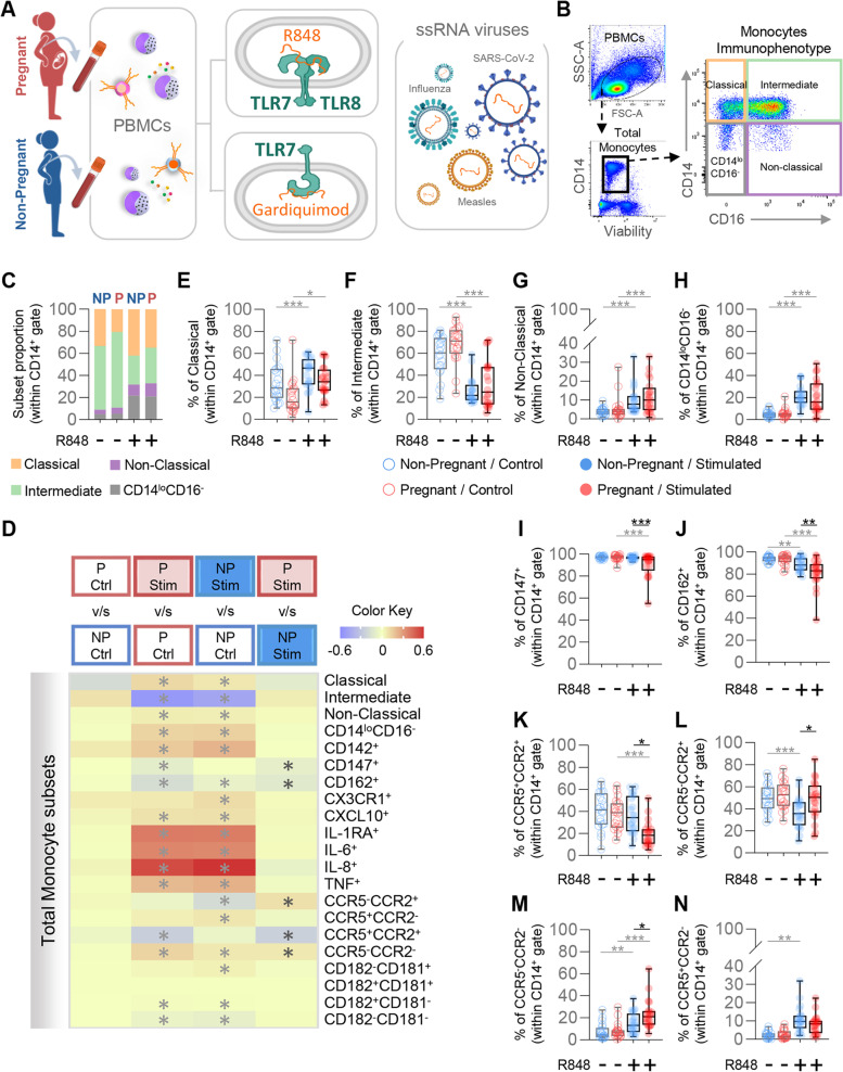
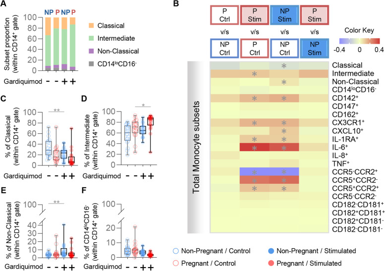
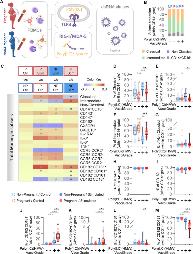
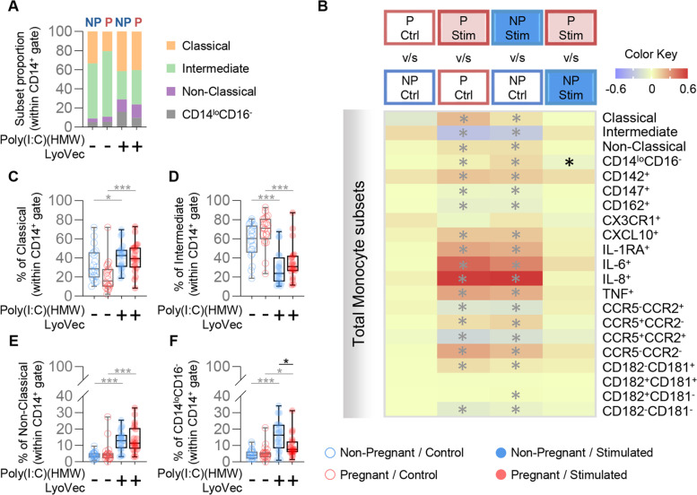
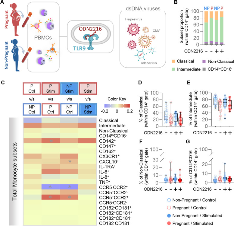
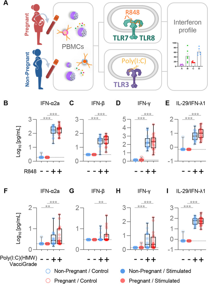
Methods: Peripheral blood was collected from third-trimester pregnant (n = 20) or non-pregnant (n = 20, controls) women. Peripheral blood mononuclear cells were isolated and exposed to R848 (TLR7/TLR8 agonist), Gardiquimod (TLR7 agonist), Poly(I:C) (HMW) VacciGrade™ (TLR3 agonist), Poly(I:C) (HMW) LyoVec™ (RIG-I/MDA-5 agonist), or ODN2216 (TLR9 agonist) for 24 h. Cells and supernatants were collected for monocyte phenotyping and immunoassays to detect specific interferons, respectively.

Results: The proportions of classical (CD14hiCD16-), intermediate (CD14hiCD16+), non-classical (CD14loCD16+), and CD14loCD16- monocytes were differentially affected between pregnant and non-pregnant women in response to TLR3 stimulation. The proportions of pregnancy-derived monocytes expressing adhesion molecules (Basigin and PSGL-1) or the chemokine receptors CCR5 and CCR2 were diminished in response to TLR7/TLR8 stimulation, while the proportions of CCR5- monocytes were increased. Such differences were found to be primarily driven by TLR8 signaling, rather than TLR7. Moreover, the proportions of monocytes expressing the chemokine receptor CXCR1 were increased during pregnancy in response to poly(I:C) stimulation through TLR3, but not RIG-I/MDA-5. By contrast, pregnancy-specific changes in the monocyte response to TLR9 stimulation were not observed. Notably, the soluble interferon response to viral stimulation by mononuclear cells was not diminished in pregnancy.

Conclusions: Our data provide insight into the differential responsiveness of pregnancy-derived monocytes to ssRNA and dsRNA, mainly driven by TLR8 and membrane-bound TLR3, which may help to explain the increased susceptibility of pregnant women to adverse outcomes resulting from viral infection as observed during recent and historic pandemics.

Keywords: Human; Infection; Innate immunity; Pregnancy; Virus.


Conflict of interest statement

The authors declare that they have no competing interests.

Figures

Fig. 1
Fig. 1
Monocyte response to TLR7/TLR8 stimulation. A Peripheral blood mononuclear cells (PBMCs) were isolated from the peripheral blood of pregnant (n = 20) and non-pregnant (n = 20) women and stimulated with R848 (TLR7/TLR8 agonist) or Gardiquimod (TLR7 agonist) for 24 h. Flow cytometry was performed to phenotype monocytes. B Flow cytometry gating strategy for phenotyping of monocyte subsets after in vitro stimulation with viral ligands. Viable monocytes were gated as live CD14+ cells from PBMCs. The expression levels of CD16 and CD14 were used to gate monocyte subsets as follows: classical (CD14hiCD16); intermediate (CD14hiCD16+); non-classical (CD14loCD16+), and CD14loCD16. C Proportions of monocyte subsets in pregnant (red) and non-pregnant (blue) women with and without R848 stimulation. D Heatmap representation of the differences in proportions of monocytes subsets from pregnant (red symbols) and non-pregnant (blue symbols) following R848 stimulation. Asterisks indicate statistically significant differences between the indicated groups. E-N Frequencies of (E) classical monocytes, (F) intermediate monocytes, (G) non-classical monocytes, (H) CD14loCD16 monocytes, (I) CD147+ monocytes, (J) CD162+ monocytes, (K) CCR5+CCR2+ monocytes, (L) CCR5CCR2+ monocytes, (M) CCR5CCR2 monocytes, and (N) CCR5+CCR2 monocytes in pregnant (red) and non-pregnant (blue) women following R848 stimulation (solid circles) or control (open circles). *p < 0.05; **p < 0.01; ***p < 0.001. ( +) Stimulated; (-) Control
Fig. 2
Fig. 2
Monocyte response to TLR7 stimulation. A Proportions of monocyte subsets in pregnant (red) and non-pregnant women (blue) with and without Gardiquimod stimulation (TLR7 agonist). B Heatmap representation of the differences in proportions of monocyte subsets from pregnant (red symbols) and non-pregnant (blue symbols) women following Gardiquimod stimulation. Asterisks indicate statistically significant differences between the indicated groups. C-F Frequencies of (C) classical monocytes (CD14hiCD16), (D) intermediate monocytes (CD14hiCD16+), (E) non-classical monocytes (CD14loCD16+), (F) CD14loCD16 monocytes in pregnant (red) and non-pregnant (blue) women following Gardiquimod stimulation (solid circles) or control (open circles). *p < 0.05; **p < 0.01. ( +) Stimulated; (-) Control
Fig. 3
Fig. 3
Monocyte response to TLR3 stimulation. A Peripheral blood mononuclear cells (PBMCs) were isolated from the peripheral blood of pregnant (n = 20) and non-pregnant (n = 20) women and stimulated with Poly(I:C) (HMW) Vaccigrade™ (poly(I:C); TLR3 agonist) or Poly(I:C) (HMW)LyoVec™ (poly(I:C)/LyoVec; RIG-I/MDA5 agonist) for 24 h. Flow cytometry was performed to phenotype monocytes. B Proportions of monocyte subsets in pregnant (red) and non-pregnant (blue) women with and without Poly(I:C) (HMW) Vaccigrade™ stimulation. C Heatmap representation of the differences in proportions of monocyte subsets from pregnant (red symbols) and non-pregnant (blue symbols) following Poly(I:C) (HMW) Vaccigrade™ stimulation. Asterisks indicate statistically significant differences between the indicated groups. (D-M) Frequencies of (D) classical monocytes (CD14hiCD16), (E) CD14loCD16 monocytes, (F) intermediate monocytes (CD14hiCD16+), (G) non-classical monocytes (CD14loCD16+), (H) CD147+ monocytes, (I) CD162+ monocytes, (J) CD182CD181+ monocytes, (K) CD182+CD181+ monocytes, (L) CD182+CD181 monocytes, and (M) CD182CD181 monocytes in pregnant (red) and non-pregnant (blue) women following Poly(I:C) stimulation (solid circles) or control (open circles). *p < 0.05; **p < 0.01; ***p < 0.001. ( +) Stimulated; (-) Control
Fig. 4
Fig. 4
Monocyte response to RIG-I/MDA-5 stimulation. A Proportions of monocyte subsets in pregnant (red) and non-pregnant women (blue) with and without Poly(I:C) (HMW) LyoVec™ stimulation. B Heatmap representation of the differences in proportions of monocyte subsets from pregnant (red symbols) and non-pregnant (blue symbols) women following 24 h Poly(I:C) (HMW) LyoVec™ stimulation. Asterisks indicate statistically significant differences between the indicated groups. C-F Frequencies of (C) classical monocytes (CD14hiCD16), (D) intermediate monocytes (CD14hiCD16+), (E) non-classical monocytes (CD14loCD16+), and (F) CD14loCD16 monocytes in pregnant (red) and non-pregnant (blue) women following Poly(I:C) (HMW) LyoVec™ stimulation (solid circles) or control (open circles). *p < 0.05; ***p < 0.001. ( +) Stimulated; (-) Control
Fig. 5
Fig. 5
Monocyte response to TLR9 stimulation. A Peripheral blood mononuclear cells (PBMCs) were isolated from the peripheral blood of pregnant (n = 20) and non-pregnant (n = 20) women with and without ODN2216 stimulation for 24 h. Flow cytometry was performed to evaluate the total monocyte subsets. B Proportions of monocyte subsets in pregnant (red) and non-pregnant (blue) women with ODN2216 stimulation. C Heatmap representation of the differences in proportions of monocyte subsets from pregnant (red symbols) and non-pregnant (blue symbols) women following ODN2216 stimulation. Asterisks indicate statistically significant differences between the indicated groups. D-G Frequencies of (D) classical monocytes (CD14hiCD16), (E) intermediate monocytes (CD14hiCD16+), (F) non-classical monocytes (CD14loCD16+), and (G) CD14loCD16 monocytes in pregnant (red) and non-pregnant (blue) women following ODN2216 stimulation (solid circles) or control (open circles). ( +) Stimulated; (-) Control
Fig. 6
Fig. 6
Interferon production by peripheral blood mononuclear cells (PBMCs) upon TLR7/TLR8 or TLR3 stimulation. A Peripheral blood samples were collected from pregnant (n = 20, indicated in red) and non-pregnant (n = 20, indicated in blue) women to isolate PBMCs for in vitro stimulation with R848 or Poly(I:C) (HMW) VacciGrade™ [Poly(I:C)]. Type-I (IFN-α2a, -β), Type-II (IFN-γ) and Type-III (IL-29/IFN-λ1) interferon concentrations were then determined in culture supernatants. B-E Log10-transformed concentrations of (B) IFN-α2a, (C) IFN-β, (D) IFN-γ, and (E) IL-29/IFN-λ1 in culture supernatants of PBMCs from pregnant (red symbols) and non-pregnant (blue symbols) women in response to R848 (solid circles) or control (open circles). F-I Log10-transformed concentrations of (F) IFN-α2a, (G) IFN-β, (H) IFN-γ, and (I) IL-29/IFN-λ1 in culture supernatants of PBMCs from pregnant (red symbols) and non-pregnant (blue symbols) women in response to Poly(I:C) (solid circles) or control (open circles). Dotted lines indicate the detection limit of each analyte. **p < 0.01, ***p < 0.001. ( +) Stimulated, (-) Control

Similar articles

Cited by

References

    1. Say L, Chou D, Gemmill A, Tuncalp O, Moller AB, Daniels J, et al. Global causes of maternal death: a WHO systematic analysis. Lancet Glob Health. 2014;2(6):e323–e333. doi: 10.1016/S2214-109X(14)70227-X. - DOI - PubMed
    1. Kourtis AP, Read JS, Jamieson DJ. Pregnancy and infection. N Engl J Med. 2014;370(23):2211–2218. doi: 10.1056/NEJMra1213566. - DOI - PMC - PubMed
    1. Gibson CS, Goldwater PN, MacLennan AH, Haan EA, Priest K, Dekker GA, et al. Fetal exposure to herpesviruses may be associated with pregnancy-induced hypertensive disorders and preterm birth in a Caucasian population. BJOG. 2008;115(4):492–500. doi: 10.1111/j.1471-0528.2007.01653.x. - DOI - PMC - PubMed
    1. Goldenberg RL, McClure EM, Saleem S, Reddy UM. Infection-related stillbirths. Lancet. 2010;375(9724):1482–1490. doi: 10.1016/S0140-6736(09)61712-8. - DOI - PMC - PubMed
    1. Kwon JY, Romero R, Mor G. New insights into the relationship between viral infection and pregnancy complications. Am J Reprod Immunol. 2014;71(5):387–390. doi: 10.1111/aji.12243. - DOI - PMC - PubMed