

Pachychoroid neovasculopathy has clinical properties that differ from conventional neovascular age-related macular degeneration
- PMID: 37149627
- PMCID: PMC10164122
- DOI: 10.1038/s41598-023-33936-z
Pachychoroid neovasculopathy has clinical properties that differ from conventional neovascular age-related macular degeneration
Abstract
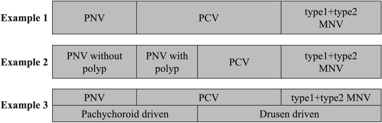
To determine the clinical properties of pachychoroid neovasculopathy (PNV) that differ from conventional neovascular age-related macular degeneration (nAMD) and suggest that they are different clinical entities. To accomplish this, we reviewed the medical records of 100 consecutive patients diagnosed with nAMD. All of the patients were Japanese, and their mean age was 75.5 years. There were 72 men and 28 women. For the bilateral cases, only the right eye was analyzed. An eye was diagnosed with PNV when a macular neovascularization (MNV) was detected just above the dilated choroidal vessels. The Indocyanine green angiographic (ICGA) and en face optical coherence tomographic (OCT) images were used to assess the vertical symmetry of the medium and large choroidal vessels. The subfoveal choroidal thickness (SCT) was also measured manually in the OCT images. After reclassification, there were 29 (29%) patients with typical nAMD (25 with type 1 MNV, 4 with type 2 MNV), 43 (43%) with PNV, 21 (21%) with polypoidal choroidal vasculopathy, and 7 (7%) with retinal angiomatous proliferation. Of the 43 PNV, 17 (39.5%) had polypoidal lesions and 26 (60.5%) had no polypoidal lesions. The percentage of eyes with vertical asymmetry of the medium and large choroidal vessels was significantly greater in the 35 PNV (81.4%) than in the 16 non-PNV (28.1%; P < 0.01) cases. The mean SCT was significantly thicker in the PNV eyes than in the non-PNV eyes (298 ± 96 μm vs. 228 ± 82 μm; P < 0.01). The response of PNV to anti-vascular endothelial growth factor treatments was better than that of non-PNV eyes [higher dry macula rate after the loading period (90.9% vs. 59.1%), fewer total number of injections (11.0 ± 2.9 vs. 13.4 ± 3.2), and longer treatment intervals for the anti-VEGF therapy (8.4 ± 3.1 vs. 13.4 ± 3.2 weeks) at 2 years (all P < 0.01)]. These differences in the morphology and response to anti-VEGF treatments suggest that PNV is a separate clinical entity to conventional nAMD.
© 2023. The Author(s).
Conflict of interest statement
Dr. Kuranami has nothing to disclose. Dr. Maruko (R) has nothing to disclose. Dr. Maruko (I) reported grants from JSPS KAKENHI (Grant Number JP20K09781); personal fees from Alcon, Bayer, Canon, Chugai, Nidek, Nikon, Novartis, Santen, Senju, and Topcon outside the submitted work. Dr. Hasegawa reported personal fees from Bayer, Novartis, Alcon, Santen, Kowa, Senju, R E medical, Nikon, JFC, Otsuka and Boehringer Ingelheim outside the submitted work. Dr. Iida reported consulting fees from Bayer, Novartis, Chugai, and Boehringer Ingelheim; personal fees from AMO, Alcon, Bayer, Canon, HOYA, Nikon, Nidek, Novartis, Otsuka, Pfizer, Santen, Senju, and Topcon outside the submitted work.
Figures


References
Publication types
MeSH terms
Substances
LinkOut - more resources
Full Text Sources
Medical
Research Materials
