. 2023 Oct;43(10):1633-1647.
doi: 10.1177/0271678X231173175. Epub 2023 May 7.

Calcium transients in nNOS neurons underlie distinct phases of the neurovascular response to barrel cortex activation in awake mice

Affiliations

Calcium transients in nNOS neurons underlie distinct phases of the neurovascular response to barrel cortex activation in awake mice

Sung Ji Ahn et al. J Cereb Blood Flow Metab. 2023 Oct.

Abstract

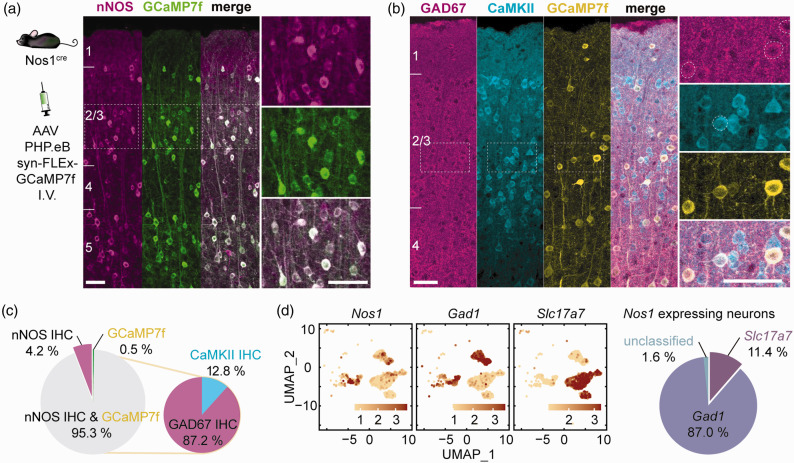
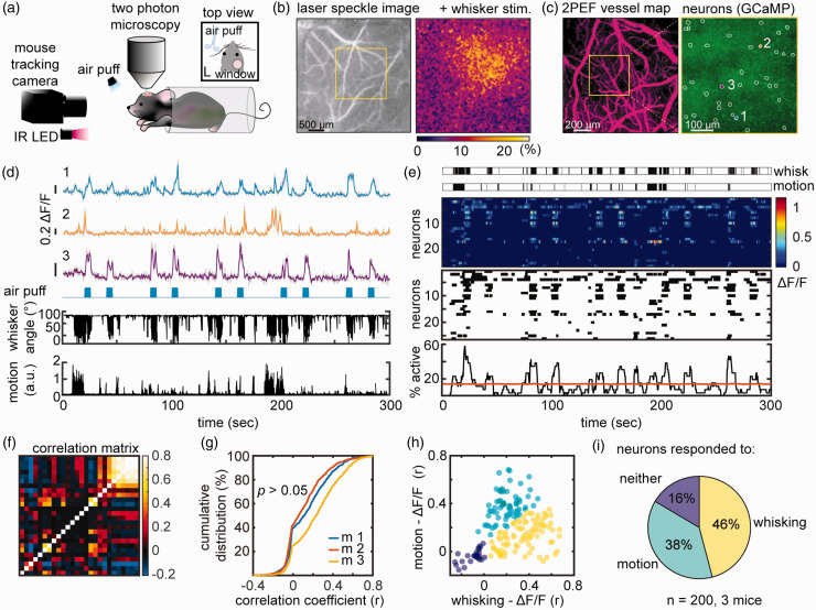
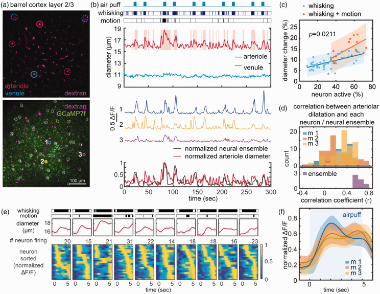
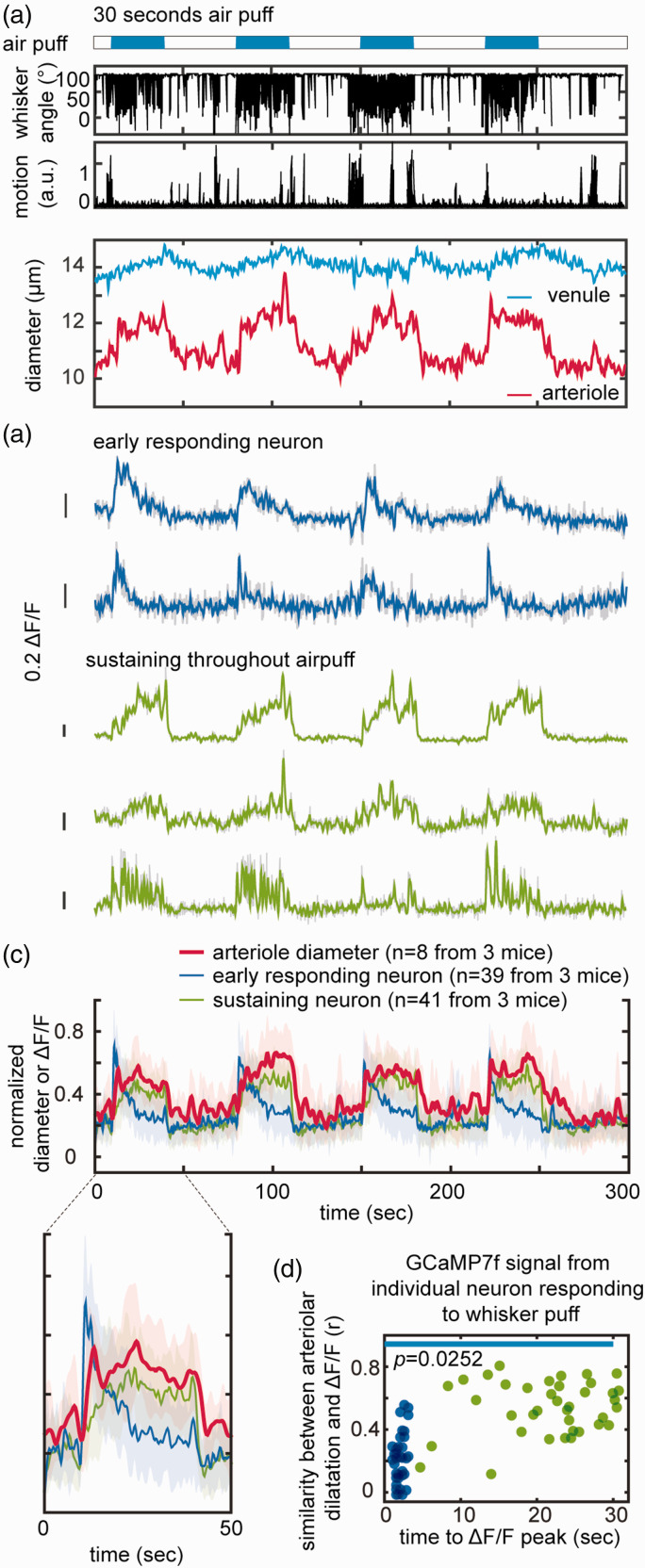
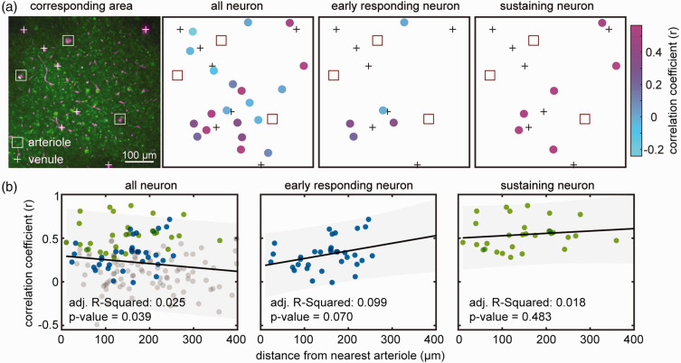
Neuronal nitric oxide (NO) synthase (nNOS), a Ca2+ dependent enzyme, is expressed by distinct populations of neocortical neurons. Although neuronal NO is well known to contribute to the blood flow increase evoked by neural activity, the relationships between nNOS neurons activity and vascular responses in the awake state remain unclear. We imaged the barrel cortex in awake, head-fixed mice through a chronically implanted cranial window. The Ca2+ indicator GCaMP7f was expressed selectively in nNOS neurons using adenoviral gene transfer in nNOScre mice. Air-puffs directed at the contralateral whiskers or spontaneous motion induced Ca2+ transients in 30.2 ± 2.2% or 51.6 ± 3.3% of nNOS neurons, respectively, and evoked local arteriolar dilation. The greatest dilatation (14.8 ± 1.1%) occurred when whisking and motion occurred simultaneously. Ca2+ transients in individual nNOS neurons and local arteriolar dilation showed various degrees of correlation, which was strongest when the activity of whole nNOS neuron ensemble was examined. We also found that some nNOS neurons became active immediately prior to arteriolar dilation, while others were activated gradually after arteriolar dilatation. Discrete nNOS neuron subsets may contribute either to the initiation or to the maintenance of the vascular response, suggesting a previously unappreciated temporal specificity to the role of NO in neurovascular coupling.

Keywords: 2-photon microscopy; Functional hyperemia; GCaMP7f; neurovascular coupling; nitric oxide.


Conflict of interest statement

Declaration of conflicting interestsThe author(s) declared the following potential conflicts of interest with respect to the research, authorship, and/or publication of this article: CI serves on the Scientific Advisory Board of Broadview Ventures. The other authors have no conflicts to declare.

Figures

Figure 1.
Figure 1.
Viral gene transfer-induced expression of GCaMP7f in nNOS neurons of NOS1cre mice. (a) GCaMP7f was expressed in NOS1cre mice using systemic administration of an AAV-PHP.eB viral vector. The localization of GCaMP7f expression in nNOS neurons in the cortical layers was verified by immunohistochemistry (IHC). Right panels are enlargements of the dashed squares in the left panel (n = 2,744 neurons in 5 mice). (b) GCaMP7f expression in inhibitory (GAD67) or excitatory (CaMKII) neurons of nNOScre mice treated with the AAV-PHP.eB viral vector. (c) Neurons (%) expressing nNOS, GCaMP7f or both, and, among GCaMP7f positive neurons, those expressing GAD67 or CaMKII (n = 690 neurons in 3 mice) and (d) Single-cell RNAseq data (mousebrain.org/development/downloads.html) showing expression of Nos1 both in excitatory (Slc17a7) and inhibitory (Gad1) neurons (%), confirming the finding by immunohistochemistry in b. Scale bar = 50µm throughout.
Figure 2.
Figure 2.
In vivo imaging of nNOS neurons in awake, head-fixed mice during air-puff whisker stimulation. (a) Experimental setup for 2PEF imaging during whisker air-puff stimulation, which includes an infrared (IR) camera for tracking whisker and upper limb movements. (b) Localization of the hemodynamic response evoked by whisker stimulation in the contralateral whisker barrel cortex by laser speckle imaging. (c) Left panel: 2PEF map of pial vessels (within the yellow square in b) imaged at 5x. Right panel: GCaMP7f expressing nNOS neurons in layer 2/3 (outlined in white) imaged at 25x. The Ca2+ activity of neurons labelled 1, 2 and 3 is shown in (d). (d) Ca2+ activity (ΔF/F see methods) in neurons 1, 2 and 3 during 10 air-puffs each lasting 5 sec (blue marks). Whisker displacement and limb motion are also displayed. Notice that neuron 3 responds to air-puffs, while neuron 2 mainly to motion. Neuron 1 exhibits mixed responses. (e) Representative raster plots of ΔF/F (top), onset and termination of neuronal activity (middle), and proportion of simultaneously active cells over time (neuronal ensemble, bottom) from the neuronal population shown in (c, right panel). Each row represents a single cell. Ensemble activity above the orange line is statistically significant. (f) Correlation matrix quantifying functional connectivity between each neuron and every other neuron shows a broad range of correlations (Spearman’s rho). (g) Cumulative distribution of correlation coefficients across all animals shows that all mice had similar distribution (nested one-way ANOVA) and (h) scatter plot of the correlation coefficient between each neuron ΔF/F and whisker activity (x-axis) or motion (y-axis) showing three populations of nNOS neurons each responding predominantly to whisking, motion or neither and (i) Pie chart summarizing data in (h) from all mice. Data were clustered using spectral clustering.
Figure 3.
Figure 3.
Microvascular responses associated with nNOS Ca2+ activity during 5 sec whisker puffs and grooming motion. (a) 2PEF map of arterioles and venules (top) and of both vessel and GCaMP7f-expressing nNOS neurons (outlined in white, bottom). Vessels analyzed in b are circled in solid red or blue, and neurons are numbered (1,2,3) (b) Arterioles, but not venules, dilate in response to whisker puffs, spontaneous whisking or motion (grooming). Representative Ca2+ transients in neuron 1, 2, and 3 (middle). Overlay of normalized neural ensemble activity and arteriolar diameter changes highlighting the close correspondence between arteriolar and neural responses (bottom). (c) Linear relationship between the magnitude of the ensemble activity and arteriolar dilatation in response to whisking or whisking and spontaneous motion. Notice that with whisking+motion the slope of the relationship is significantly greater than with whisking alone (multiple comparison of one-way analysis of covariance models). (d) The distribution of correlation coefficients between arteriolar dilatation and nNOS Ca2+ transients for individual neurons does not differ in the 3 mice studied (p>0.05; nested one-way ANOVA)(top). The strongest correlation is observed between ensemble activity and arteriolar dilatation (bottom). (e) Representative arteriolar diameter changes relative to the timing of the peak Ca2+ activity in individual neuron during 10 consecutive 5 sec whisker air-puffs shown in (b). Notice that in some neurons peak activity occur earlier than in other neurons and (f) Average of ten raster plots in 3 mice showing early and late active nNOS neurons. Shading indicates standard deviation.
Figure 4.
Figure 4.
Microvascular responses associated with nNOS Ca2+ activity during 30 sec whisker puffs. (a) Representative dataset from four 30-seconds whisker air-puff showing the corresponding dilatation of arterioles and venules. (b) Representative pattern of Ca2+ (ΔF/F) transients showing early responding neurons (blue) and neurons with sustained activity throughout the stimulus (green). (c) Average of normalized Ca2+ transients and corresponding arteriolar dilatation (8 datasets in 3 mice). The enlarged graph for a single air-puff shows neurons active prior to the arteriolar dilatation (early responding neurons; blue) and neurons active during the dilatation (sustaining neurons; green) and Continued.(d) plot of the time to peak of Ca2+ (ΔF/F) transients, and similarity of the time course of neuronal and arteriolar responses. Two distinct nNOS neuronal populations can be identified: one rapidly decaying and preceding the arteriolar dilatation (blue; early responding neurons), and the other exhibiting a time course similar to the arteriolar dilation (green; sustaining neurons). The blue line indicates that duration of air-puff. Statistical significance was calculated as in Figure 3(c).
Figure 5.
Figure 5.
Spatial relationship of nNOS neurons responsive to air-puff and arterioles. (a) Representative 2PEF map of arterioles (square outline) and venules (+ sign) and GCaMP7f-expressing nNOS neurons. The schematic maps on the right show individual neurons color coded for the correlation of their Ca2+ response with that arteriolar dilatation. Notice that the highly correlated neurons do not cluster near the arterioles and (b) Linear regression analysis between distance of individual neuron from arterioles and correlation of their Ca2+ response with that arteriolar dilatation (n = 170 neurons from 3 mice). There is no relationship between the distance from the nearest arteriole and correlation of Ca2+ transients with arteriolar dilatation.

Similar articles

Cited by

References

    1. Gorelick PB, Furie KL, Iadecola C, American Heart Association/American Stroke Association et al.. Defining optimal brain health in adults: a presidential advisory from the American heart association/American stroke association. Stroke 2017; 48: e284–e303. - PMC - PubMed
    1. Schaeffer S, Iadecola C. Revisiting the neurovascular unit. Nat Neurosci 2021; 24: 1198–1209. - PMC - PubMed
    1. Iadecola C, Yang G, Ebner TJ, et al.. Local and propagated vascular responses evoked by focal synaptic activity in cerebellar cortex. J Neurophysiol 1997; 78: 651–659. - PubMed
    1. Longden TA, Dabertrand F, Koide M, et al.. Capillary K(+)-sensing initiates retrograde hyperpolarization to increase local cerebral blood flow. Nat Neurosci 2017; 20: 717–726. - PMC - PubMed
    1. Chen BR, Kozberg MG, Bouchard MB, et al.. A critical role for the vascular endothelium in functional neurovascular coupling in the brain. J Am Heart Assoc 2014; 3: e000787. - PMC - PubMed

Publication types

LinkOut - more resources