. 2023 May 30;42(5):112455.
doi: 10.1016/j.celrep.2023.112455. Epub 2023 May 5.

A tug of war between DCC and ROBO1 signaling during commissural axon guidance

Affiliations

A tug of war between DCC and ROBO1 signaling during commissural axon guidance

Brianna Dailey-Krempel et al. Cell Rep. .

Abstract

Dynamic and coordinated axonal responses to changing environments are critical for establishing neural connections. As commissural axons migrate across the CNS midline, they are suggested to switch from being attracted to being repelled in order to approach and to subsequently leave the midline. A molecular mechanism that is hypothesized to underlie this switch in axonal responses is the silencing of Netrin1/Deleted in Colorectal Carcinoma (DCC)-mediated attraction by the repulsive SLIT/ROBO1 signaling. Using in vivo approaches including CRISPR-Cas9-engineered mouse models of distinct Dcc splice isoforms, we show here that commissural axons maintain responsiveness to both Netrin and SLIT during midline crossing, although likely at quantitatively different levels. In addition, full-length DCC in collaboration with ROBO3 can antagonize ROBO1 repulsion in vivo. We propose that commissural axons integrate and balance the opposing DCC and Roundabout (ROBO) signaling to ensure proper guidance decisions during midline entry and exit.

Keywords: CP: Developmental biology; CP: Neuroscience; Netrin1/DCC; SLIT/ROBO; alternative splicing; axon guidance; midline switch; silencing model.


Conflict of interest statement

Declaration of interests The authors declare no competing interests.

Figures

Figure 1.
Figure 1.. Two distinct Dcc isoforms are expressed during commissural axon guidance
(A) Alternative splicing between exons 16 and 17 generates two Dcc isoforms that differ in 60 nt (shaded in gray). NOVA family of splicing factors bind Dcc pre-mRNA and promote DccL production. (B and C) Dcc isoform expression measured by semi-quantitative and quantitative RT-PCR, respectively. Both isoforms are expressed during commissural neurogenesis and axon guidance (E11–E15 in rats, equivalent to E9.5 to E13.5 in mice. Rat embryos were used to facilitate microdissection of the DSC). Data in (C) were collected from three embryos and are presented as mean ± SD. (D and E) DCC isoforms have distinct abilities to stimulate ERK1/2 phosphorylation upon Netrin1 stimulation (250 ng/mL for the indicated durations). (E) Quantification of ERK1/2 phosphorylation in (D). Data were collected from three independent experiments and are presented as mean ± SD. Two-way ANOVA was performed with a Tukey post hoc test (**p < 0.01).
Figure 2.
Figure 2.. Generation of CRISPR-Cas9-engineered mouse models for Dcc isoforms
(A) Design of guide RNAs (gRNAs) for Dcc isoforms. Targeted nucleotides of the gRNAs are underlined, and the alternative splice sites are in red. (B) Genomic mutations in two independent founder lines for each Dcc isoform. Deleted nucleotides are in lighter gray and inserted nucleotides in lower case. (C) Only one isoform is produced in Dcc isoform mutants, measured by semi-quantitative RT-PCR. Both isoforms are expressed in wild-type (WT) controls at E11.5. (D) Anti-DCC staining of transverse spinal cord sections from WT and Dcc isoform mutant embryos at E11.5. DCC is present in both pre- and post-crossing axonal segments (yellow arrows). Brackets indicate the floorplate (FP) area. Dcc is also expressed in ipsilateral-projecting interneurons and motor neurons. Scale bar, 100 μm.
Figure 3.
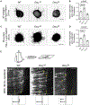
Figure 3.. Expressing only one of two DCC isoforms differentially affects commissural axon outgrowth and guidance
(A and C) Axon outgrowth from dorsal spinal cord (DSC) explants in response to bath-applied Netrin1 in 3D collagen matrix (A) and to Netrin1 coated on 2D surface (C), respectively (Netrin1 concentration and culture period as indicated). Under both conditions, axon outgrowth was increased in DccLSE mutants but was reduced in DccSSE mutants. (B and D) Quantification of axon outgrowth in (A) and (C), respectively; 3–5 explants were examined for each embryo. Data were normalized to WT controls and are presented as mean ± SD. One-way ANOVA with a Dunnett post hoc test; p values and animal numbers as indicated. (E) Axon projection around the midline in cultured whole embryos. The spinal cord was cut open at the dorsal midline and flattened in an open-book configuration to allow visualization of the FP area. A Gfp reporter was electroporated unilaterally into the DSC. Within 36 h of culturing (E10 to the equivalent of E11.5), WT axons were able to enter and exit the FP and make a sharp turn on the contralateral side (dashed lines indicate the FP boundaries). DccLSE axons entered the FP normally, but the majority stalled within or at the contralateral boundary of the FP (two examples are highlighted with arrowheads). By contrast, most DccSSE axons had not reached or entered the FP by the end of culturing (examples highlighted by arrowheads). Scale bars, 100 μm.
Figure 4.
Figure 4.. Expressing only one of two DCC isoforms differentially disrupts midline entry and exit
(A) DiI tracing of spinal commissural axons from WT controls and different mutants in an open-book orientation. At E11.5, WT axons had projected across the FP and made a sharp turn upon exit. Most DccLSE axons stalled within the FP, and the axons were present in bundles (arrowheads). Only a small number of axons had exited the FP and were present as single axons (arrows). Many DccSSE axons stalled before entering the FP at E11.5 (arrowheads). In DccLSE; DccSSE transheterozygotes, the axons entered and exited the midline normally. A heterozygous Robo3 KO allele facilitated midline exit when it was introduced into DccLSE mutants. Robo3+/− by itself does not cause significant guidance defects.,, Scale bar, 100 μm. (B and C) Quantification of midline entry (B) and exit (C), respectively, by DiI tracing at E11.5. The signals from four boxed areas were measured with ImageJ. The ratio between areas 2 and 1 represents midline entry, and the ratio between areas 4 and 3 represents midline exit. Data were normalized to WT controls and are presented as mean ± SD. One-way ANOVA with a Tukey post hoc test; p values and animal numbers as indicated.
Figure 5.
Figure 5.. Expressing only one of two DCC isoforms differentially affects the ventral commissure size
(A) Ventral projection and midline crossing of commissural axons examined by anti-ROBO3 staining of transverse spinal cord sections at E11.5. Bottom panels show close-up of the FP. The ventral commissure was thicker in DccLSE mutants but was thinner in DccSSE mutants at E11.5. Transheterozygous DccLSE; DccSSE mutants had normal-sized ventral commissure. Reducing Robo3 dose decreased the commissure size in both Dcc isoform mutants. Scale bars, 100 μm. (B) Quantification of ventral commissure size by anti-ROBO3 staining. The commissure size is quantified as the ratio between the thickness of the ventral commissure and the height of the FP. Data were normalized to WT controls and are presented as mean ± SD. One-way ANOVA with a Tukey post hoc test; p values and animal numbers as indicated; ns, not significant.
Figure 6.
Figure 6.. ROBO1 localization is unaffected in Dcc isoform mutants and ROBO1 binds both DCC isoforms
(A) Anti-ROBO1 and anti-ROBO2 staining of transverse spinal cord sections at E11.5. In both WT and Dcc isoform mutants, ROBO1 and ROBO2 are present at low levels in pre-crossing axonal domains but become highly upregulated post crossing. Robo1/2 are also expressed in ipsilateral-projecting interneurons and motor neurons. Scale bar, 100 μm. (B) CoIP between DCC isoforms and ROBO1. Hemagglutinin (HA)-tagged DCC (L, long; S, short) and V5-tagged ROBO1 were coexpressed in COS-1 cells in the absence or presence of Netrin1 and SLIT2N stimulation (250 ng/mL each for 30 min). (C and D) Quantification of DCC and ROBO1 interaction from immunoblotting (IB) with anti-HA (C) and anti-V5 (D). Data were collected from three independent experiments and are presented as mean ± SD. Two-way ANOVA was performed with a Tukey post hoc test (not significant for all data points). Both DCC isoforms interacted with ROBO1 and at comparable levels, with or without ligand stimulation.
Figure 7.
Figure 7.. Netrin1/DCC and SLIT/ROBO1 signaling antagonize each other during midline entry and exit
(A) Axon projection and midline crossing in compound Netrin1 and Robo1 KO mutants, examined by anti-ROBO3, anti-TAG1, and anti-NF staining of transverse spinal cord sections at E11.5. Netrin1−/− null mutants exhibited severe guidance defects in the spinal cord, including misprojections into the roof plate, ventricular zone, and motor columns (highlighted with yellow arrowheads), abnormal exit from the CNS (yellow arrow), and an almost complete absence of the ventral commissure (bracket). Removing Robo1 allowed more axons to cross the midline in Netrin1−/− null background (the ventral commissure indicated by the bracket is thicker in Netrin1−/−; Robo1−/− embryos), but other guidance errors, indicated by yellow arrowheads and yellow arrows, persisted. (B) Quantification of the ventral commissure size in (A). Anti-ROBO3 was used as it does not label any motor axons. (C and D) Effect of bath-applied SLIT2N (250 ng/mL) on DSC axon outgrowth in the absence and presence of Netrin1, respectively. Explants were cultured in 3D collagen matrix for 40 h without Netrin1 in (C) and for 24 h with 250 ng/mL bath-applied Netrin1 in (D). SLIT2N repressed WT axon outgrowth regardless of whether Netrin1 was present but did not inhibit the growth from Robo1/2 double KO explants. (E and F) Quantification of axon outgrowth in (C) and (D), respectively. (G) DiI tracing of spinal commissural axons in an open-book orientation in compound Netrin1 and Robo1 KO mutants. Most Robo1−/− axons stalled within and at the contralateral boundary of the FP at E12.5. Introduction of a heterozygous Netrin1 KO allele into Robo1−/− mutant background allowed more axons to exit. Scale bars, 100 μm. (H) Quantification of midline exit in (G). See Figure 4C for description of quantification. Data were normalized to WT controls and are presented as mean ± SD. One-way ANOVA with a Tukey post hoc test in (B) and (H), two-way ANOVA with a Tukey post hoc test in (E) and (F); p values and animal numbers as indicated; ns, not significant; 5–10 sections, 3–5 explants, and 3–5 DiI injection sites were quantified for each embryo.

References

    1. Serafini T, Kennedy TE, Galko MJ, Mirzayan C, Jessell TM, and Tessier-Lavigne M (1994). The netrins define a family of axon outgrowth-promoting proteins homologous to C. elegans UNC-6. Cell 78, 409–424. - PubMed
    1. Brose K, Bland KS, Wang KH, Arnott D, Henzel W, Goodman CS, Tessier-Lavigne M, and Kidd T (1999). Slit proteins bind Robo receptors and have an evolutionarily conserved role in repulsive axon guidance. Cell 96, 795–806. - PubMed
    1. Butler SJ, and Dodd J (2003). A role for BMP heterodimers in roof plate-mediated repulsion of commissural axons. Neuron 38, 389–401. - PubMed
    1. Augsburger A, Schuchardt A, Hoskins S, Dodd J, and Butler S (1999). BMPs as mediators of roof plate repulsion of commissural neurons. Neuron 24, 127–141. - PubMed
    1. Charron F, Stein E, Jeong J, McMahon AP, and Tessier-Lavigne M (2003). The morphogen sonic hedgehog is an axonal chemoattractant that collaborates with netrin-1 in midline axon guidance. Cell 113, 11–23. - PubMed

Publication types