

Malignant melanoma associated with a plaque-type blue nevus of the cheek: a case report
- PMID: 37150529
- PMCID: PMC10165239
- DOI: 10.7181/acfs.2023.00024
Malignant melanoma associated with a plaque-type blue nevus of the cheek: a case report
Abstract
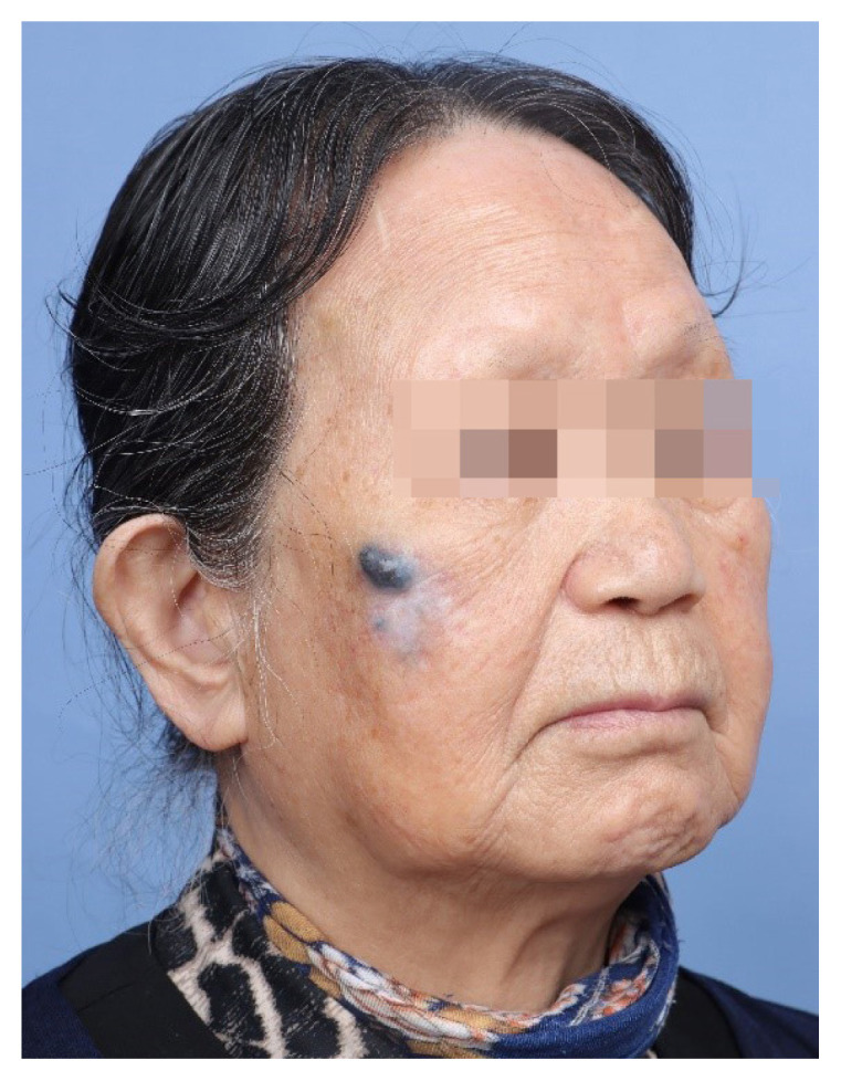
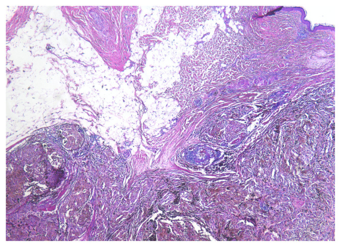
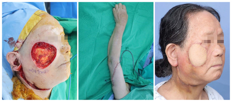
Blue nevi, which are characterized by collections of pigment-producing melanocytes in the dermis, have a variety of clinicopathological characteristics. Plaque-type blue nevus (PTBN) is a variant of blue nevi. PTBN presents at birth or arises in early childhood, and it shows a combination of the features found in common blue nevus and cellular blue nevus. It is typically found on the dorsal surface of the hands and feet or on the head and neck, and it is usually benign and stable over time. However, reports have occasionally described malignant melanomas developing in or associated with a PTBN. Malignant blue nevi are most commonly found on the scalp. We report the case of an 88-year-old woman with a malignant melanoma associated with a PTBN of the cheek.
Keywords: Blue nevus; Case reports; Malignant melanoma.
Conflict of interest statement
No potential conflict of interest relevant to this article was reported.
Figures
Similar articles
-
A malignant melanoma associated with a blue nevus of the lip.Ann Dermatol. 2010 Feb;22(1):119-24. doi: 10.5021/ad.2010.22.1.119. Epub 2010 Feb 28. Ann Dermatol. 2010. PMID: 20548900 Free PMC article.
-
Malignant melanoma arising in a plaque-type blue nevus.Pathol Int. 2012 Nov;62(11):749-53. doi: 10.1111/pin.12000. Pathol Int. 2012. PMID: 23121606
-
Melanoma arising from a plaque-type blue naevus with subcutaneous cellular nodules of the scalp.Clin Exp Dermatol. 2018 Mar;43(2):164-167. doi: 10.1111/ced.13287. Epub 2017 Oct 15. Clin Exp Dermatol. 2018. PMID: 29034495
-
Atypical Spitz Tumor Arising on a Congenital Linear Plaque-Type Blue Nevus: A Case Report With a Review of the Literature on Plaque-Type Blue Nevus.Am J Dermatopathol. 2015 Dec;37(12):915-9. doi: 10.1097/DAD.0000000000000282. Am J Dermatopathol. 2015. PMID: 25943242 Free PMC article. Review.
-
Malignant blue nevus. Case report and literature review.J Am Acad Dermatol. 1988 Oct;19(4):712-22. doi: 10.1016/s0190-9622(88)70227-3. J Am Acad Dermatol. 1988. PMID: 3053804 Review.
Cited by
-
Clinical benefit with tebentafusp in a patient with GNAQ mutant metastatic blue nevus-associated melanoma.J Immunother Cancer. 2024 Nov 17;12(11):e009609. doi: 10.1136/jitc-2024-009609. J Immunother Cancer. 2024. PMID: 39551602 Free PMC article.
References
-
- Fistarol SK, Itin PH. Plaque-type blue nevus of the oral cavity. Dermatology. 2005;211:224–33. - PubMed
-
- Murali R, McCarthy SW, Scolyer RA. Blue nevi and related lesions: a review highlighting atypical and newly described variants, distinguishing features and diagnostic pitfalls. Adv Anat Pathol. 2009;16:365–82. - PubMed
Publication types
LinkOut - more resources
Full Text Sources
