

An update on selected cutaneous (myo) fibroblastic mesenchymal tumors
- PMID: 37150655
- PMCID: PMC10602371
- DOI: 10.1053/j.semdp.2023.04.018
An update on selected cutaneous (myo) fibroblastic mesenchymal tumors
Abstract
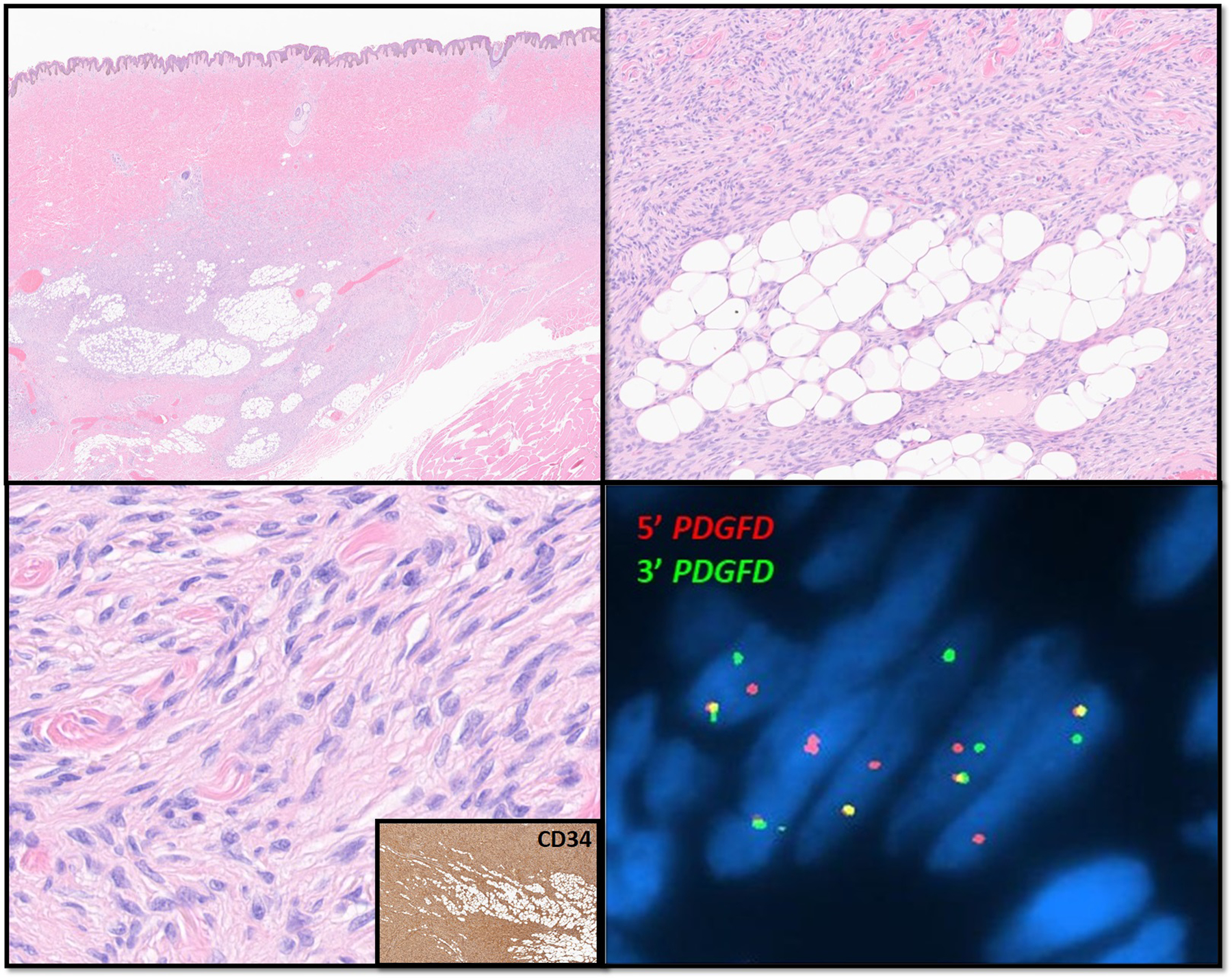
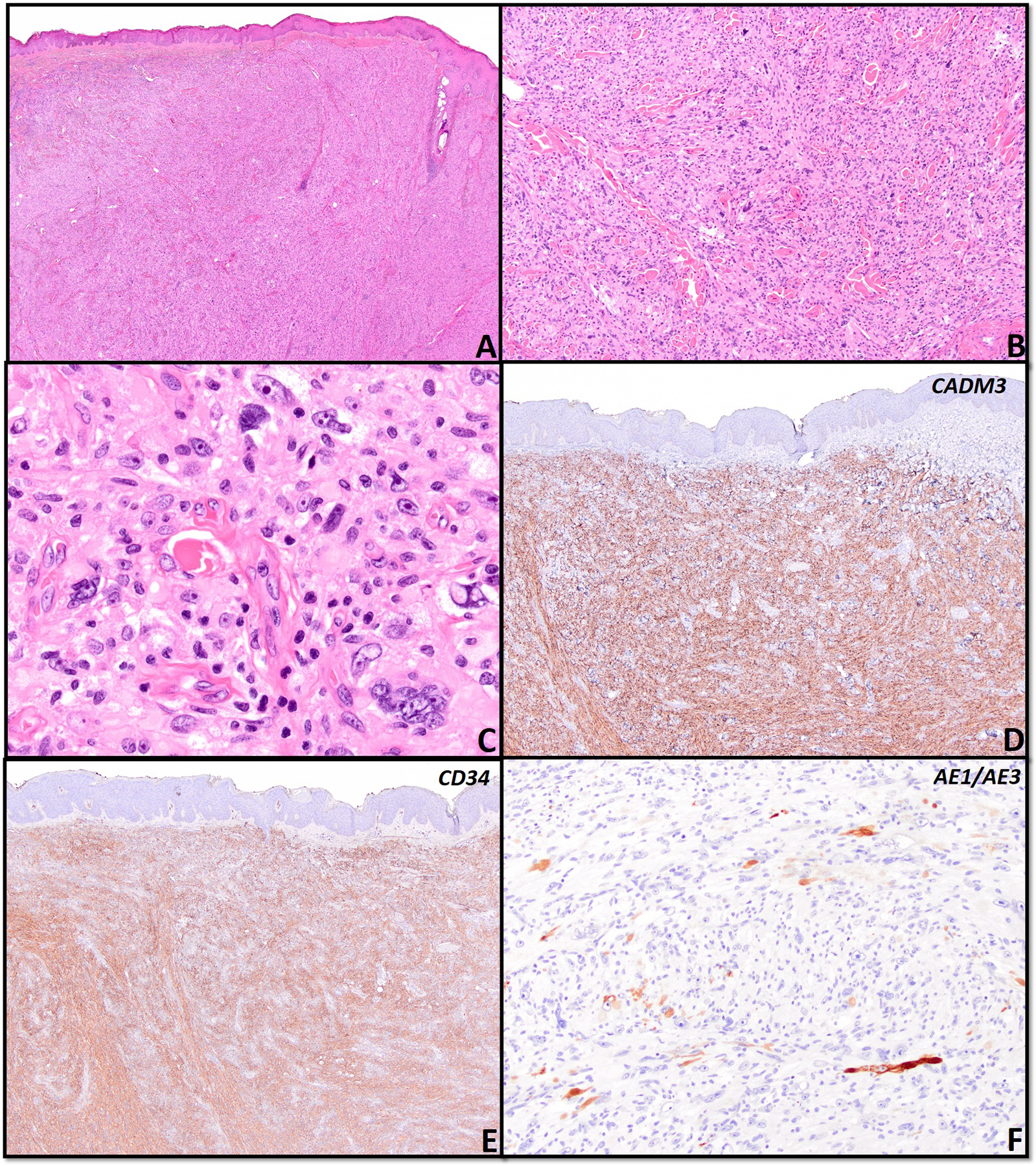
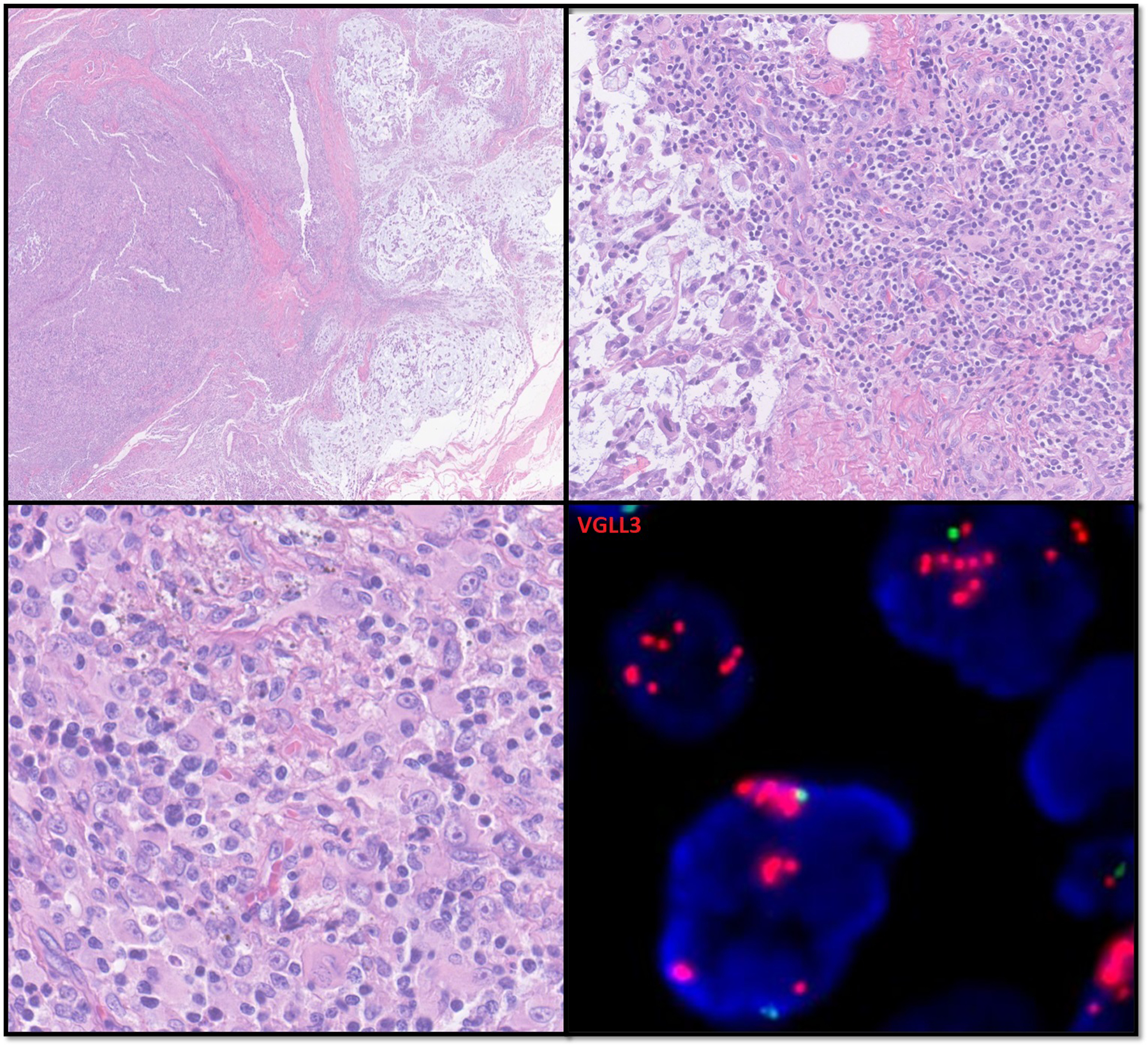
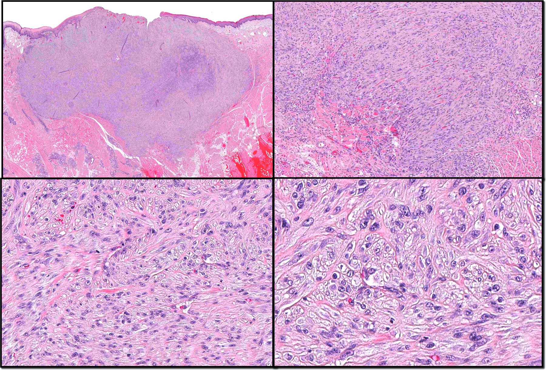
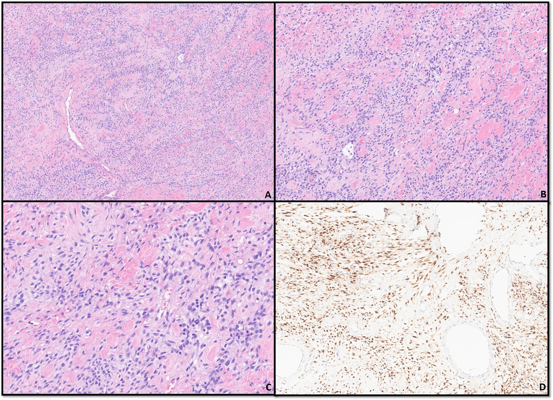
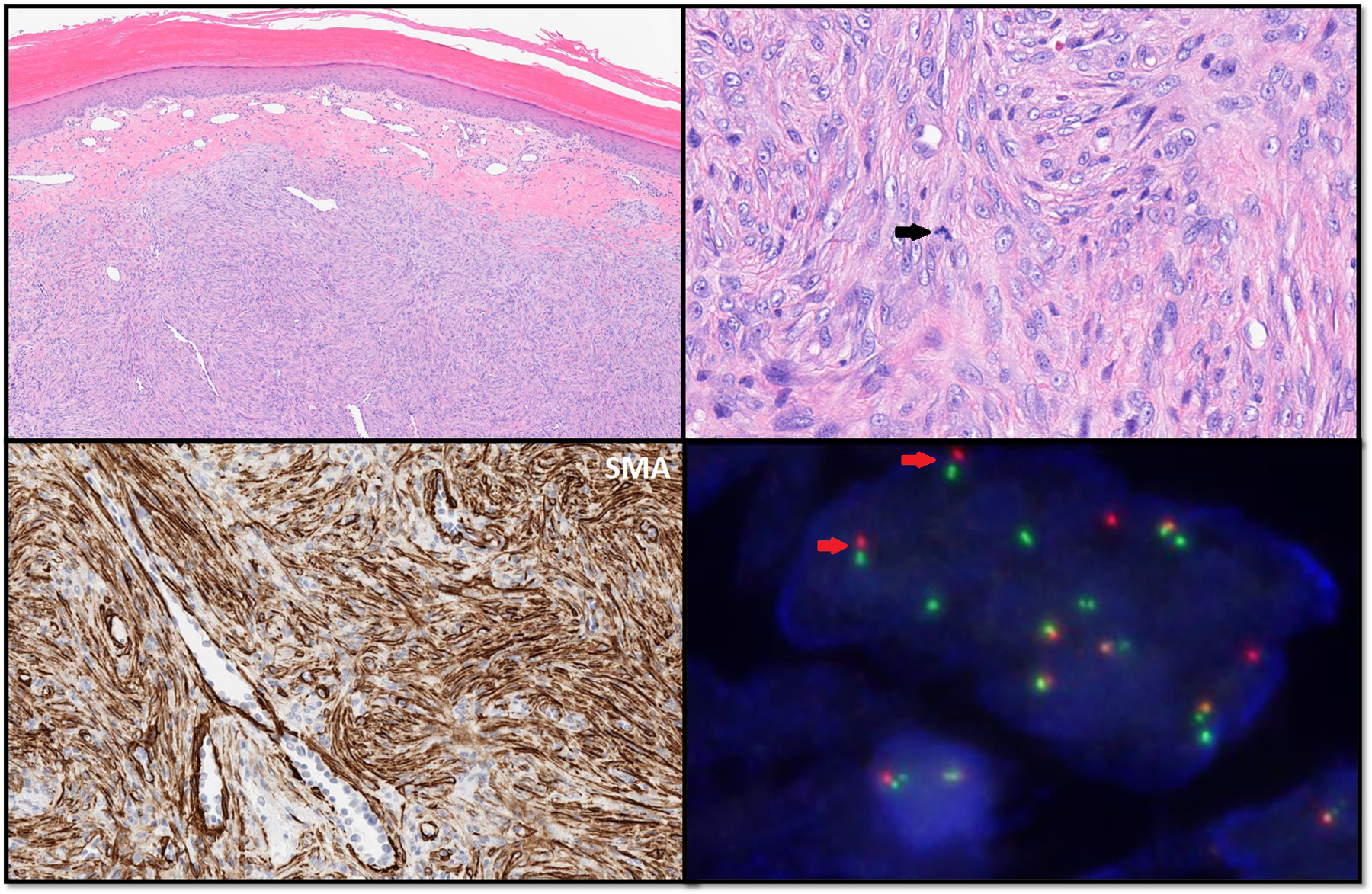
Cutaneous (myo)fibroblastic tumors constitute a group of tumors with overlapping clinicopathological features and variable biologic behavior. In the present review we focus on the histomorphology, immunohistochemical profile and molecular background of the following entities: dermatofibrosarcoma protuberans (DFSP), CD34-positive fibroblastic tumor (SCD34FT), myxoinflammatory sarcoma (MIFS), low-grade myofibroblastic sarcoma, solitary fibrous tumor and nodular fasciitis. Although some of these entities typically arise in deep-seated locations, they may occasionally present as cutaneous/superficial tumors and might be challenging to recognize. This review covers in depth the latest advances in molecular diagnostics and immunohistochemical markers that have significantly facilitated the correct classification and diagnosis of these neoplasms.
Keywords: Dermatofibrosarcoma protuberans; Low-grade myofibroblastic sarcoma; Myxoinflammatory fibroblastic sarcoma; Nodular fasciitis; Solitary fibrous tumor; Superficial CD34 fibroblastic tumor.
Copyright © 2023. Published by Elsevier Inc.
Conflict of interest statement
Declaration of Competing Interest The authors declare that they have no known competing financial interests or personal relationships that could have appeared to influence the work reported in this paper.
Figures
Similar articles
-
PRAME immunohistochemistry in soft tissue tumors and mimics: a study of 350 cases highlighting its imperfect specificity but potentially useful diagnostic applications.Virchows Arch. 2023 Aug;483(2):145-156. doi: 10.1007/s00428-023-03606-6. Epub 2023 Jul 21. Virchows Arch. 2023. PMID: 37477762
-
Fibroblastic and Myofibroblastic Soft-Tissue Tumors: Imaging Spectrum and Radiologic-Pathologic Correlation.Radiographics. 2023 Aug;43(8):e230005. doi: 10.1148/rg.230005. Radiographics. 2023. PMID: 37440448
-
Soft Tissue Special Issue: Fibroblastic and Myofibroblastic Neoplasms of the Head and Neck.Head Neck Pathol. 2020 Mar;14(1):43-58. doi: 10.1007/s12105-019-01104-3. Epub 2020 Jan 16. Head Neck Pathol. 2020. PMID: 31950474 Free PMC article. Review.
-
Apolipoprotein D in CD34-positive and CD34-negative cutaneous neoplasms: a useful marker in differentiating superficial acral fibromyxoma from dermatofibrosarcoma protuberans.Mod Pathol. 2008 Jan;21(1):31-8. doi: 10.1038/modpathol.3800971. Epub 2007 Sep 21. Mod Pathol. 2008. PMID: 17885669
-
Recent advances in the diagnosis, classification and molecular pathogenesis of cutaneous mesenchymal neoplasms.Histopathology. 2022 Jan;80(1):216-232. doi: 10.1111/his.14450. Histopathology. 2022. PMID: 34958499 Review.
References
-
- Choi Joon Hyuk MD, PhD; Ro Jae Y. MD, PhD. Recent Advances in Fibroblastic/Myofibroblastic Tumors. AJSP: Reviews & Reports 22(2):p 124–134, March/April 2017. | DOI: 10.1097/PCR.0000000000000190 - DOI
-
- Slack JC, Bründler MA, Nohr E, McIntyre JB, Kurek KC (May 2021). “Molecular Alterations in Pediatric Fibroblastic/Myofibroblastic Tumors: An Appraisal of a Next Generation Sequencing Assay in a Retrospective Single Centre Study”. Pediatric and Developmental Pathology. 24 (5)421. doi:10.1177/10935266211015558. - DOI - PubMed
Publication types
MeSH terms
Substances
Grants and funding
LinkOut - more resources
Full Text Sources
Medical
