. 2023 May 8;42(1):116.
doi: 10.1186/s13046-023-02674-5.

Convergence of YAP/TAZ, TEAD and TP63 activity is associated with bronchial premalignant severity and progression

Affiliations

Convergence of YAP/TAZ, TEAD and TP63 activity is associated with bronchial premalignant severity and progression

Boting Ning et al. J Exp Clin Cancer Res. .

Abstract

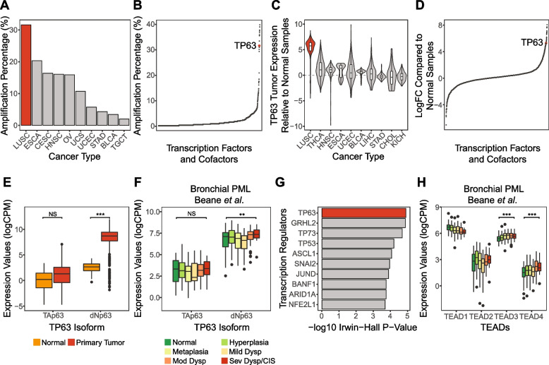
Background: Bronchial premalignant lesions (PMLs) are composed primarily of cells resembling basal epithelial cells of the airways, which through poorly understood mechanisms have the potential to progress to lung squamous cell carcinoma (LUSC). Despite ongoing efforts that have mapped gene expression and cell diversity across bronchial PML pathologies, signaling and transcriptional events driving malignancy are poorly understood. Evidence has suggested key roles for the Hippo pathway effectors YAP and TAZ and associated TEAD and TP63 transcription factor families in bronchial basal cell biology and LUSC. In this study we examine the functional association of YAP/TAZ, TEADs and TP63 in bronchial epithelial cells and PMLs.

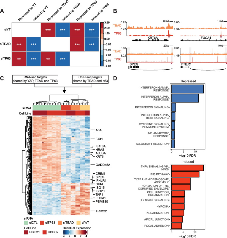
Methods: We performed RNA-seq in primary human bronchial epithelial cells following small interfering RNA (siRNA)-mediated depletion of YAP/TAZ, TEADs or TP63, and combined these data with ChIP-seq analysis of these factors. Directly activated or repressed genes were identified and overlapping genes were profiled across gene expression data obtained from progressive or regressive human PMLs and across lung single cell RNA-seq data sets.

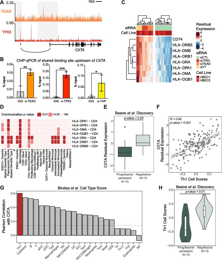
Results: Analysis of genes regulated by YAP/TAZ, TEADs, and TP63 in human bronchial epithelial cells revealed a converged transcriptional network that is strongly associated with the pathological progression of bronchial PMLs. Our observations suggest that YAP/TAZ-TEAD-TP63 associate to cooperatively promote basal epithelial cell proliferation and repress signals associated with interferon responses and immune cell communication. Directly repressed targets we identified include the MHC Class II transactivator CIITA, which is repressed in progressive PMLs and associates with adaptive immune responses in the lung. Our findings provide molecular insight into the control of gene expression events driving PML progression, including those contributing to immune evasion, offering potential new avenues for lung cancer interception.

Conclusions: Our study identifies important gene regulatory functions for YAP/TAZ-TEAD-TP63 in the early stages of lung cancer development, which notably includes immune-suppressive roles, and suggest that an assessment of the activity of this transcriptional complex may offer a means to identify immune evasive bronchial PMLs and serve as a potential therapeutic target.

Keywords: Bronchial premalignant lesions; CIITA; Immune evasion; MHCII; TAZ; TEAD; TP63; YAP.


Conflict of interest statement

X.V., S.M., M.E.L., and J.B. have received funding from sponsored research with Janssen Pharmaceuticals. Additionally, A.S. and M.E. are employees of Janssen Pharmaceuticals. All other authors declare no competing interests.

Figures

Fig. 1
Fig. 1
TP63 is associated human LUSC carcinogenesis and early lung cancer progression. a TP63 amplification frequency in TCGA samples by cancer types. The top 10 cancer types ranked by TP63 amplification frequencies are shown. b Transcription factors ranked by amplification frequencies in the TCGA LUSC samples. The vertical axis showed the amplification frequency for each TF in TCGA LUSC, and the horizontal axis shows the ranking of TFs by the amplification frequency. c TP63 expression z-scores in TCGA primary tumor samples relative to normal samples by cancer types. The top 10 cancer types ranked by TP63 overexpression are shown. d Transcription factors ranked by logFC comparing TCGA LUSC tumor to normal samples. The vertical axis shows the logFC of each TF and the horizontal axis shows the ranking of TFs by the logFC. ef Boxplots show the TP63 isoform (TAp63 and dNp63) expression levels between normal and primary tumor samples in (e) TCGA LUSC and (f) across bronchial PML histological grades in Beane et al. (Mild/Mod/Sev Dysp = Mild/Moderate/Severe Dysplasia; CIS = carcinoma in situ). *p < 0.05, **p < 0.01, ***p < 0.001. g Top 10 TFs responsible for genes upregulated in LUSC versus normal samples based on BART cancer. h Boxplots show the TEADs expression levels across bronchial PML histological grades in Beane et al. Only significant increase with higher histological grades are marked. ***p < 0.001
Fig. 2
Fig. 2
YAP, TP63 and TEADs associate and show common genomic binding regions in HBECs. a Venn diagram shows peak overlaps between YAP, TEAD and TP63 chromatin binding domains in HBECs. b Distribution of YAP/TEAD/TP63 ChIP-seq signal around ± 2 kb of YAP and YAP/TEAD overlapped peak regions (N = 4817 and 735). c Top transcription factor binding motifs enriched in the YAP/TEAD overlapped peak regions in HBECs. P-values were calculated by HOMER. d YAP, TEAD and TP63 ChIP-seq tracks shows the co-binding at the promoter regions of Hippo or TP63 canonical target genes. Overlapped peak regions are highlighted with red strips below. e Proximity ligation assays (PLA) demonstrating close association between YAP, TEAD and TP63 in proliferating HBECs. Representative images of association between the respective factors are shown, along with quantification showing the average number of PLA foci per cell across a field of view. The average of five fields of view (FOV) for each condition with standard error of mean is depicted. Unpaired t-test **p < 0.01, *** p < 0.001, ****p < 0.0001. f Western blot showing TEAD and TP63 co-immunoprecipitation in HBECS
Fig. 3
Fig. 3
YAP, TP63 and TEADs co-regulate genes associated with carcinogenesis pathways in HBECs. a Correlation plot summarizes the GSEA results of genes associated with YAP/TAZ, TEAD and TP63 siRNA treatments in HBECs. Rank lists were generated by arranging genes in order of t-statistics for their association with siRNA treatment comparing to the controls in HBECs. Genes significantly up or down-regulated with the siRNA treatments were used as gene sets (absolute logFC > 0.5 and FDR < 0.05). **p < 0.01, ***p < 0.001. b TEAD and TP63 ChIP-seq tracks shows the representative co-binding associated TEAD-TP63 direct target genes. Overlapped peak regions are shown in red strips. The grey rectangles indicate the fragment regions in pcHi-C and the black arcs show the long-range interactions between distal elements and promoter regions. Only the direct target genes are plotted. c Heatmap of gene expression significantly altered in siYAP/TAZ, siTEAD and siTP63 treatment (absolute logFC > 0.5 and FDR < 0.05). Genes annotated on the right are associated with interferon response pathways or shown to be canonical target genes of Hippo or TP63 pathways. d Top enriched functional pathways associated with the TEAD-TP63 direct repressed (top) and induced (bottom) target genes
Fig. 4
Fig. 4
TEAD-TP63 direct target genes are associated with human bronchial PML progressive pathology and early immune evasion. a Correlation plot shows the relationship between the expression levels of transcription factors and metagene scores of TEAD-TP63 direct induced and repressed target genes (calculated with GSVA) in the Beane et al. Discovery cohort. The color and the size of the circles indicate the Pearson correlation coefficients. ***Pearson correlation, FDR < 0.005. b The metagene scores of TEAD-TP63 direct repressed (left) and induced (right) target gene sets across human bronchial PML data by histological grades in the Beane et al. Discovery cohort (Mild/Mod/Sev Dysp = Mild/Moderate/Severe Dysplasia; CIS = carcinoma in situ). c Enrichment plot for TEAD-TP63 direct repressed target genes among genes ranked by tstatistics comparing the regressive PML samples to the progressive/persistent ones of the Proliferative subtypes in the Beane et al. Discovery cohort (positive t-statistics indicate upregulation among the regressive lesions). d Bubble plot shows the enrichment of TEAD-TP63 direct repressed and induced target gene sets among human bronchial PML co-expressed gene modules. The color and the size of the squares indicate the odds ratio. ***Fisher’s exact test p-value < 0.001. e Violin plots show the summarized expression of TEAD-TP63 direct repressed (left) and induced (right) target genes (calculated using AUCell) in the healthy human airway scRNA-seq data from Deprez et al. and human lung scRNA-seq data from Travaglini et al. ***one-tail Wilconxon test, p-value < 0.001
Fig. 5
Fig. 5
CIITA associates with bronchial PML progressive pathology and tracks with MHC Class II gene expression and the presence of Th1 T cells. a TEAD and TP63 ChIP-seq tracks showing the co-binding peaks associated with CIITA. Overlapped peak regions are shown with red strips below. The grey rectangles indicate the fragment regions in pcHi-C and the black arc depicts the interactions between distal elements and CIITA promoter. b Proliferating HBECs were lysed and ChIP-qPCR was performed using TEAD, TP63, YAP or IGG control antibodies examining the peak region identified by ChIP-seq as overlapping for TEAD and TP63 binding upstream of CIITA. The average % input from three experiments is shown -/ + standard error of mean. Unpaired t-test, *p < 0.01, **p < 0.001, ***p < 0.0001. c. Heatmap of significantly repressed MHC Class II gene expression (FDR < 0.01) in siYAP/TAZ, siTEAD and siTP63 treatment in HBECs. d Heatmap of communication significance levels between MHC Class II genes in basal cell and binding partners in immune cells in the human lung scRNA-seq data from Travaglini et al. The ligand-receptor pairs that involve MHC Class II gene expression in the basal cells are plotted as the row, and the immune cell types that the basal cells are communicating to are plotted as the columns. The color of heatmap reflects the significance levels of the cell–cell communication based on CellChat. e Expression levels of CIITA in progressive/persistent and regressive PML samples of the Proliferative subtype in the Beane et al. Discovery cohort. f Scatter plots show the Pearson correlation between the expression level of CIITA and Th1 scores (calculated using GSVA based on genes from Bindea et al.) in the Beane et al. Discovery cohort. g Immune cell-type ranked by their Pearson correlation coefficients with CIITA expression level in the Beane et al. Discovery cohort. The dashed line indicates the Pearson correlation coefficient that reaches p-value = 0.05. h Th1 cell scores in progressive/persistent and regressive PML samples of the Proliferative subtype in the Beane et al. Discovery cohort
Fig. 6
Fig. 6
Model for YAP/TAZ-TEAD-TP63 transcriptional complexes in bronchial PMLs. YAP/TAZ, TEAD and TP63 associate on genomic regions that control gene expression important for the growth of bronchial basal cells and the evasion of adaptive immune responses. CITIIA, a key regulator of MHCII genes, is repressed by YAP/TAZ-TEAD-TP63, and CITIIA repression is associated with decreased CD8 T cell recruitment and decreased Th1 T cell responses that track with PML progression

References

    1. Siegel RL, Miller KD, Fuchs HE, Jemal A. Cancer statistics, 2022. CA Cancer J Clin. 2022;72(1):7–33. doi: 10.3322/caac.21708. - DOI - PubMed
    1. Auerbach O, Stout AP, Hammond EC, Garfinkel L. Changes in bronchial epithelium in relation to cigarette smoking and in relation to lung cancer. N Engl J Med. 1961;265:253–267. doi: 10.1056/NEJM196108102650601. - DOI - PubMed
    1. Campbell JD, Mazzilli SA, Reid ME, Dhillon SS, Platero S, Beane J, et al. The Case for a Pre-Cancer Genome Atlas (PCGA) Cancer Prev Res (Phila) 2016;9(2):119–124. doi: 10.1158/1940-6207.CAPR-16-0024. - DOI - PubMed
    1. Beane J, Campbell JD, Lel J, Vick J, Spira A. Genomic approaches to accelerate cancer interception. Lancet Oncol. 2017;18(8):e494–e502. doi: 10.1016/S1470-2045(17)30373-X. - DOI - PMC - PubMed
    1. Beane JE, Mazzilli SA, Campbell JD, Duclos G, Krysan K, Moy C, et al. Molecular subtyping reveals immune alterations associated with progression of bronchial premalignant lesions. Nat Commun. 2019;10(1):1856. doi: 10.1038/s41467-019-09834-2. - DOI - PMC - PubMed

MeSH terms