. 2023 Jul 3;146(7):2975-2988.
doi: 10.1093/brain/awad145.

Data-driven neuropathological staging and subtyping of TDP-43 proteinopathies

Affiliations

Data-driven neuropathological staging and subtyping of TDP-43 proteinopathies

Alexandra L Young et al. Brain. .

Abstract

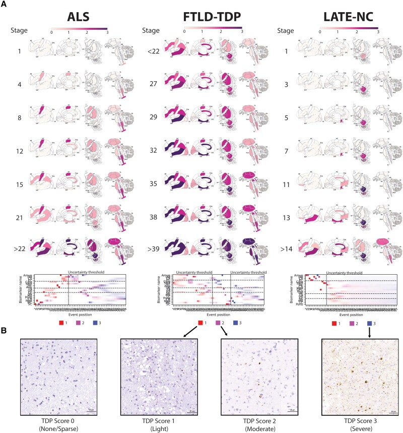
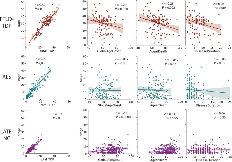
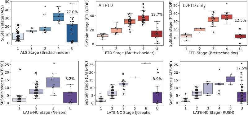
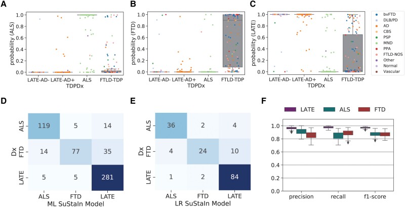
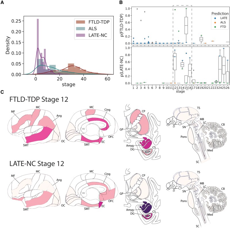
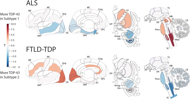
TAR DNA-binding protein-43 (TDP-43) accumulation is the primary pathology underlying several neurodegenerative diseases. Charting the progression and heterogeneity of TDP-43 accumulation is necessary to better characterize TDP-43 proteinopathies, but current TDP-43 staging systems are heuristic and assume each syndrome is homogeneous. Here, we use data-driven disease progression modelling to derive a fine-grained empirical staging system for the classification and differentiation of frontotemporal lobar degeneration due to TDP-43 (FTLD-TDP, n = 126), amyotrophic lateral sclerosis (ALS, n = 141) and limbic-predominant age-related TDP-43 encephalopathy neuropathologic change (LATE-NC) with and without Alzheimer's disease (n = 304). The data-driven staging of ALS and FTLD-TDP complement and extend previously described human-defined staging schema for ALS and behavioural variant frontotemporal dementia. In LATE-NC individuals, progression along data-driven stages was positively associated with age, but negatively associated with age in individuals with FTLD-TDP. Using only regional TDP-43 severity, our data driven model distinguished individuals diagnosed with ALS, FTLD-TDP or LATE-NC with a cross-validated accuracy of 85.9%, with misclassifications associated with mixed pathological diagnosis, age and genetic mutations. Adding age and SuStaIn stage to this model increased accuracy to 92.3%. Our model differentiates LATE-NC from FTLD-TDP, though some overlap was observed between late-stage LATE-NC and early-stage FTLD-TDP. We further tested for the presence of subtypes with distinct regional TDP-43 progression patterns within each diagnostic group, identifying two distinct cortical-predominant and brainstem-predominant subtypes within FTLD-TDP and a further two subcortical-predominant and corticolimbic-predominant subtypes within ALS. The FTLD-TDP subtypes exhibited differing proportions of TDP-43 type, while there was a trend for age differing between ALS subtypes. Interestingly, a negative relationship between age and SuStaIn stage was seen in the brainstem/subcortical-predominant subtype of each proteinopathy. No subtypes were observed for the LATE-NC group, despite aggregating individuals with and without Alzheimer's disease and a larger sample size for this group. Overall, we provide an empirical pathological TDP-43 staging system for ALS, FTLD-TDP and LATE-NC, which yielded accurate classification. We further demonstrate that there is substantial heterogeneity amongst ALS and FTLD-TDP progression patterns that warrants further investigation in larger cross-cohort studies.

Keywords: amyotrophic lateral sclerosis; frontotemporal lobar degeneration; limbic-predominant age-related TDP-43 encephalopathy; machine learning; neuropathological staging.


Conflict of interest statement

O.H. has acquired research support (for the institution) from ADx, AVID Radiopharmaceuticals, Biogen, Eli Lilly, Eisai, Fujirebio, GE Healthcare, Pfizer and Roche. In the past 2 years, he has received consultancy/speaker fees from AC Immune, Amylyx, Alzpath, BioArctic, Biogen, Cerveau, Fujirebio, Genentech, Novartis, Roche and Siemens. L.E. receives consulting fees from Biogen, PTC Therapeutics, Apellis Pharmaceuticals and Edgewise Therapeutics. D.W. has served as a paid consultant to Eli Lilly, GE Healthcare and Qynapse. He serves on a DSMB for Functional Neuromodulation. He is a site investigator for a clinical trial sponsored by Biogen. R.O. has received research support from Avid Radiopharmaceuticals, Janssen Research & Development, Roche, Quanterix and Optina Diagnostics, and has given lectures in symposia sponsored by GE Healthcare. He is an editorial board member of Alzheimer’s Research & Therapy and the European Journal of Nuclear Medicine and Molecular Imaging. All other authors report no competing interests.

Figures

Figure 1
Figure 1
Progression patterns of TDP-43 proteinopathies. (A) Inferred trajectory of regional TDP-43 progression based on individuals with a primary pathological diagnosis of ALS (left), a primary pathological diagnosis of FTLD-TDP (middle), or a secondary or tertiary pathological diagnosis of LATE-NC. Colours within each brain region represent the cumulative sum of probabilities (0–1) summed across three stages of severity, light pathology (1) in red, moderate pathology (2) in purple and severe pathology (3) in blue (see key at the bottom of each column). Only stages with reduced event ordering uncertainty (e.g. those represented by at least five individual donors) are shown. Brain schematics were generated using: https://github.com/AllenInstitute/hba_brain_schematic. Below the brains are positional variance diagrams. Each box represents the degree of certainty that a given brain region (y-axis) has reached a given severity stage (red, light; purple, moderate; blue, severe) at a given SuStaIn stage (x-axis). (B) Representative micrographs for TDP-43 scores of 0, 1, 2 and 3, respectively. ALS = amyotrophic lateral sclerosis; Amyg = amygdala; Ang = angular gyrus; CB = cerebellum; Cing = anterior cingulate; CP = caudate/putamen; CS = CA1/subiculum; DG = dentate gyrus; EC = entorhinal cortex; FTLD-TDP = frontotemporal lobar degeneration due to TDP-43; GP = globus pallidus; LATE-NC = limbic-predominant age-related TDP-43 encephalopathy neuropathologic change; LC = locus coeruleus; MB = midbrain; MC = motor cortex; MF = middle frontal gyrus; Med = medulla; OC = occipital cortex; OFC = orbitofrontal cortex; SC = spinal cord; SMT = superior and middle temporal gyrus; SN = substantia nigra; TS = thalamus.
Figure 2
Figure 2
Relationships between age and disease progression. The relationship between total TDP-43 pathology, age of disease onset, age at death and disease duration with total TDP-43 pathologic burden (as measured with SuStaIn stage) across all three diagnostic categories. SuStaIn stage was strongly related with total TDP-43 pathology across all diagnostic groups. LATE-NC showed a significant positive relationship between SuStaIn stage and age (at onset and at death), whereas FTLD-TDP showed a significant negative relationship with age and disease duration. ALS = amyotrophic lateral sclerosis; FTLD-TDP = frontotemporal lobar degeneration due to TDP-43; LATE-NC = limbic-predominant age-related TDP-43 encephalopathy neuropathologic change.
Figure 3
Figure 3
Comparison between SuStaIn-inferred TDP-43 pathological progression and previous staging systems. Individuals are staged based on various proposed staging criteria (Supplementary Fig. 2). TDP-43 pathology was averaged within regions belong to each stage. If an individual shows TDP-43 pathology (1+) in an advanced stage before showing TDP-43 pathology in a previous stage, they are considered to be ‘out-of-stage’, represented by ‘U’ for ‘unclassifiable’. SuStaIn stage generally showed good correspondence with previous pathological staging systems. ALS = amyotrophic lateral sclerosis; (bv)FTD = (behavioural variant) frontotemporal dementia; FTLD-TDP = frontotemporal lobar degeneration due to TDP-43; LATE-NC = limbic-predominant age-related TDP-43 encephalopathy neuropathologic change.
Figure 4
Figure 4
Pathology-based disease classification. All three diagnostic models were applied to all individuals regardless of TDP-type or genetic group, and the probability of the maximum likelihood stage was recorded. This probability represents a proxy for how well the individual’s regional TDP-43 pattern fit the model trajectory for LATE-NC/ALS/FTLD-TDP. Box plots in AC show the distribution of probabilities for each of the three diagnostic models (x-axis), stratified by pathological diagnosis (TDPDx on the x-axis), for (A) the ALS model, (B) the FTLD-TDP model and (C) the LATE-NC model. Note that each graph (AC) includes a probability for every subject. Note also that probabilities were derived using 10-fold cross-validation for within-diagnosis assessments (e.g. ALS cases tested using the ALS model) to avoid over-fitting. Each individual is coloured in accordance with their clinical diagnosis. Generally, individuals showed a high probability in models trained on their diagnosis, and a low probability in others. (D) A confusion matrix showing agreement between pathological diagnosis and maximum likelihood subtype model. True pathological diagnosis labels are represented on the y-axis, while predicted labels are shown on the x-axis. Individual pathological profiles tended to agree best with models fit to their clinical diagnostic group, but this was not true in all cases. (E) A logistic regression model was trained on the probabilities, plus maximum likelihood SuStaIn stage and age at death, using 100 iterations of train/test splits. The confusion matrix shows the average agreement between pathological diagnosis and predicted subtype in the 100 left-out test groups. (F) Distribution of classification statistics for performance of the maximum likelihood model across the 100 train/test splits, stratified by pathological diagnosis. See Supplementary Table 3 for further statistics and comparison with the maximum likelihood model. AD = Alzheimer's disease; ALS = amyotrophic lateral sclerosis; bvFTD = behavioural variant frontotemporal dementia; DLB/PD = dementia with Lewy bodies/Parkinson's disease; CBS = corticobasal syndrome; Dx = diagnosis; FTD = frontoptemporal dementia; FTLD-NOS = frontotemporal lobar degeneration not otherwise specified; LATE-AD+ = Alzheimer’s individuals with amygdalar but not medullar TDP-43 pathology; LATE-AD− = non-Alzheimer’s individuals with amygdalar but not medullar TDP-43 pathology; MND = motor neuron disease; PPA = primary progressive aphasia; PSP = progressive supranuclear palsy.
Figure 5
Figure 5
Comparing LATE-NC and FTLD-TDP. (A) Histograms showing the proportion of individuals assigned to each stage, separately for FTLD-TDP, ALS and LATE-NC models/individuals. Nearly all LATE-NC individuals were assigned to Stages 16 or lower, while nearly all FTLD-individuals were assigned to Stages 16 or higher. (B) SuStaIn tends to confuse LATE-NC and FTLD-TDP, particularly the stages of the crossover noted in A. The top plot represents the probability of being classified as FTLD-TDP among LATE-NC individuals, whereas the bottom plot represents the probability of being classified as LATE-NC for FTLD-TDP individuals. For both sets of patients, probability of misclassification is very low, except between Stages 12–16. (C) Comparison of FTLD-TDP and LATE-NC at Stage 12, with colours representing the amount of pathology using the same scale as Fig. 1, ranging from white (no pathology) to red (light pathology) to purple (moderate pathology) to blue (severe pathology). Brain schematics were generated using: https://github.com/AllenInstitute/hba_brain_schematic. ALS = amyotrophic lateral sclerosis; Amyg = amygdala; Ang = angular gyrus; CB = cerebellum; Cing = anterior cingulate; CP = caudate/putamen; CS = CA1/subiculum; DG = dentate gyrus; EC = entorhinal cortex; FTD = frontotemporal dementia; FTLD-TDP = frontotemporal lobar degeneration due to TDP-43; GP = globus pallidus; LATE-NC = limbic-predominant age-related TDP-43 encephalopathy neuropathologic change; LC = locus coeruleus; MB = midbrain; MC = motor cortex; MF = middle frontal gyrus; Med = medulla; OC = occipital cortex; OFC = orbitofrontal cortex; SC = spinal cord; SMT = superior and middle temporal gyrus; SN = substantia nigra; TS = thalamus.
Figure 6
Figure 6
Subtypes of primary TDP-43 proteinopathies. T-maps showing regions that are significantly different between ALS subtypes (top) and FTLD-TDP subtypes (bottom), after controlling for SuStaIn stage and multiple comparisons. A positive t-value (red) indicates more severe TDP-43 pathology in Subtype 1 and a negative t-value (blue) indicates more severe TDP-43 pathology in Subtype 2. Brain schematics were generated using: https://github.com/AllenInstitute/hba_brain_schematic. ALS = amyotrophic lateral sclerosis; Amyg = amygdala; Ang = angular gyrus; CB = cerebellum; Cing = anterior cingulate; CP = caudate/putamen; CS = CA1/subiculum; DG = dentate gyrus; EC = entorhinal cortex; FTLD-TDP = frontotemporal lobar degeneration due to TDP-43; GP = globus pallidus; LC = locus coeruleus; MB = midbrain; MC = motor cortex; MF = middle frontal gyrus; Med = medulla; OC = occipital cortex; OFC = orbitofrontal cortex; SC = spinal cord; SMT = superior and middle temporal gyrus; SN = substantia nigra; TS = thalamus.

Update of

Comment in

References

    1. Nelson PT, Dickson DW, Trojanowski JQ, et al. . Limbic-predominant age-related TDP-43 encephalopathy (LATE): consensus working group report. Brain. 2019;142:1503–1527. - PMC - PubMed
    1. Brettschneider J, Del Tredici K, Toledo JB, et al. . Stages of pTDP-43 pathology in amyotrophic lateral sclerosis. Ann Neurol. 2013;74:20–38. - PMC - PubMed
    1. Brettschneider J, Del Tredici K, Irwin DJ, et al. . Sequential distribution of pTDP-43 pathology in behavioral variant frontotemporal dementia (bvFTD). Acta Neuropathol. 2014;127:423–439. - PMC - PubMed
    1. Josephs KA, Murray ME, Whitwell JL, et al. . Staging TDP-43 pathology in Alzheimer’s disease. Acta Neuropathol. 2014;127:441–450. - PMC - PubMed
    1. Nag S, Yu L, Wilson RS, Chen EY, Bennett DA, Schneider JA. TDP-43 pathology and memory impairment in elders without pathologic diagnoses of AD or FTLD. Neurology. 2017;88:653–660. - PMC - PubMed

Publication types

MeSH terms

Substances

Supplementary concepts