Intraparenchymal meningioma in the parieto-occipital region: A case report of a diagnostically challenging tumor
- PMID: 37151446
- PMCID: PMC10159323
- DOI: 10.25259/SNI_131_2023
Intraparenchymal meningioma in the parieto-occipital region: A case report of a diagnostically challenging tumor
Abstract
Background: Intraparenchymal meningioma is a rare entity of one of the most common brain tumors. It is challenging to diagnose preoperatively due to the vague clinical presentation and absence of stereotypical radiological features. These atypical features might mislead the differential to favor high-grade gliomas or brain metastasis.
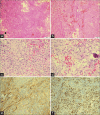
Case description: We describe a case of a 46-year-old male who presented with vertigo, right-sided sensorineural hearing loss, and bilateral blurred vision. Contrast-enhanced magnetic resonance imaging of the brain revealed a large parieto-occipital contrast-enhanced mass with a multi-loculated cystic component and diffusion restriction but without dural attachment. A gross total reaction was achieved, and the histopathological results yielded a World Health Organization Grade I meningioma diagnosis. The patient exhibited no signs of recurrence after 2 years of follow-up.
Conclusion: Intraparenchymal meningiomas are difficult to identify without histopathological assessment. We emphasize the importance of considering this diagnosis when outlining an initial differential as it may direct management planning. Total surgical resection is the best treatment modality for such cases; however, radiotherapy is a valuable option. The prognosis of intraparenchymal meningiomas is generally favorable.
Keywords: Differential diagnosis; Intraparenchymal; Meningioma; Subcortical meningioma; Transitional meningioma.
Copyright: © 2023 Surgical Neurology International.
Conflict of interest statement
There are no conflicts of interest.
Figures


Similar articles
-
Atypical Growth Pattern of an Intraparenchymal Meningioma.Case Rep Radiol. 2016;2016:7985402. doi: 10.1155/2016/7985402. Epub 2016 Sep 26. Case Rep Radiol. 2016. PMID: 27752384 Free PMC article.
-
Intraparenchymal Meningioma: Clinical, Radiologic, and Histologic Review.World Neurosurg. 2016 Aug;92:23-30. doi: 10.1016/j.wneu.2016.04.098. Epub 2016 May 4. World Neurosurg. 2016. PMID: 27155381 Review.
-
Giant Intraparenchymal Meningioma in a Female Child: Case Report and Literature Review.Cancer Manag Res. 2021 Feb 25;13:1989-1997. doi: 10.2147/CMAR.S294224. eCollection 2021. Cancer Manag Res. 2021. PMID: 33658857 Free PMC article.
-
Intraparenchymal meningioma mimicking cavernous malformation: a case report and review of the literature.J Med Case Rep. 2014 Dec 29;8:467. doi: 10.1186/1752-1947-8-467. J Med Case Rep. 2014. PMID: 25547419 Free PMC article. Review.
-
Benign Transformation of Atypical Meningioma: A Rare Histopathological Phenomenon at Recurrence.Case Rep Surg. 2024 Dec 1;2024:4118914. doi: 10.1155/cris/4118914. eCollection 2024. Case Rep Surg. 2024. PMID: 39649036 Free PMC article.
Cited by
-
Clinical and radiological presentation of meningiomas.Indian J Ophthalmol. 2025 May 1;73(5):691-694. doi: 10.4103/IJO.IJO_672_24. Epub 2025 Apr 24. Indian J Ophthalmol. 2025. PMID: 40272297 Free PMC article.
-
A large occipital meningioma with minimal symptoms in a 40-year old female: a rare case report from Syria.Ann Med Surg (Lond). 2025 Apr 15;87(6):3870-3873. doi: 10.1097/MS9.0000000000003249. eCollection 2025 Jun. Ann Med Surg (Lond). 2025. PMID: 40486610 Free PMC article.
References
-
- Curnes JT. MR imaging of peripheral intracranial neoplasms: Extraaxial vs Intraaxial masses. J Comput Assist Tomogr. 1987;11:932–7. - PubMed
-
- Cushing H, Eisenhardt L. Meningiomas: Their classification, regional behavior, life history, and surgical end result. Bull Med Libr Assoc. 1938;111:735.
-
- Goyal LK, Suh JH, Mohan DS, Prayson RA, Lee J, Barnett GH. Local control and overall survival in atypical meningioma: A retrospective study. Int J Radiat Oncol Biol Phys. 2000;46:57–61. - PubMed
Publication types
LinkOut - more resources
Full Text Sources
