. 2023 Apr 3;122(14):143701.
doi: 10.1063/5.0142405.

Femtosecond laser preparation of resin embedded samples for correlative microscopy workflows in life sciences

Affiliations

Femtosecond laser preparation of resin embedded samples for correlative microscopy workflows in life sciences

Carles Bosch et al. Appl Phys Lett. .

Abstract

Correlative multimodal imaging is a useful approach to investigate complex structural relations in life sciences across multiple scales. For these experiments, sample preparation workflows that are compatible with multiple imaging techniques must be established. In one such implementation, a fluorescently labeled region of interest in a biological soft tissue sample can be imaged with light microscopy before staining the specimen with heavy metals, enabling follow-up higher resolution structural imaging at the targeted location, bringing context where it is required. Alternatively, or in addition to fluorescence imaging, other microscopy methods, such as synchrotron x-ray computed tomography with propagation-based phase contrast or serial blockface scanning electron microscopy, might also be applied. When combining imaging techniques across scales, it is common that a volumetric region of interest (ROI) needs to be carved from the total sample volume before high resolution imaging with a subsequent technique can be performed. In these situations, the overall success of the correlative workflow depends on the precise targeting of the ROI and the trimming of the sample down to a suitable dimension and geometry for downstream imaging. Here, we showcase the utility of a femtosecond laser (fs laser) device to prepare microscopic samples (1) of an optimized geometry for synchrotron x-ray tomography as well as (2) for volume electron microscopy applications and compatible with correlative multimodal imaging workflows that link both imaging modalities.


Conflict of interest statement

Yes, J.L., A.C.D., R.C., and H.S. are employees of Carl Zeiss Microscopy GmbH, the manufacturer of the femtosecond laser and of the Zeiss Crossbeam FIB-SEM employed and evaluated in this study. The authors have no further relevant interest to disclose.

Figures

FIG. 1.
FIG. 1.
Correlative microscopy workflows incorporating samples processed using a femtosecond laser.
FIG. 2.
FIG. 2.
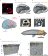
Preparation of targeted cylindrical sample for synchrotron x-ray imaging. (a–c) Mouse brain olfactory bulb tissue with a fluorescently labeled MOR174/9 glomerulus was imaged ex vivo with two-photon microscopy, stained, embedded, and imaged with lab x-ray CT (LXRT). Both two-photon (a) and LXRT (b) datasets were warped to the same space making it possible to identify the MOR174/9 glomerulus in the stained and embedded sample (c). (d) A targeted region of interest (magenta circle) was defined so it would contain the MOR174/9 glomerulus (crosshairs) and their associated projection neurons. Other non-targeted regions of interest could be also programmed in the remaining specimen (blue circles). (e) and (f) The sample was then mounted on a standard SEM pin (e) and the excess tissue was milled with the fs laser, leaving the carved pillars (f). (g) The milled pillar (orange) was mounted individually and imaged with parallel-beam synchrotron x-ray computed tomography with propagation-based phase contrast (SXRT) at the ID19 beamline of the European Synchrotron Radiation Facility. This x-ray tomography technique provides near-μm detail nondestructively in heavy metal-stained, epoxy resin-embedded samples of the biological soft tissue. The CMI approach allowed warping the 2p data to the LXRT (gray volume) and to this new SXRT dataset. This panel shows the actual position of the milled pillar (orange, segmented from SXRT data) within the original sample volumes before milling (gray, segmented from LXRT data). (h)–(i) Warping the 2p data onto the SXRT dataset revealed the position of the fluorescently labeled MOR174/9 glomerulus in the resin-embedded pillar. (i) As previously reported, the SXRT data can provide a subcellular biological context across the imaged landscapes, such as an apical dendrite of a mitral cell (black arrowheads) evolving in straight trajectory toward the genetically identified MOR174/9 glomerulus (circled with a dashed yellow line). onl, olfactory nerve layer; gl, glomerular layer; epl, external plexiform layer; mcl, mitral cell layer; ipl, internal plexiform layer; gcl, granule cell layer.
FIG. 3.
FIG. 3.
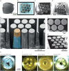
Production of pillar arrays. Multiple pillars can be extracted from a sample blockface of ∼1 mm2. The process involves imaging the blockface with scanning electron microscopy (a) and configuring the desired milling plan (b). The pillars milled present sharp edges and can, therefore, be densely packed (c)–(f). Finally, engraving of pillar IDs is possible following a similar procedure (g). (h)–(i) Pillars can then be separated and individually mounted (h) and embedded with appropriate materials to make them suitable for the intended follow-up imaging technique (i).
FIG. 4.
FIG. 4.
Multiple pillars can be extracted from neighboring locations within a 1 × 1 mm2 blockface. (a)–(d) The milling angle achieved in multiple neighboring samples enabled dense packing of pillar footprints, leaving <200 μm between the blockfaces at the sample surface, yet still consistently recovering pillars 500 μm tall. (e)–(g) Further improvements in the steepness of the milling process might allow for even tighter region of interest (ROI) packing density in a 1 mm2 blockface.

References

    1. Tordoff B., Hartfield C., Holwell A. J. et al., “ The LaserFIB: New application opportunities combining a high-performance FIB-SEM with femtosecond laser processing in an integrated second chamber,” Appl. Microsc. 50, 24 (2020).10.1186/s42649-020-00044-5 - DOI - PMC - PubMed
    1. Barnett R., Mueller S., Hiller S. et al., “ Rapid production of pillar structures on the surface of single crystal CMSX-4 superalloy by femtosecond laser machining,” Opt. Lasers Eng. 127, 105941 (2020).10.1016/j.optlaseng.2019.105941 - DOI
    1. Gu A., Terada M., Stegmann H. et al., in IEEE International Symposium on the Physical and Failure Analysis of Integrated Circuits (IPFA) ( IEEE, 2022), pp. 1–5.
    1. Sugioka K. and Cheng Y., “ Ultrafast lasers—Reliable tools for advanced materials processing,” Light. 3, e149 (2014).10.1038/lsa.2014.30 - DOI
    1. Le Harzic R., Huot N., Audouard E. et al., “ Comparison of heat-affected zones due to nanosecond and femtosecond laser pulses using transmission electronic microscopy,” Appl. Phys. Lett. 80, 3886–3888 (2002).10.1063/1.1481195 - DOI

LinkOut - more resources