Case Reports
. 2023 Apr 20:13:1071403.
doi: 10.3389/fonc.2023.1071403. eCollection 2023.

Advanced diffuse hepatic angiosarcoma treated successfully with TACE and targeted immunotherapy: A case report

Affiliations
Case Reports

Advanced diffuse hepatic angiosarcoma treated successfully with TACE and targeted immunotherapy: A case report

Yucheng Lin et al. Front Oncol. .

Abstract

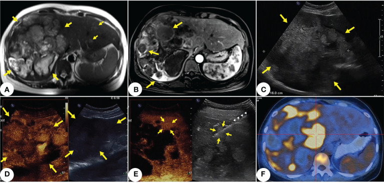
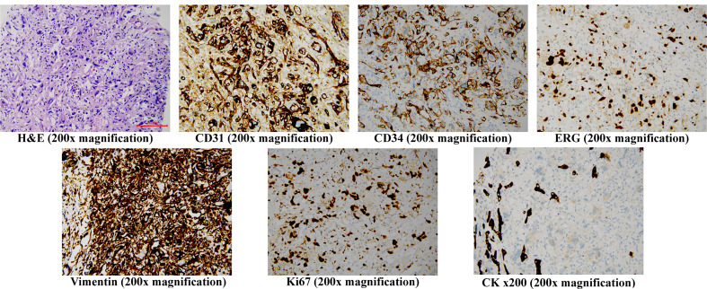
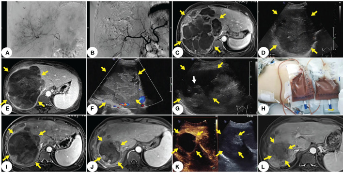
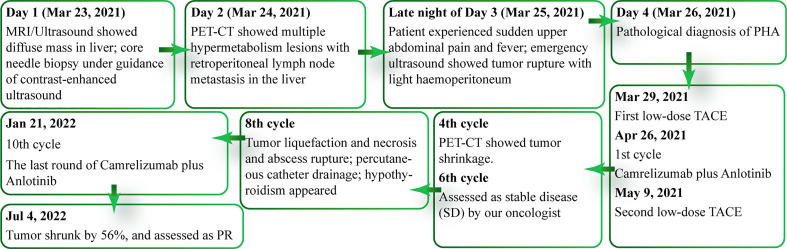
Primary hepatic angiosarcoma (PHA), a rare soft tissue tumor, accounts for only 2% of all liver malignancies. Pathologically challenging, PHA is difficult to be distinguished from other malignancies with ultrasound, Computed Tomography (CT), or Magnetic Resonance Imaging (MRI). Due to late diagnosis and resistance against traditional chemotherapy and/or radiotherapy, only 3% of PHA patients can survive up to two years after diagnosis. To our best knowledge, this case report presents the first case of an advanced diffuse PHA with ruptured hemorrhage that has been effectively treated with TACE and Anlotinib plus Camrelizumab. So far, the patient has received 10 cycles of treatment and is faring well. Latest MRI results show that the tumor has shrunk by 56% and can be assessed as a partial response (PR). This case report includes our experience in treating such a advanced malignancy, and we hope that larger studies on advanced PHA can better quantify the potential benefit.

Keywords: anlotinib; camrelizumab; hepatic angiosarcoma; immunotargeted therapy; interventional treatment (TACE).


Conflict of interest statement

The authors declare that the research was conducted in the absence of any commercial or financial relationships that could be construed as a potential conflict of interest.

Figures

Figure 1
Figure 1
Primary hepatic angiosarcoma (PHA) in a 42-years-old woman. (A) T2-weighted axial MRI shows multiple variable-sized lesions with bright T2 signal intensity are located in both hepatic lobes. Mass lesions of PHA reveal a markedly heterogeneous architecture, with focal areas of high intensity along with septum-like or rounded areas of low intensity (indicated by yellow arrows). (B) Some lesions show annular enhancement at the edges; large masses show ruptures and hemorrhages (indicated by yellow arrows). (C) Ultrasound shows a 16cm inhomogeneous hypoechoic lesion in the right hepatic lobe (indicated by yellow arrows). Multiple masses scatter within both hepatic lobes. (D) Contrast-enhanced ultrasound shows the lesion (indicated by yellow arrows) in ill-defined, peripheral irregular isoenhancement, and necrotic and hemorrhagic areas within the tumor. (E) After selecting a target lesion with multimodal enhanced imaging, (indicated by yellow arrows), we performed core needle biopsy (indicated by white triangles) under the guidance of contrast-enhanced ultrasound. (F) PET-CT shows multiple hypermetabolism lesions with retroperitoneal lymph node metastasis in the liver.
Figure 2
Figure 2
Histopathology staining of the core needle liver biopsy specimen H&E (200x magnification) shows cells with pleomorphic nuclei and frequent mitotic figures, solid nodules of similar atypical spindled endothelial cells and focal areas of with epithelioid morphology. Immunohistochemistry (200x magnification) shows the cells to be positive for CD31, CD34, ERG, Vimentin and Ki67, but negative for CK, H&E, hematoxylin and eosin.
Figure 3
Figure 3
The main treatment and imaging of this case. (A) Tumor imaging stains visible under DSA before TACE. (B) No tumor staining under DSA after TACE. (C) MRI one month after TACE shows no significant tumor progression. (D) Ultrasound two months after TACE shows slightly shrunk tumor. (E) MRI after the 8th cycle of treatment shows that the onset of tumor liquefaction, and necrosis and disintegration. (F) Ultrasound clearly shows tumor liquefaction and necrosis. (G) Ultra- sound-guided percutaneous catheter punctures into the pus and necrosis at the internal drainage site of the tumor (white arrow indicates the end of the drainage catheter). (H) A large amount of brown serous pus fluid drained with percutaneous catheter from the liquefied area of the thorax and tumor. (I) MRI after thoracic and abdominal drainage. (J) MRI at 15 months after diagnosis of PHA. (K) Contrast-enhanced ultrasound image at 15 months after diagnosis of PHA shows significant shrinkage of the main lesion without enhancement and no new lesions in the liver. (L) MRI at two years after diagnosis of PHA shows that shrunk lesions, and no activity or new lesion.
Figure 4
Figure 4
Timeline of patient's clinical course.

References

    1. Mani H, Van Thiel DH. Mesenchymal tumors of the liver. Clin Liver Dis (2001) 5(1):219–57, viii. doi: 10.1016/s1089-3261(05)70162-8 - DOI - PubMed
    1. Matthaei H, Krieg A, Schmelzle M, Boelke E, Poremba C, Rogiers X, et al. . Long-term survival after surgery for primary hepatic sarcoma in adults. Arch Surg (2009) 144(4):339–44. doi: 10.1001/archsurg.2009.30 - DOI - PubMed
    1. Cazorla A, Félix S, Delabrousse E, Valmary-Degano S. [Primary hepatic angiosarcoma: a retrospective analysis of eight cases]. Ann Pathol (2014) 34(6):462–8. doi: 10.1016/j.annpat.2014.10.004 - DOI - PubMed
    1. Yi L-L, Zhang J-X, Zhou S-G, Wang J, Huang Y-Q, Li J, et al. . CT and MRI studies of hepatic angiosarcoma. Clin Radiol (2019) 74(5):406.e1–8. doi: 10.1016/j.crad.2018.12.013 - DOI - PubMed
    1. Wang L, Lv K, Chang XY, Xia Y, Yang ZY, Jiang YX, et al. . Contrast-enhanced ultrasound study of primary hepatic angiosarcoma: a pitfall of non-enhancement. Eur J Radiol (2012) 81(9):2054–9. doi: 10.1016/j.ejrad.2011.06.026 - DOI - PubMed

Publication types