

Acetabular rim extension using a personalized titanium implant for treatment of hip dysplasia in dogs: short-term results
- PMID: 37152693
- PMCID: PMC10157081
- DOI: 10.3389/fvets.2023.1160177
Acetabular rim extension using a personalized titanium implant for treatment of hip dysplasia in dogs: short-term results
Abstract
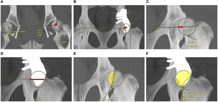
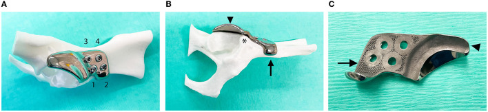
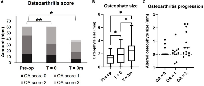
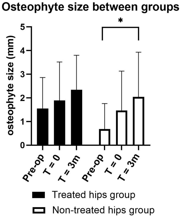
Hip dysplasia (HD) is a common orthopedic problem in young dogs. To decrease the laxity of the hip joint related to HD, the surgical treatments are recommended to increase femoral head coverage. ACEtabular rim eXtension (ACE-X) using a personalized 3-dimensional printed titanium shelf implant is a new surgical treatment to increase femoral head coverage and decrease laxity of the dysplastic hip joint, however, the efficacy is less know. Client-owned dogs older than 6 months with clinical signs of coxofemoral joint subluxation and radiographic evidence of HD with no or mild osteoarthritis (OA) were included. The Norberg angle (NA), linear percentage of femoral head overlap (LFO), and percentage of femoral head coverage (PC) were investigated radiographically and with computed tomography (CT) before and after surgery. OA was graded (scores 0-3) according to the maximum osteophyte size measured on CT. In addition, joint laxity (Ortolani) test results, gait analysis, and the Helsinki chronic pain index (HCPI) questionnaire were obtained at preoperative, immediately postoperative and at 1.5- and 3-month evaluations. Acetabular rim extension was performed in 61 hips of 34 dogs; NA, LFO, and PC were significantly higher immediately postoperatively and at the 1.5- and 3-month follow-up examinations compared with preoperative values (p < 0.05). Osteophyte size gradually increased over time (p < 0.05). The OA score significantly increased between preoperatively and directly postoperatively, and between preoperatively and at 3-month follow-up (p < 0.05). The laxity test normalized in 59 out of 61 hips after surgery, and the HCPI questionnaire showed that the pain score decreased significantly at 1.5 and 3 months, postoperatively. The force plate showed no significant improvement during the 3 months follow-up. Although pain reduction by the implant was unclear in short-term results, a personalized shelf implant significantly increased femoral head coverage and eliminated subluxation of the dysplastic hip joint. Further studies are required to study the long-term efficacy of gait, chronic pain, and progression of osteoarthritis.
Keywords: 3D printed implant; acetabular rim extension; dog; femoral head coverage; hip dysplasia; shelf arthroplasty.
Copyright © 2023 Kwananocha, Magré, Willemsen, Weinans, Sakkers, How, Verseijden, Tryfonidou, van der Wal and Meij.
Conflict of interest statement
The authors declare that the research was conducted in the absence of any commercial or financial relationships that could be construed as a potential conflict of interest.
Figures
Similar articles
-
Outcome One Year after Acetabular Rim Extension Using a Customized Titanium Implant for Treating Hip Dysplasia in Dogs.Animals (Basel). 2024 Aug 17;14(16):2385. doi: 10.3390/ani14162385. Animals (Basel). 2024. PMID: 39199919 Free PMC article.
-
Pilot study on the use of an off-the-shelf 3D-printed titanium acetabular rim extension implant for treating canine hip dysplasia.Res Vet Sci. 2025 Aug;191:105668. doi: 10.1016/j.rvsc.2025.105668. Epub 2025 Apr 29. Res Vet Sci. 2025. PMID: 40328200
-
Patient-specific 3D-printed shelf implant for the treatment of hip dysplasia tested in an experimental animal pilot in canines.Sci Rep. 2022 Feb 22;12(1):3032. doi: 10.1038/s41598-022-06989-9. Sci Rep. 2022. PMID: 35194117 Free PMC article.
-
Periacetabular osteotomy in the treatment of severe acetabular dysplasia. Surgical technique.J Bone Joint Surg Am. 2006 Mar;88 Suppl 1 Pt 1:65-83. doi: 10.2106/JBJS.E.00887. J Bone Joint Surg Am. 2006. PMID: 16510801 Review.
-
Physical Rehabilitation for the Management of Canine Hip Dysplasia: 2021 Update.Vet Clin North Am Small Anim Pract. 2022 May;52(3):719-747. doi: 10.1016/j.cvsm.2022.01.012. Vet Clin North Am Small Anim Pract. 2022. PMID: 35465906 Review.
Cited by
-
Outcome One Year after Acetabular Rim Extension Using a Customized Titanium Implant for Treating Hip Dysplasia in Dogs.Animals (Basel). 2024 Aug 17;14(16):2385. doi: 10.3390/ani14162385. Animals (Basel). 2024. PMID: 39199919 Free PMC article.
-
Digitally Designed Bone; A 3D-patient-specific Allograft Shelf for Severe Adolescent Hip Dysplasia: From Digital Design to Clinical Reality-A Conceptual Case Report.J Am Acad Orthop Surg Glob Res Rev. 2025 Jul 17;9(7):e24.00382. doi: 10.5435/JAAOSGlobal-D-24-00382. eCollection 2025 Jul 1. J Am Acad Orthop Surg Glob Res Rev. 2025. PMID: 40680264 Free PMC article.
-
Psychometric Testing and Validation of the Italian Version of the Helsinki Chronic Pain Index (I-HCPI) in Dogs with Pain Related to Osteoarthritis.Animals (Basel). 2023 Dec 25;14(1):83. doi: 10.3390/ani14010083. Animals (Basel). 2023. PMID: 38200814 Free PMC article.
References
-
- Wang Y, Prentice LF, Vitetta L, Wluka AE, Cicuttini FM. The effect of nutritional supplements on osteoarthritis. Altern Med Rev. (2004) 9:275–96. - PubMed
LinkOut - more resources
Full Text Sources
Miscellaneous