. 2023 Apr 21:27:409-428.
doi: 10.1016/j.bioactmat.2023.04.004. eCollection 2023 Sep.

Double-network hydrogel enhanced by SS31-loaded mesoporous polydopamine nanoparticles: Symphonic collaboration of near-infrared photothermal antibacterial effect and mitochondrial maintenance for full-thickness wound healing in diabetes mellitus

Affiliations

Double-network hydrogel enhanced by SS31-loaded mesoporous polydopamine nanoparticles: Symphonic collaboration of near-infrared photothermal antibacterial effect and mitochondrial maintenance for full-thickness wound healing in diabetes mellitus

Qing-Song Deng et al. Bioact Mater. .

Abstract

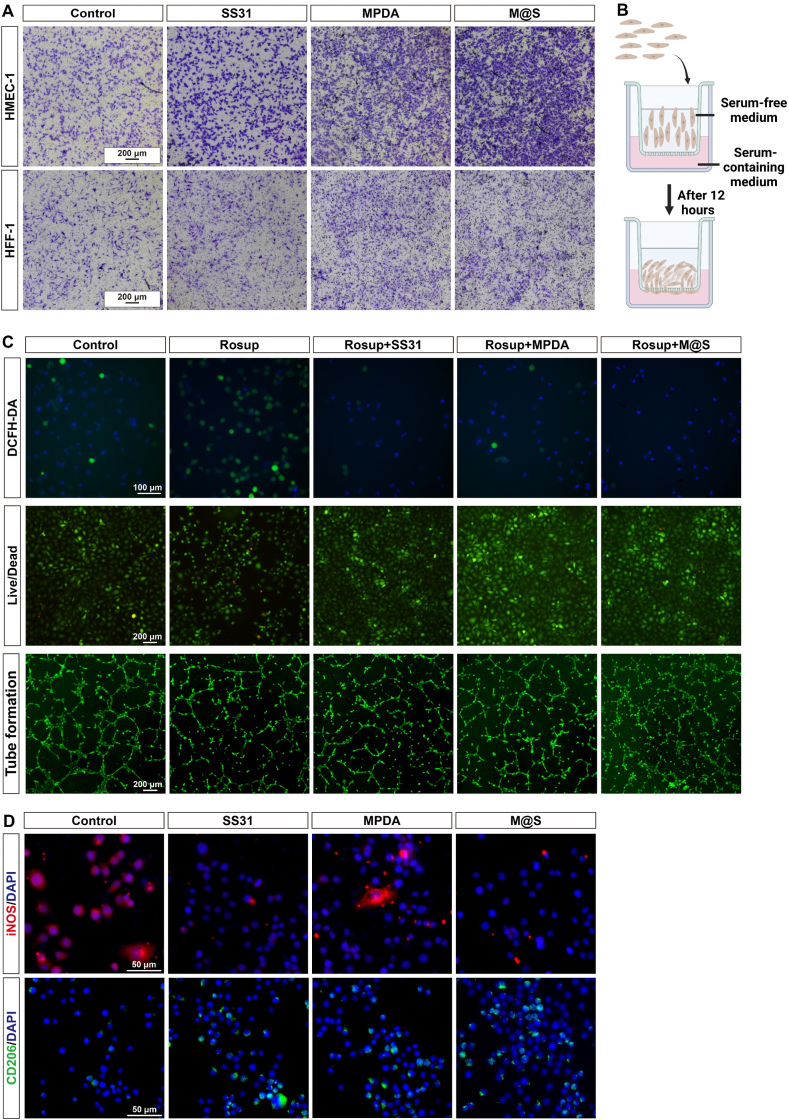
Diabetic wound healing has become a serious healthcare challenge. The high-glucose environment leads to persistent bacterial infection and mitochondrial dysfunction, resulting in chronic inflammation, abnormal vascular function, and tissue necrosis. To solve these issues, we developed a double-network hydrogel, constructed with pluronic F127 diacrylate (F127DA) and hyaluronic acid methacrylate (HAMA), and enhanced by SS31-loaded mesoporous polydopamine nanoparticles (MPDA NPs). As components, SS31, a mitochondria-targeted peptide, maintains mitochondrial function, reduces mitochondrial reactive oxygen species (ROS) and thus regulates macrophage polarization, as well as promoting cell proliferation and migration, while MPDA NPs not only scavenge ROS and exert an anti-bacterial effect by photothermal treatment under near-infrared light irradiation, but also control release of SS31 in response to ROS. This F127DA/HAMA-MPDA@SS31 (FH-M@S) hydrogel has characteristics of adhesion, superior biocompatibility and mechanical properties which can adapt to irregular wounds at different body sites and provide sustained release of MPDA@SS31 (M@S) NPs. In addition, in a diabetic rat full thickness skin defect model, the FH-M@S hydrogel promoted macrophage M2 polarization, collagen deposition, neovascularization and wound healing. Therefore, the FH-M@S hydrogel exhibits promising therapeutic potential for skin regeneration.

Keywords: Diabetic wound healing; Mesoporous polydopamine nanoparticles (MPDA NPs); Mitochondrial function maintenance; Photothermal antibacterial; SS31.


Conflict of interest statement

The authors declare that they have no known competing financial interests or personal relationships that could have appeared to influence the work reported in this paper.

Figures

Image 1
Graphical abstract
Fig. 1
Fig. 1
Characteristics of M@S NPs. (A) Schematic illustration of the synthesis of M@S NPs and release of SS31. This illustration was created with BioRender.com. (B, C) Particle size distribution of SS31, MPDA NPs, and M@S NPs. (D) Zeta potential of SS31, MPDA NPs, and M@S NPs. (E, F, G) TEM images of SS31, MPDA NPs, and M@S NPs. (H) Molecular docking study between SS31 and four different redox states of PDA NPs monomers (DA1, DA2, DA3, and DA4). (I) Molecular dynamics simulation panoramic view and local interaction of SS31–DA1 complex and SS31–DA4 complex.
Fig. 2
Fig. 2
Characteristics of FH, FH–SS31, FH–MPDA, and FH–M@S hydrogels. (A) Schematic illustration of the elements of the four types of hydrogels. This illustration was created with BioRender.com. (B) Photographs of the hydrogel precursor solutions and the corresponding hydrogels. (C) Photographs of the compression process: initial state, 30% compression, 60% compression, and recovery. (D) Compressive stress–strain curve of the four types of hydrogels. (E) Elastic modulus of the four types of hydrogels. Data are expressed as mean ± SD (n = 3). (F) Rheological performance of the hydrogels, after exposure to 405 nm ultraviolet light.
Fig. 3
Fig. 3
Photothermal effect of the four types of hydrogels. (A) Diagram of the photothermal process. This illustration was created with BioRender.com. (B) Laser intensity–dependent temperature change curves of FH-M@S hydrogel with NIR (0.1, 0.3, 0.5, and 0.7 W/cm2). (C) “On−off” temperature change of FH–M@S hydrogel under 0.7 W/cm2 laser irradiation for 5 min. (D) Photothermal stability of FH–M@S hydrogel over four consecutive photothermal heating (0.7 W/cm2) and natural cooling cycles. (E) Temperature change curves of the four types of hydrogels. (F) Infrared thermal images and central temperature of FH and FH–M@S hydrogels irradiated by 0.7 W/cm2 laser at 0 s, 60 s, 180 s, and 300 s.
Fig. 4
Fig. 4
In vitro antibacterial properties of M@S NPs under NIR irradiation (808 nm, 0.7 W/cm2). (A) Schematic diagram showing the photothermal antibacterial process. This illustration was created with BioRender.com. (B) Photographs of bacterial colony plate counting after exposure to NIR for 10 min.
Fig. 5
Fig. 5
Targeted uptake and mechanisms of action of SS31. (A) Schematic diagram showing the mechanism of SS31. This illustration was created with BioRender.com. (B) Representative confocal images of cellular uptake of SS31-FITC. SS31-FITC is shown in green, and mitochondria are shown in red. Scale bar, 100 μm. (C) Fluorescence microscopic images of JC-10 assay to measure mitochondrial membrane potential in Rosup-stimulated cells after different treatments. The red fluorescence represents JC-10 aggregates and the green fluorescence represents JC-10 monomers. Scale bar, 100 μm. (D) SS31 (purple sticks) docked into the binding site of cardiolipin (green sticks).
Fig. 6
Fig. 6
Exploring the biological functions of SS31. (A) Volcano plot visualization of the differentially-expressed genes in response to SS31 treatment. (B, C, D, E) The results of GO, KEGG, Reactome, and WikiPathways pathway enrichment analysis after RNA sequencing (RNA-seq). (F, G) The relative mRNA expression level of pro-inflammatory cytokines (IL-1β and TNF-α) and anti-inflammatory cytokines (IL-4 and IL-13) after treatment with SS31. Data are expressed as mean ± SD (n = 3). **, p < 0.01 compared with the control group, and ***, p < 0.001 compared with the control group.
Fig. 7
Fig. 7
Biological effects of M@S NPs in vitro. (A) Evaluation of migration of HMEC-1 and HFF-1 treated with different substrates for 12 h. Scale bar, 200 μm. (B) Schematic diagram showing performance of the transwell assay. This illustration was created with BioRender.com. (C) Fluorescence images of intracellular ROS analysis with DCFH-DA in Rosup-stimulated cells after different treatments. Scale bar, 100 μm. Fluorescence images of the Live/Dead assay revealed the anti-apoptosis effect of Rosup stimulation in cells after different treatments. Scale bar, 200 μm. Tube-formation assay of HMEC-1 in Rosup-stimulated cells after different treatments. Scale bar, 200 μm. (D) Fluorescence images of macrophage polarization after different treatments (red: iNOS; green: CD206; blue: cell nuclei). Scale bar, 50 μm.
Fig. 8
Fig. 8
Biocompatibility and biological effects of FH–M@S hydrogel in vitro. (A) Live/Dead staining of HMEC-1 and HFF-1 with different hydrogels after co-culture for 3 days. Scale bar, 200 μm. (B) Fluorescence images of intracellular ROS analysis with DCFH-DA in Rosup-stimulated cells after co-culture with different hydrogels. Scale bar, 100 μm. (C) Schematic diagram showing performance of the co-culture hydrogel and cells. This illustration was created with BioRender.com. (D) Flow cytometric analysis of M1 macrophage (F4/80+ and CD86+) and M2 macrophage (F4/80+ and CD206+) polarization after co-culture with different hydrogels.
Fig. 9
Fig. 9
FH–M@S hydrogel accelerated wound healing in vivo. (A) Representative photographs of diabetic cutaneous wounds of control, FH, FH–SS31, FH–MPDA, and FH–M@S hydrogel groups at days 0, 5, 7, and 14 after surgery. (B) Quantitative analysis of the unhealed area relative to the original area at day 5, day 7, and day 14 after surgery. Data are expressed as mean ± SD (n = 3). *, p < 0.05 compared with the control group, ****, p < 0.0001 compared with the control group. (C) Schematic diagram showing the experimental procedure in vivo. This illustration was created with BioRender.com.
Fig. 10
Fig. 10
Histological staining and immunofluorescence (IF) to evaluate wound healing. (A) H&E staining images of wound sections at day 14 after surgery. Scale bar, 1 mm. (B) Histological images of Masson's trichrome staining of wound sections at low and high magnification. Low magnification scale bar = 200 μm and high magnification scale bar = 50 μm. (C) 3D reconstructions of blood vessels from micro-CT at day 7 after surgery. (D) IF co-staining for α-SMA (green), CD31 (red) and DAPI (blue). Scale bar, 500 μm.
Fig. 11
Fig. 11
IF to evaluate macrophage polarization and expression of pro-inflammatory cytokines in wound healing. (A) IF staining of CD86 (red), CD206 (green), and DAPI (blue). Scale bar, 50 μm. (B) IF staining of IL-1β (red), TNF-α (red), IL-6 (red), and DAPI (blue). Scale bar, 50 μm.
Fig. 12
Fig. 12
Schematic illustration of FH–M@S hydrogel promoting diabetic wound healing. Illustration credit: Lina Cao.

References

    1. Guo L., Niu X., Chen X., Lu F., Gao J., Chang Q. 3D direct writing egg white hydrogel promotes diabetic chronic wound healing via self-relied bioactive property. Biomaterials. 2022;282 - PubMed
    1. Hu B., Gao M., Boakye-Yiadom K.O., Ho W., Yu W., Xu X., Zhang X.Q. An intrinsically bioactive hydrogel with on-demand drug release behaviors for diabetic wound healing. Bioact. Mater. 2021;6(12):4592–4606. - PMC - PubMed
    1. Lou D., Luo Y., Pang Q., Tan W.Q., Ma L. Gene-activated dermal equivalents to accelerate healing of diabetic chronic wounds by regulating inflammation and promoting angiogenesis. Bioact. Mater. 2020;5(3):667–679. - PMC - PubMed
    1. Khan A.U.R., Huang K., Khalaji M.S., Yu F., Xie X., Zhu T., Morsi Y., Jinzhong Z., Mo X. Multifunctional bioactive core-shell electrospun membrane capable to terminate inflammatory cycle and promote angiogenesis in diabetic wound. Bioact. Mater. 2021;6(9):2783–2800. - PMC - PubMed
    1. Li Y., Xu T., Tu Z., Dai W., Xue Y., Tang C., Gao W., Mao C., Lei B., Lin C. Bioactive antibacterial silica-based nanocomposites hydrogel scaffolds with high angiogenesis for promoting diabetic wound healing and skin repair. Theranostics. 2020;10(11):4929–4943. - PMC - PubMed