

Structural gray matter alterations in glioblastoma and high-grade glioma-A potential biomarker of survival
- PMID: 37152811
- PMCID: PMC10162111
- DOI: 10.1093/noajnl/vdad034
Structural gray matter alterations in glioblastoma and high-grade glioma-A potential biomarker of survival
Abstract
Background: Patients with glioblastoma (GBM) and high-grade glioma (HGG, World Health Organization [WHO] grade IV glioma) have a poor prognosis. Consequently, there is an unmet clinical need for accessible and noninvasively acquired predictive biomarkers of overall survival in patients. This study evaluated morphological changes in the brain separated from the tumor invasion site (ie, contralateral hemisphere). Specifically, we examined the prognostic value of widespread alterations of cortical thickness (CT) in GBM/HGG patients.
Methods: We used FreeSurfer, applied with high-resolution T1-weighted MRI, to examine CT, evaluated prior to standard treatment with surgery and chemoradiation in patients (GBM/HGG, N = 162, mean age 61.3 years) and 127 healthy controls (HC; 61.9 years mean age). We then compared CT in patients to HC and studied patients' associated changes in CT as a potential biomarker of overall survival.
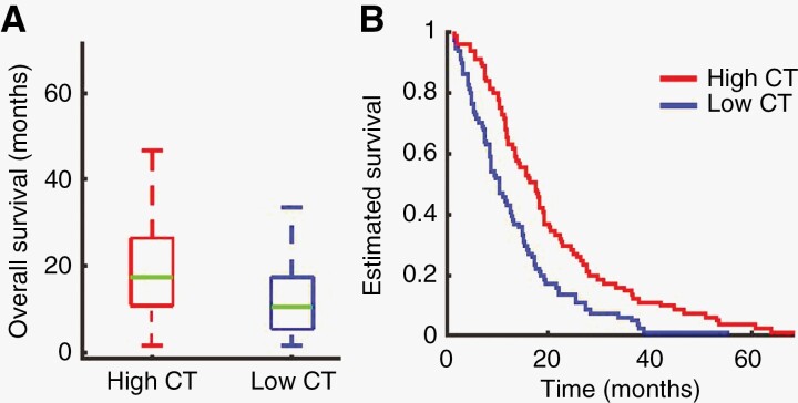
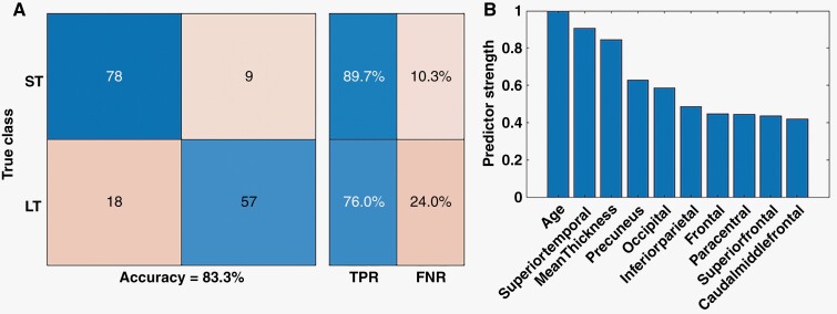
Results: Compared to HC cases, patients had thinner gray matter in the contralesional hemisphere at the time of tumor diagnosis. patients had significant cortical thinning in parietal, temporal, and occipital lobes. Fourteen cortical parcels showed reduced CT, whereas in 5, it was thicker in patients' cases. Notably, CT in the contralesional hemisphere, various lobes, and parcels was predictive of overall survival. A machine learning classification algorithm showed that CT could differentiate short- and long-term survival patients with an accuracy of 83.3%.
Conclusions: These findings identify previously unnoticed structural changes in the cortex located in the hemisphere contralateral to the primary tumor mass. Observed changes in CT may have prognostic value, which could influence care and treatment planning for individual patients.
Keywords: biomarker of GBM and HGG; brain tumor; cortical thickness; glioblastoma and high-grade glioma; overall survival.
© The Author(s) 2023. Published by Oxford University Press, the Society for Neuro-Oncology and the European Association of Neuro-Oncology.
Conflict of interest statement
E.C.L. has equity in Neurolutions, Inner Cosmos. E.C.L., D.S.M., and C.D.H. have equity in Sora Neuroscience. E.C.L, J.J.L., and J.S.S. have patent interests licensed to Sora Neuroscience. D.S.M. has equity in Radiologics, Inc. Washington University has equity in Neurolutions. B.L and E.C.L. have filed a provisional patent on the techniques described in this manuscript. All other authors declared that they had no conflicts of interest to their authorship or the publication of this article.
Figures

Similar articles
-
ACTC1 as an invasion and prognosis marker in glioma.J Neurosurg. 2017 Feb;126(2):467-475. doi: 10.3171/2016.1.JNS152075. Epub 2016 Apr 15. J Neurosurg. 2017. PMID: 27081897
-
Narrative review of palliative hypofractionated radiotherapy for high grade glioma.Ann Palliat Med. 2021 Jan;10(1):846-862. doi: 10.21037/apm-20-1246. Epub 2020 Sep 22. Ann Palliat Med. 2021. PMID: 33040565 Review.
-
Association between small heat shock protein B11 and the prognostic value of MGMT promoter methylation in patients with high-grade glioma.J Neurosurg. 2016 Jul;125(1):7-16. doi: 10.3171/2015.5.JNS142437. Epub 2015 Nov 6. J Neurosurg. 2016. PMID: 26544773
-
The prognostic value of clinical factors and cancer stem cell-related markers in gliomas.Dan Med J. 2014 Oct;61(10):B4944. Dan Med J. 2014. PMID: 25283629
-
Radiotherapy of high-grade gliomas: current standards and new concepts, innovations in imaging and radiotherapy, and new therapeutic approaches.Chin J Cancer. 2014 Jan;33(1):16-24. doi: 10.5732/cjc.013.10217. Chin J Cancer. 2014. PMID: 24384237 Free PMC article. Review.
Cited by
-
Predicting survival in glioblastoma with multimodal neuroimaging and machine learning.J Neurooncol. 2023 Sep;164(2):309-320. doi: 10.1007/s11060-023-04439-8. Epub 2023 Sep 5. J Neurooncol. 2023. PMID: 37668941 Free PMC article.
-
Cortical thickness deviations as biomarker for subtyping and prognosis in pediatric brainstem tumors.Sci Rep. 2025 Apr 16;15(1):13132. doi: 10.1038/s41598-025-95017-7. Sci Rep. 2025. PMID: 40240399 Free PMC article.
-
Structural changes in eloquent cortex secondary to glioma in sensorimotor area.Hum Brain Mapp. 2024 Jun 1;45(8):e26723. doi: 10.1002/hbm.26723. Hum Brain Mapp. 2024. PMID: 38864296 Free PMC article.
-
Mapping glioma's impact on cognition: Insights from macrostructure, microstructure, and beyond.Neurooncol Adv. 2025 Jan 8;7(1):vdaf003. doi: 10.1093/noajnl/vdaf003. eCollection 2025 Jan-Dec. Neurooncol Adv. 2025. PMID: 39911704 Free PMC article.
References
-
- Stupp R, Hegi ME, Mason WP, et al. Effects of radiotherapy with concomitant and adjuvant temozolomide versus radiotherapy alone on survival in glioblastoma in a randomised phase III study: 5-year analysis of the EORTC-NCIC trial. Lancet Oncol. 2009;10(5):459–466. - PubMed
-
- Julkunen V, Niskanen E, Koikkalainen J, et al. Differences in cortical thickness in healthy controls, subjects with mild cognitive impairment, and Alzheimer’s disease patients: a longitudinal study. J Alzheimers Dis. 2010;21(4):1141–1151. - PubMed