

Published Erratum
doi: 10.1016/j.radcr.2023.03.027.
eCollection 2023 Jun.
Erratum: Spontaneous transverse colon volvulus in a patient with Duchenne muscular dystrophy: An unreported complication
Affiliations
- PMID: 37153484
- PMCID: PMC10159818
- DOI: 10.1016/j.radcr.2023.03.027
Item in Clipboard
Published Erratum
Erratum: Spontaneous transverse colon volvulus in a patient with Duchenne muscular dystrophy: An unreported complication
Radiol Case Rep.
.
Abstract
[This corrects the article DOI: 10.1016/j.radcr.2022.12.062.][This corrects the article DOI: 10.1016/j.radcr.2023.03.026.].
Keywords: Chronic constipation; Duchenne muscular dystrophy; Gastrointestinal; Transverse colon volvulus.
© 2023 The Authors.
Figures
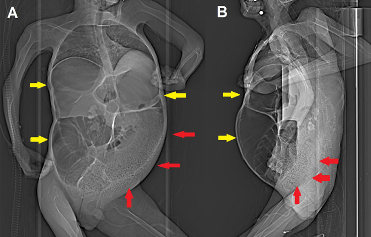
Anteroposterior (A) and lateral (B) scout images obtained before contrast-enhanced CT already show extensive bowel dilatation (yellow arrows) and left colon fecaloma (red arrows).
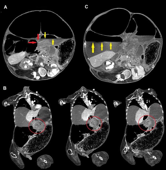
Contrast-enhanced CT scans of the abdomen demonstrating volvulus of the transverse colon. Axial scans (A-C) show an abrupt caliber change (“beak sign,” red arrows, commonly seen in many types of mechanical ileum) representing the site of the occlusion, and proximal air-fluid level (yellow arrows). Coronal view (B), obtained with multiplanar reconstructions (MPR), demonstrates the same loop twisting around the long axis of its meso (“whirl sign,” red circles); most of the right colon is displaced medially, as well as jejunal loops, due to the partial involvement of the root of the mesentery. Significant right colic dilatation with atony is present; the caliber of the caecum measures more than 17 cm and an evident air-fluid level is identified (yellow arrows).
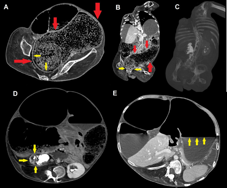
Axial (A, D-E) and coronal (B, C) contrast-enhanced CT scans demonstrate an extensive recto-sigmoid fecaloma measuring more than 30 × 10 cm (red arrows); chronic thickening of rectal walls is visible (yellow arrows), as well as compressive bilateral grade III hydronephrosis with staghorn stones and thinning of renal parenchyma, frequently seen in DMD patients (C and D, yellow arrows). Dilatation with atonia and an evident air-fluid level also involves the stomach (E, yellow arrows), further witnessing the multifocal nature of gastrointestinal motility disorders in patients with DMD.
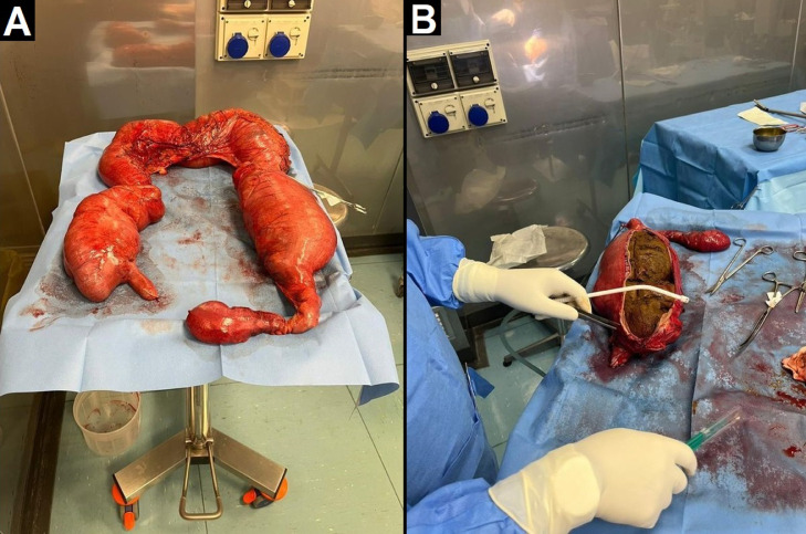
Pictures from the operating room showing subtotal colectomy. Multiple ischemic areas are visible, especially at the level of the transverse colon, where focal perforation with initial fecal leakage is present. There is also distension of the caecum and descending colon (A), the latter containing a huge fecaloma (B).
Erratum for
-
Spontaneous transverse colon volvulus in a patient with Duchenne muscular dystrophy: An unreported complication.Radiol Case Rep. 2023 Jan 18;18(3):1306-1310. doi: 10.1016/j.radcr.2022.12.062. eCollection 2023 Mar. Radiol Case Rep. 2023. PMID: 36698720 Free PMC article.
References
-
- Bushby KM. Genetic and clinical correlations of Xp21 muscular dystrophy. J Inherit Metab Dis. 1992;15(4):551–564. - PubMed
-
- Hoffman EP, Brown RH, Jr, Kunkel LM. Dystrophin: the protein product of the Duchenne muscular dystrophy locus. Cell. 1987;51(6):919–928. - PubMed
-
- Mercuri E, Bönnemann CG, Muntoni F. Muscular dystrophies. Lancet. 2019;394(10213):2025–2038. - PubMed
Publication types
LinkOut - more resources
Full Text Sources
