. 2023 Jun 23;133(1):e1-e16.
doi: 10.1161/CIRCRESAHA.122.322213. Epub 2023 May 8.

Downregulation of FKBP5 Promotes Atrial Arrhythmogenesis

Affiliations

Downregulation of FKBP5 Promotes Atrial Arrhythmogenesis

Xiaolei Wang et al. Circ Res. .

Abstract

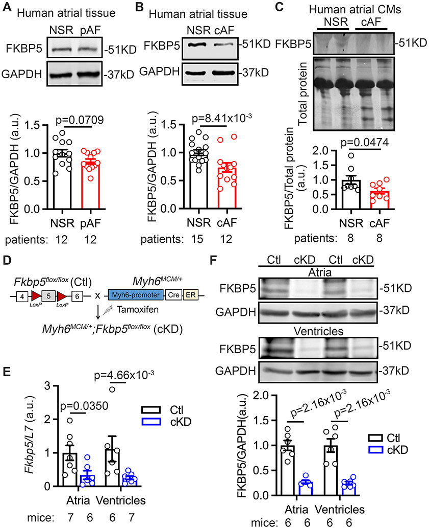
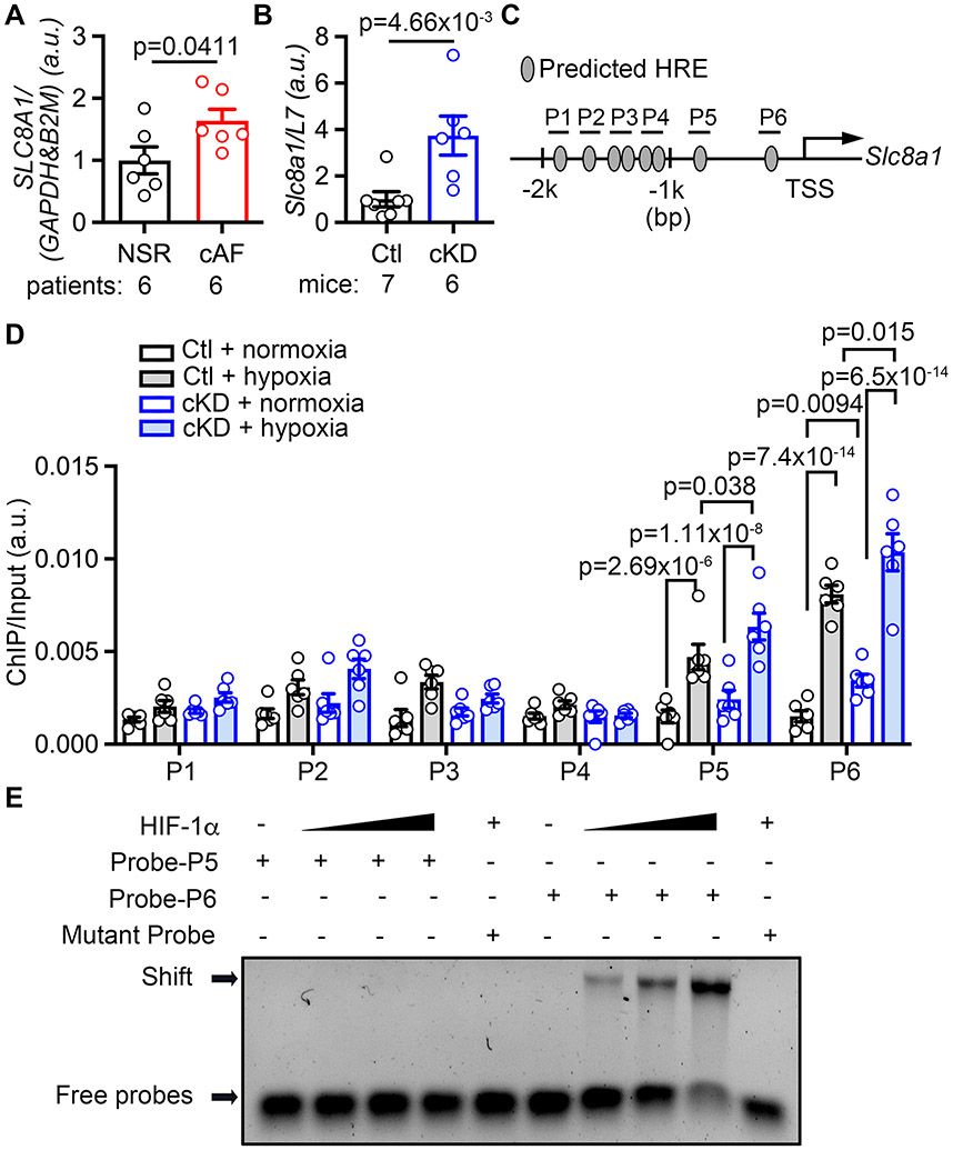
Background: Atrial fibrillation (AF), the most common arrhythmia, is associated with the downregulation of FKBP5 (encoding FKBP5 [FK506 binding protein 5]). However, the function of FKBP5 in the heart remains unknown. Here, we elucidate the consequences of cardiomyocyte-restricted loss of FKBP5 on cardiac function and AF development and study the underlying mechanisms.

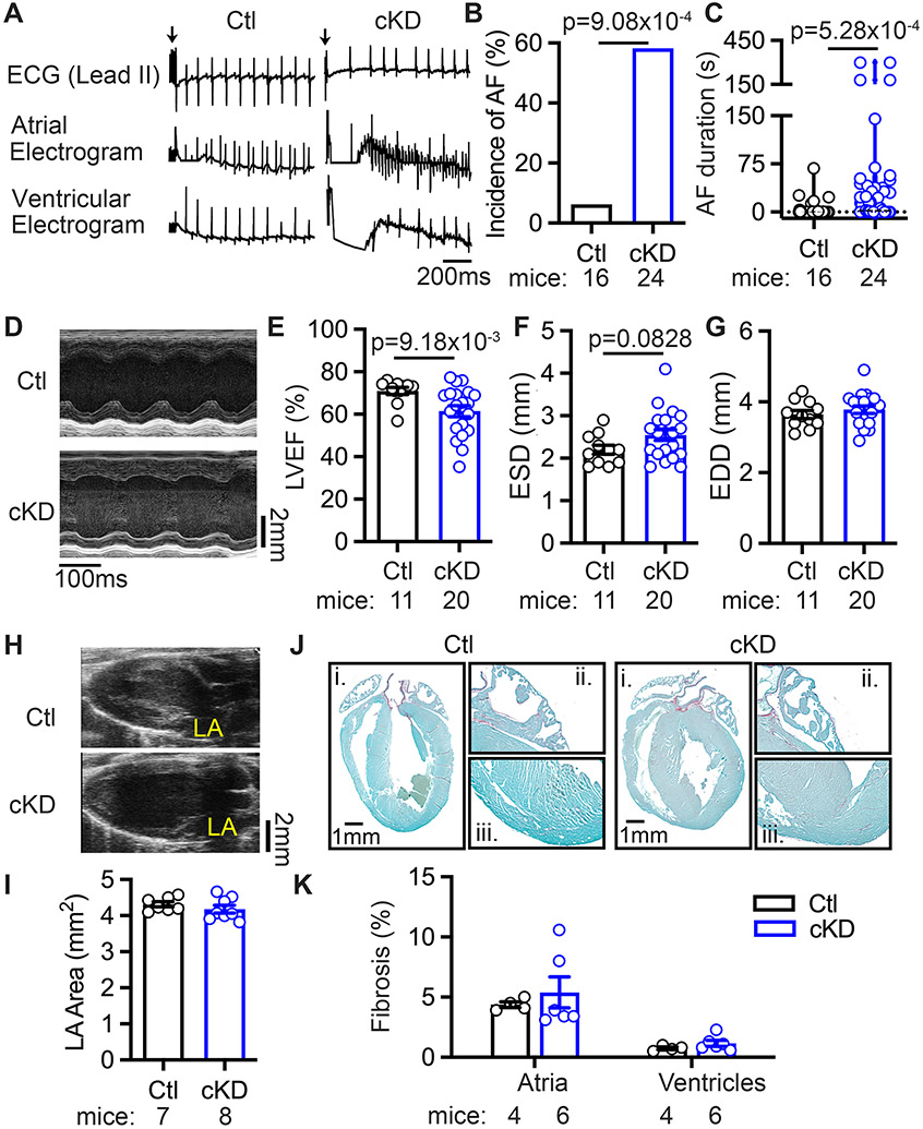
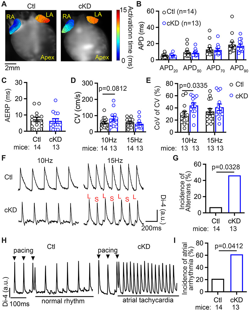
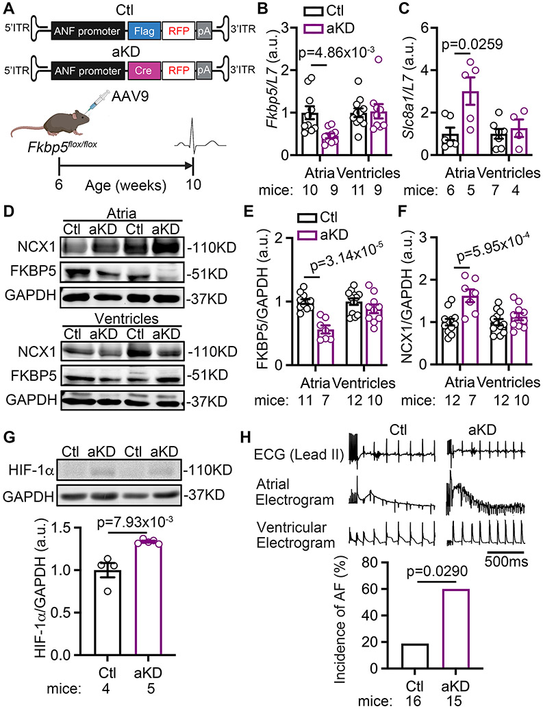
Methods: Right atrial samples from patients with AF were used to assess the protein levels of FKBP5. A cardiomyocyte-specific FKBP5 knockdown mouse model was established by crossbreeding Fkbp5flox/flox mice with Myh6MerCreMer/+ mice. Cardiac function and AF inducibility were assessed by echocardiography and programmed intracardiac stimulation. Histology, optical mapping, cellular electrophysiology, and biochemistry were employed to elucidate the proarrhythmic mechanisms due to loss of cardiomyocyte FKBP5.

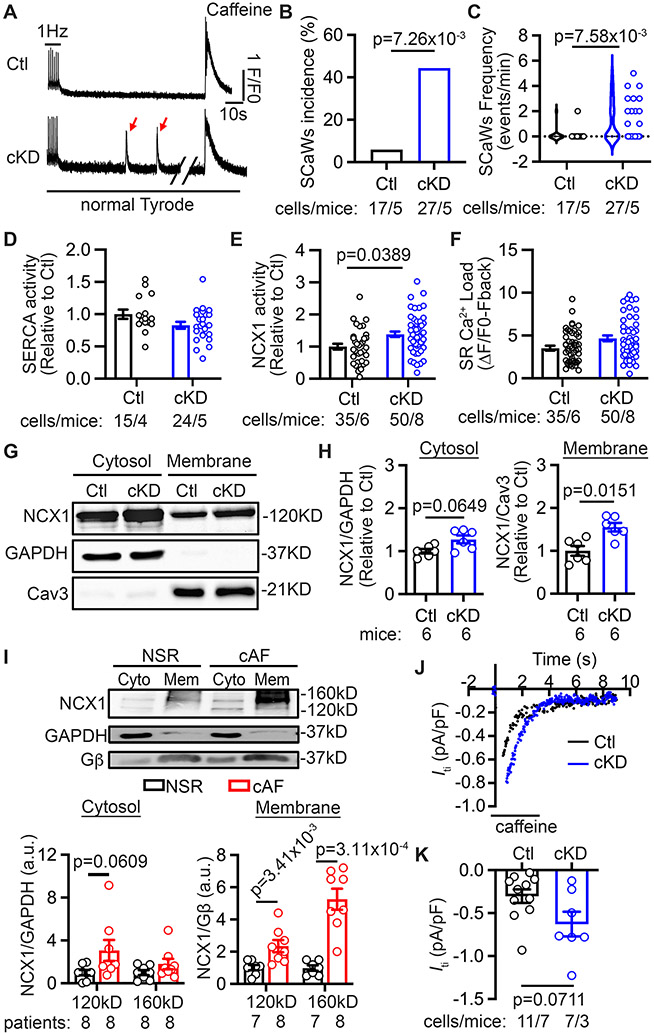
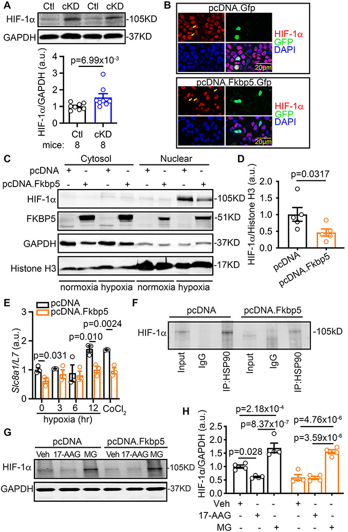
Results: FKBP5 protein levels were lower in the atrial lysates of patients with paroxysmal AF or long-lasting persistent (chronic) AF. Cardiomyocyte-specific knockdown mice exhibited increased AF inducibility and duration compared with control mice. Enhanced AF susceptibility in cardiomyocyte-specific knockdown mice was associated with the development of action potential alternans and spontaneous Ca2+ waves, and increased protein levels and activity of the NCX1 (Na+/Ca2+-exchanger 1), mimicking the cellular phenotype of chronic AF patients. FKBP5-deficiency enhanced transcription of Slc8a1 (encoding NCX1) via transcription factor hypoxia-inducible factor 1α. In vitro studies revealed that FKBP5 negatively modulated the protein levels of hypoxia-inducible factor 1α by competitively interacting with heat-shock protein 90. Injections of the heat-shock protein 90 inhibitor 17-AAG normalized protein levels of hypoxia-inducible factor 1α and NCX1 and reduced AF susceptibility in cardiomyocyte-specific knockdown mice. Furthermore, the atrial cardiomyocyte-selective knockdown of FKBP5 was sufficient to enhance AF arrhythmogenesis.

Conclusions: This is the first study to demonstrate a role for the FKBP5-deficiency in atrial arrhythmogenesis and to establish FKBP5 as a negative regulator of hypoxia-inducible factor 1α in cardiomyocytes. Our results identify a potential molecular mechanism for the proarrhythmic NCX1 upregulation in chronic AF patients.

Keywords: animals; atrial fibrillation; electrophysiology; mice.


Conflict of interest statement

Disclosures None.

Figures

Figure 1.
Figure 1.. Reduced FKBP5 protein levels in patients with AF and FKBP5-cKD mice.
(A-B) Representative Western blots and quantification of FKBP5 protein in atrial tissue of paroxysmal AF (pAF, A) and chronic AF (cAF, B) compared with NSR patients. (C) Representative Western blots and quantification of FKBP5 protein in atrial cardiomyocytes (CMs) of cAF patients compared with NSR patients. (D) The schematic diagram showed the development of the FKBP5-cKD and Ctl mice. (E) mRNA levels of Fkbp5 in atria and ventricles of Ctl and cKD mice. (F) Western blots and quantification of FKBP5 protein levels in atria and ventricles of Ctl and cKD mice. p-values were determined using unpaired Student’s t-test in A, B, and C, and Mann-Whitney test in E and F.
Figure 2.
Figure 2.. CM-specific FKBP5-deficiency enhances AF susceptibility.
(A) Representative simultaneous recordings of surface ECG and intracardiac electrograms in Ctl and cKD mice, suggestive of sinus rhythm in Ctl and AF in cKD mice after pacing. (B) The incidence and (C) the duration of pacing-induced AF in Ctl and cKD mice. (D) Representative M-mode echocardiography recording in Ctl and cKD mice. (E-G) The quantification of LVEF%, ESD, and EDD in Ctl and cKD mice. (H) Representative long-axis echocardiography recording to assess left atria (LA) in Ctl and cKD mice. (I) The quantification of LA area. (J) Representative Picrosirius Red staining in whole hearts (i), atria (ii) and ventricles (iii) of Ctl and cKD mice. (K) The quantification of fibrosis areas in atria and ventricles of Ctl and cKD mice. p-values were determined with Fisher’s exact test in B, Mann-Whitney test in C and E, and unpaired Student’s t-test in F.
Figure 3.
Figure 3.. CM-specific FKBP5-deficiency promotes arrhythmogenic alternans.
(A) Representative activation maps in Ctl and cKD mice. (B) Quantification of APD and (C) AERP at 10 Hz pacing. (D) CV at 10 Hz and 15 Hz pacing. (E) Coefficient of variation (CoV) of CV at 10 Hz and 15 Hz pacing. (F) Representative examples of AP alternans in cKD mice evoked by 15 Hz pacing, but not by 10 Hz pacing. (G) Incidence of AP alternans. (H) Representative traces of normal rhythm in Ctl and atrial tachycardia in cKD mice after pacing. (I) Incidence of pacing-induced atrial arrhythmia ex vivo. p-values were determined with unpaired Student’s t-test in D (with Welch’s correction) and E, and Fisher’s exact test in G and I.
Figure 4.
Figure 4.. CM-specific FKBP5-deficiency leads to the increased activity of NCX1.
(A) Representative traces of the 1 Hz-pacing induced Ca2+ transients (CaTs), followed by baseline recording and the caffeine (10 mmol/L) induced CaTs in atrial CMs of Ctl and cKD mice. Red arrows pointed to the spontaneous Ca2+ waves (SCaWs) in the atrial CM of cKD mice. (B) Incidence and (C) frequency of SCaWs in atrial CMs of Ctl and cKD mice. (D) Quantification of relative SERCA activity. (E) Quantification of relative NCX function. (F) SR Ca2+ load. (G) Western blots and (H) quantification of NCX1 in cytosol- and membrane-fractions of atrial tissue of Ctl and cKD mice. GAPDH and caveolin 3 (Cav3) were used as control for the cytosol- and membrane-fractions, respectively. (I) Western blots and quantification of NCX1 in cytosol (Cyto)- and membrane (Mem)-fractions of atrial tissue of NSR and cAF patients. GAPDH and Gβ were used as control for the cytosol- and membrane-fractions, respectively. (J) Representative recording of Iti elicited by the rapid application caffeine in atrial CMs. (K) Quantification of the current density of Iti. p-values were determined with Fisher’s exact test in B, Mann-Whitney test in C, H, and I, and multilevel mixed model in E and K.
Figure 5.
Figure 5.. CM-specific FKBP5-deficiency enhances the HIF-1α-mediated transcription of Slc8a1.
(A-B) Increased SLC8A1 mRNA level in atrial tissue of cAF patients (A) and cKD mice (B). (C) Schematic diagram showed the 6 HREs in the promoter region (P1-P6) of cardiac Slc8a1. (D) ChIP-qPCR showed that the interaction between HIF-1α and the P5- and P6-regions of Slc8a1-promoter were increased in cKD tissue, which was further exacerbated under hypoxia. (E) EMSA results confirmed the direct binding between HIF-1α protein and the P6-probe. p-values were determined with Mann-Whitney test in A and B, and two-way ANOVA with Tukey’s comparison in D.
Figure 6.
Figure 6.. FKBP5 negatively regulates HIF-1α.
(A) Increased HIF-1α protein level in the atria of cKD mice revealed by Western blots. (B) Immunostaining of HIF-1α in the HEK 293 cells transfected with pcDNA.Gfp (control) or pcDNA.Fkbp5.Gfp. (C) Representative Western blots with the cytosol- or nuclear-fractions of H9C2 cells transfected with pcDNA (control) or pcDNA.Fkbp5 (FKBP5-OE) vectors. (D) Relative level of the nuclear HIF-1α protein normalized to Histone H3 in H9C2 cells. (E) Relative mRNA levels of Slc8a1 in H9C2 cells treated with pcDNA vector (control) or pcDNA.Fkbp5 (FKBP5-OE). (F) Co-immunoprecipitation of HSP90 (IP) and HIF-1α (Western blots) in H9C2 cells treated with pcDNA (control) or pcDNA.Fkbp5 (FKBP5-OE) vectors. (G) Representative Western blots and (H) quantification of HIF-1α protein levels in the pcDNA or pcDNA.Fkbp5 vector transfected H9C2 cells, in the presence of vehicle (Veh), 17-AAG (HSP90 inhibitor), or MG (MG-132, proteasome inhibitor). p-values were determined with Mann-Whitney test in A and D, Shapiro-Wilk test and the unpaired Student’s t-test in E, and two-way ANOVA with Tukey’s comparison in H.
Figure 7.
Figure 7.. HIF-1α inhibition prevents AF inducibility in FKBP5 cKD mice.
(A) Timeline of HIF-1α inhibition studies in cKD mice. (B-C) Representative long-axis echocardiography images (B) and quantification (C) of left atrial (LA) area in cKD mice treated with vehicle (control) or 17-AAG. (D-F) Representative M-mode echocardiography images (D) and quantification of LVEF% (E) and diameters (F) of left ventricles in cKD mice treated with vehicle (control) or 17-AAG. (G-H) Representative Western blots (G) and quantification (H) of protein levels of NCX1, HIF-1α and HSP90 in atria of cKD mice treated with vehicle (control) or 17-AAG. (I-K) Representative recordings of surface and intracardiac electrograms (I) and the incidence (J) and duration (K) of pacing-induced AF in cKD mice treated with vehicle (control) or 17-AAG. p-values were determined with Mann-Whitney test in H and K, and Fisher’s exact test in J.
Figure 8.
Figure 8.. Atrial CM-selective knockdown of FKBP5 enhances AF susceptibility.
(A) Schematic diagram showed the designs to knockdown FKBP5 in an atrial CM-selective manner using AAV9-ANF-Cre virus injected Fkbp5flox/flox mice (aKD). AAV9-ANF-Flag virus injected Fkbp5flox/flox mice used as control (Ctl) (B) Relative mRNA levels of Fkbp5 and Slc8a1 in atria and ventricles of Ctl and mice. (C) Representative Western blots and (D) quantification of FKBP5 and NCX1 proteins in atria and ventricles of Ctl and aKD mice. (E) Representative Western blots and quantification of HIF-1α protein level in the atria of Ctl and aKD mice. (F) Representative recording and the incidence of pacing-induced AF in Ctl and aKD mice. p-values were determined with unpaired Student’s t-test in B, Mann-Whitney test in C, E, F, and G, and Fisher’s exact test in H.

Comment in

References

    1. Jackson SL, Tong X, Yin X, George MG and Ritchey MD. Emergency Department, Hospital Inpatient, and Mortality Burden of Atrial Fibrillation in the United States, 2006 to 2014. Am J Cardiol. 2017;120:1966–1973. - PMC - PubMed
    1. Schnabel RB, Yin X, Gona P, Larson MG, Beiser AS, McManus DD, Newton-Cheh C, Lubitz SA, Magnani JW, Ellinor PT, et al. 50 year trends in atrial fibrillation prevalence, incidence, risk factors, and mortality in the Framingham Heart Study: a cohort study. Lancet. 2015;386:154–62. - PMC - PubMed
    1. Chiang DY, Zhang M, Voigt N, Alsina KM, Jakob H, Martin JF, Dobrev D, Wehrens XH and Li N. Identification of microRNA-mRNA dysregulations in paroxysmal atrial fibrillation. International journal of cardiology. 2015;184C:190–197. - PMC - PubMed
    1. Cioffi DL, Hubler TR and Scammell JG. Organization and function of the FKBP52 and FKBP51 genes. Curr Opin Pharmacol. 2011;11:308–13. - PMC - PubMed
    1. Zannas AS, Balsevich G and Gassen NC. The emerging role of FKBP5 in the regulation of metabolism and body weight. Surg Obes Relat Dis. 2016;12:1560–1561. - PubMed

Publication types

Substances