

Three-dimensional structural analysis of papillary thyroid carcinoma nuclei with serial block-face scanning electron microscopy (SBF-SEM)
- PMID: 37154498
- PMCID: PMC11551838
- DOI: 10.1111/pin.13329
Three-dimensional structural analysis of papillary thyroid carcinoma nuclei with serial block-face scanning electron microscopy (SBF-SEM)
Abstract
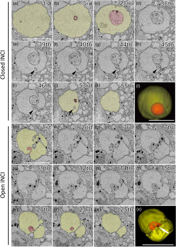
Nuclear morphology of carcinoma cells is critical for the pathological diagnosis of papillary thyroid carcinoma (PTC). However, three-dimensional architecture of PTC nuclei is still elusive. In this study, we analyzed the three-dimensional ultrastructure of PTC nuclei using serial block-face scanning electron microscopy which takes advantage of the high-throughput acquisition of serial electron microscopic images and three-dimensional reconstruction of subcellular structures. En bloc-stained and resin-embedded specimens were prepared from surgically removed PTCs and normal thyroid tissues. We acquired two-dimensional images from serial block-face scanning electron microscopy and reconstructed three-dimensional nuclear structures. Quantitative comparisons showed that the nuclei of carcinoma cells were larger and more complex than those of normal follicular cells. The three-dimensional reconstruction of carcinoma nuclei divided intranuclear cytoplasmic inclusions into "open intranuclear cytoplasmic inclusions" connecting to cytoplasm outside the nucleus and "closed intranuclear cytoplasmic inclusions" without that connection. Cytoplasm with abundant organelles was observed in open inclusions, but closed inclusions contained fewer organelles with or without degeneration. Granules with a dense core were only observed in closed inclusions. Our observations suggested that open inclusions originate from nuclear invaginations, and disconnection from cytoplasm leads to closed inclusions.
Keywords: intranuclear cytoplasmic inclusion; nuclear invagination; papillary thyroid carcinoma; serial block-face scanning electron microscopy; three-dimension; thyroid gland; ultrastructure.
© 2023 The Authors. Pathology International published by Japanese Society of Pathology and John Wiley & Sons Australia, Ltd.
Conflict of interest statement
None declared.
Figures


Similar articles
-
New insights into intranuclear inclusions in thyroid carcinoma: Association with autophagy and with BRAFV600E mutation.PLoS One. 2019 Dec 16;14(12):e0226199. doi: 10.1371/journal.pone.0226199. eCollection 2019. PLoS One. 2019. PMID: 31841566 Free PMC article.
-
Studies on intranuclear inclusions and nuclear grooves in papillary thyroid cancer by light, scanning electron and transmission electron microscopy.Acta Cytol. 1996 May-Jun;40(3):417-22. doi: 10.1159/000333892. Acta Cytol. 1996. PMID: 8669172
-
Intranuclear inclusions in hepatocellular carcinoma contain autophagy-associated proteins and correlate with prolonged survival.J Pathol Clin Res. 2019 Jul;5(3):164-176. doi: 10.1002/cjp2.129. Epub 2019 Apr 8. J Pathol Clin Res. 2019. PMID: 30859721 Free PMC article.
-
beta-Catenin expression in thyroid follicular lesions: potential role in nuclear envelope changes in papillary carcinomas.Endocr Pathol. 2004 Winter;15(4):329-37. doi: 10.1385/ep:15:4:329. Endocr Pathol. 2004. PMID: 15681857 Review.
-
Potential pitfalls for false suspicion of papillary thyroid carcinoma: a cytohistologic review of 22 cases.Diagn Cytopathol. 2012 May;40 Suppl 1:E74-9. doi: 10.1002/dc.21726. Epub 2011 May 11. Diagn Cytopathol. 2012. PMID: 21563322 Review.
References
-
- LiVolsi VA. Papillary thyroid carcinoma: an update. Mod Pathol. 2011;24(S2):S1–9. - PubMed
-
- Papotti M, Manazza AD, Chiarle R, Bussolati G. Confocal microscope analysis and tridimensional reconstruction of papillary thyroid carcinoma nuclei. Virchows Arch. 2004;444(4):350–5. - PubMed
-
- Sjöstrand FS. Ultrastructure of retinal rod synapses of the guinea pig eye as revealed by three‐dimensional reconstructions from serial sections. J Ultrastruct Res. 1958;2:122–70. - PubMed
-
- Stevens JK, Davis TL, Friedman N, Sterling P. A systematic approach to reconstructing microcircuitry by electron microscopy of serial sections. Brain Res Rev. 1980;2(1–3):265–93. - PubMed
MeSH terms
LinkOut - more resources
Full Text Sources
Medical
Research Materials
