. 2023 Sep;47(3):1721-1733.
doi: 10.1007/s11259-023-10130-0. Epub 2023 May 8.

Generation of a novel three-dimensional scaffold-based model of the bovine endometrium

Affiliations

Generation of a novel three-dimensional scaffold-based model of the bovine endometrium

M C Díez et al. Vet Res Commun. 2023 Sep.

Abstract

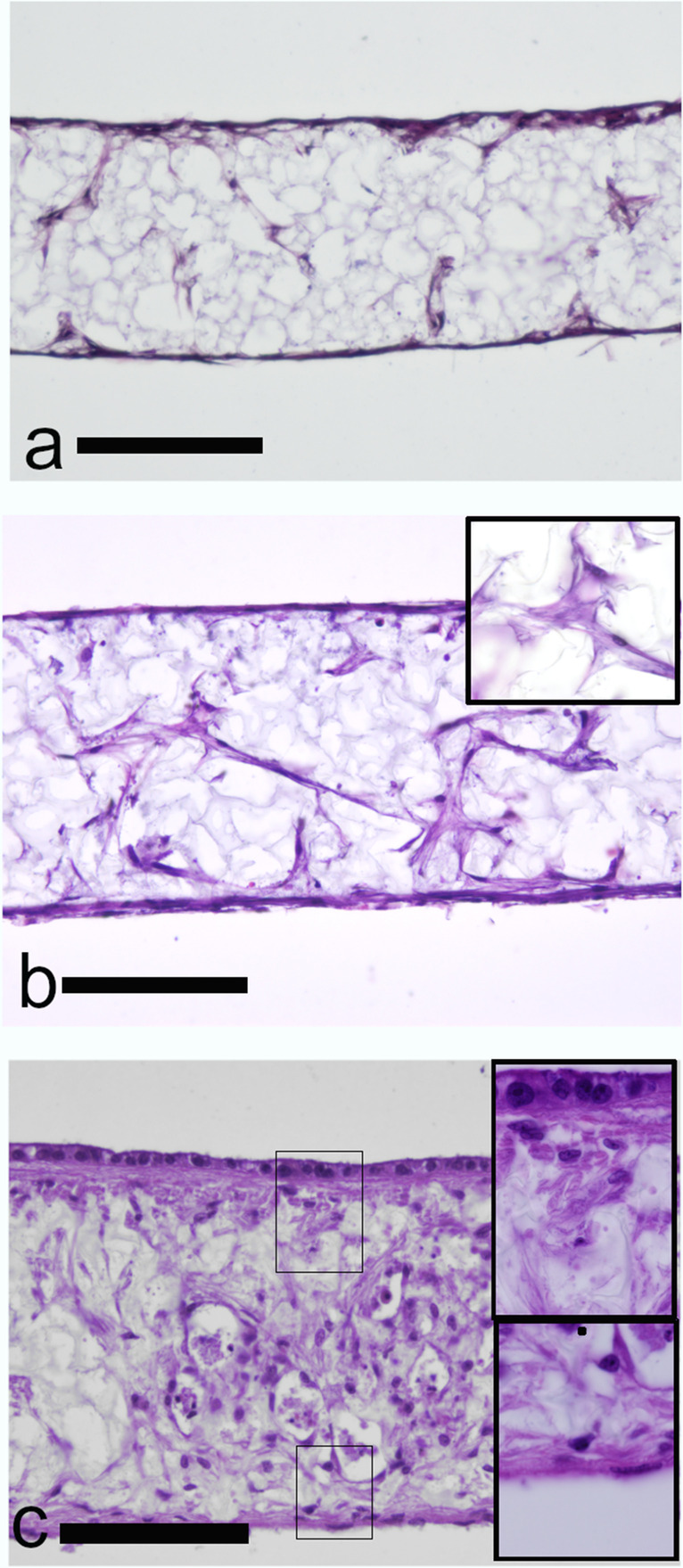
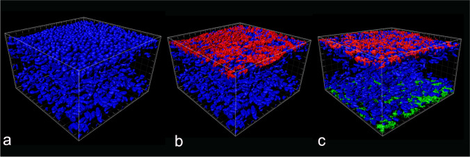
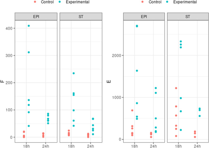
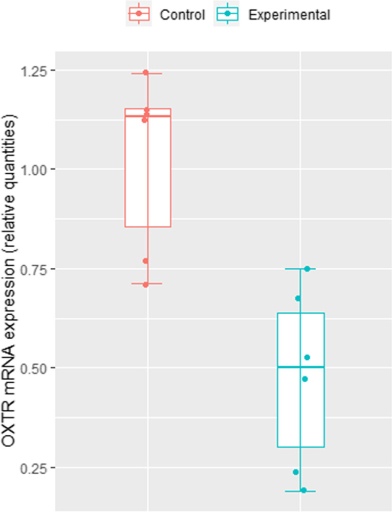
Bovine in vitro endometrial models that resemble tissue function in vivo are needed to study infertility, long-term uterine alterations induced by pathogens and impact of endocrine disruptor chemicals on reproductive function and other reproductive system complications that cause high economic losses in livestock species. The present study aimed to generate an innovative, reproducible, and functional 3D scaffold-based model of the bovine endometrium structurally robust for long term-culture. We developed a multicellular model containing both endometrial epithelial and stromal cells. Epithelial cells organized to form a luminal-like epithelial layer on the surface of the scaffold. Stromal cells produced their own extracellular matrix forming a stable subepithelial compartment that physiologically resembles the normal endometrium. Both cell types released prostaglandin E2 and prostaglandin F following a treatment with oxytocin and arachidonic acid. Additionally signal pathways mediating oxytocin and arachidonic acid stimulation of prostaglandin synthesis were analyzed by real time PCR (RT-PCR). Oxytocin receptor (OXTR), prostaglandin E2 receptor 2 (EP2), prostaglandin E2 receptor 4 (EP4), prostaglandin F receptor (PTGFR), prostaglandin E synthase (PTGES), PGF-synthase (PGFS) and prostaglandin-endoperoxide synthase 2 (COX-2) expression was detected in both control and treatment groups, however, only significant changes in abundance of OXTR mRNA transcripts were found. The results obtained by this study are a step forward in bovine in vitro culture technology. This 3D scaffold-based model provides a platform to study regulatory mechanisms involved in endometrial physiology and can set the basis for a broader tool for designing and testing novel therapeutic strategies for recurrent uterine pathologies.

Keywords: Co-culture; Endometrium; Scaffold; Three-dimensional (3D) cell culture.


Conflict of interest statement

SP acts as a consultant for Reprocell Europe.

Figures

Fig. 1
Fig. 1
Diagrammatic representation of the construction of the 3D endometrial model and functional assays performed
Fig. 2
Fig. 2
Validation of subepithelial/stromal and epidermal structure in full-thickness bovine endometrial tissue equivalents. (a, b) Representative photomicrographs of haematoxylin and Eosin (H&E) stained Alvetex™ seeded with bovine endometrial fibroblast for: (a) seven days (b) fourteen days. (c) Seven days after epithelial cell seeding, a single cuboidal monolayer of epithelial cells was established
Fig. 3
Fig. 3
Expression of vimentin fibroblast marker in subepithelial stromal cells over time. Representative fluorescent confocal photomicrographs for the expression of vimentin (green) in single confocal laser microscopy section at: (a) seven days, (b) 14 days and (c) 21 days after fibroblast seeding on Alvetex™; and high-quality 3D images obtained by processing the optical section stacks with volume render and surface display parameters in microVoxel at: (d) seven days, (e) 14 days and (f) 21 days after fibroblast seeding. Nuclei of the cells were stained using DAPI (blue)
Fig. 4
Fig. 4
Extracellular matrix component type III collagen in subepithelial stromal compartment. (a) Representative fluorescence photomicrograph of type III collagen (green) expression of cross-sectional section of stromal seeded scaffolds on day 21 and (b and c) combination of fluorescence image and transmission image with confocal microscopy was used to visualized Collagen III expression (green) on Alvetex™ scaffold. Images (a) and (b) with nuclei of the cells stained with DAPI (in blue). (c) image (c) without DAPI to improve visualization of collagen III deposition over scaffold
Fig. 5
Fig. 5
Immunohistochemistry images of co-cultured stromal and epithelial cells seeded on Alvetex scaffolds on day 32 of culture. Cytokeratin (a, red) or E-cadherin (b green) expression by epithelial cells of cross sectional sections of the scaffold. Both epithelial markers are restricted to the monolayer of epithelial cells found on top of the stromal compartment. Representative fluorescent confocal photomicrographs for the expression of cytokeratin (c) and E-cadherin (d) were obtained at the top cellular monolayer of the endometrial construct. For all images, DAPI was used as nuclear stain (blue)
Fig. 6
Fig. 6
Depth-color coded projection of co-cultured stromal and epithelial cell nuclei seeded on Alvetex™ scaffolds on day 35 of culture. 3D reconstruction of confocal microscope images of epithelial and stromal cells co-cultivated on Alvetex™ scaffold for 35 days. Compensation tool during confocal images acquisition enabled to visualize cell nuclei placed in deeper positions within the scaffold. Cell nuclei have been color-coded based upon depth (DAPI stain)
Fig. 7
Fig. 7
3D rendering illustrating the distribution of epithelial and stromal cells within the co-culture on day 35 of culture. a) 3D BECs cell nuclei with DAPI stain (blue). (b and c) Cytokeratin (red stain) is localized at the epithelial cell layer in the upper surface of the 3D endometrial co-culture model. (c) Vimentin (green stain) is expressed by fibroblast located at the bottom layers
Fig. 8
Fig. 8
Prostaglandin accumulation of epithelial and stromal cell co-cultured on Alvetex™ scaffold and treated with oxytocin plus arachidonic acid. Accumulation of (a) PGF and (b) PGE following 18 h or 24 h treatment. Supernatants from the apical (EPI) and basal compartments (ST) were analyzed separately. Prostaglandin accumulation differed significantly between treated and control (P < 0.05)
Fig. 9
Fig. 9
Oxytocin receptor mRNA expression by epithelial and stromal cell co-cultured on Alvetex™ scaffold and treated with oxytocin plus arachidonic acid at 24 h. Relative mRNA expression results for OXTR in control and experimental groups when normalized with endogenous control genes SUZ12 and C2ORF29. OXTR expression was significantly decrease in the experimental group vs. the control group (p < 0.05)

References

    1. Almeida GHD, Iglesia RP, Araujo MS, Carreira ACO, Santos EXD, Calomeno CVAQ, Miglino MA. Uterine Tissue Engineering: Where We Stand and the Challenges Ahead. Tissue Eng Part B Rev. 2022;28(4):861–890. doi: 10.1089/ten.TEB.2021.0062. - DOI - PubMed
    1. Asselin E, Fortier MA. Influence of thrombin on proliferation and prostaglandin production in cultured bovine endometrial cells. J Cell Physiol. 1996;168(3):600–7. doi: 10.1002/(SICI)1097-4652(199609)168:3<600::. - DOI - PubMed
    1. Badr-Eldin SM, Aldawsari HM, Kotta S, Deb PK, Venugopala KN. Three-Dimensional In Vitro Cell Culture Models for Efficient Drug Discovery: Progress So Far and Future Prospects. Pharmaceuticals (Basel) 2022;15(8):926. doi: 10.3390/ph15080926. - DOI - PMC - PubMed
    1. Chankeaw W, Lignier S, Richard C, Ntallaris T, Raliou M, Guo Y, Plassard D, Bevilacqua C, Sandra O, Andersson G, Humblot P, Charpigny G. Analysis of the transcriptome of bovine endometrial cells isolated by laser micro-dissection (1): specific signatures of stromal, glandular and luminal epithelial cells. BMC Genomics. 2021;22(1):451. doi: 10.1186/s12864-021-07712-0. - DOI - PMC - PubMed
    1. Costello L, Darling N, Freer M, Bradbury S, Mobbs C, Przyborski S. Use of Porous Polystyrene Scaffolds to Bioengineer Human Epithelial Tissues In Vitro. Methods Mol Biol. 2021;2273:279–296. doi: 10.1007/978-1-0716-1246-0_20. - DOI - PubMed