

Ultrasound-Targeted Microbubble Disruption with Key Nanodroplets for Effective Ferroptosis in Triple-Negative Breast Cancer Using Animal Model
- PMID: 37155504
- PMCID: PMC10122866
- DOI: 10.2147/IJN.S400495
Ultrasound-Targeted Microbubble Disruption with Key Nanodroplets for Effective Ferroptosis in Triple-Negative Breast Cancer Using Animal Model
Abstract
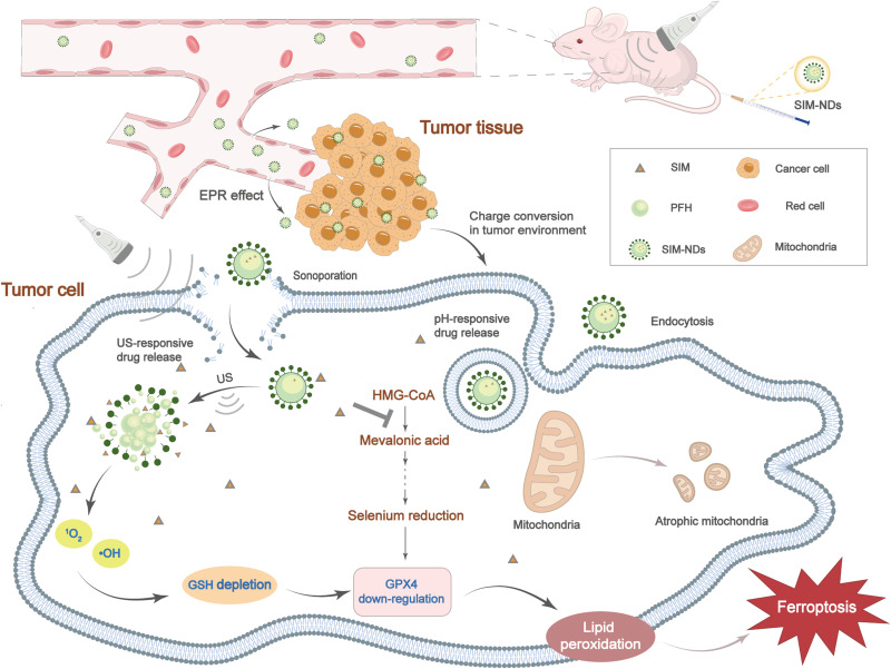
Introduction: Triple-negative breast cancer (TNBC) is known to be the most aggressive form of breast cancer. Due to its high recurrence and mortality rates, the treatment of TNBC is a significant challenge for the medical community. Besides, ferroptosis is an emerging regulatory cell death that may provide new insights into the treatment of TNBC. As a central inhibitor of the ferroptosis process, the selenoenzyme glutathione peroxidase 4 (GPX4) is its classical therapeutic target. However, inhibition of GPX4 expression is quite detrimental to normal tissues. Ultrasound contrast agents, as an emerging visualization precision treatment, may provide a solution to the existing problem.
Methods: In this study, nanodroplets (NDs) carrying simvastatin (SIM) were constructed using the homogeneous/emulsification method. Then, the characterization of SIM-NDs was systematically evaluated. Meanwhile, in this study, the ability of SIM-NDs combined with ultrasound-targeted microbubble disruption (UTMD) to initiate ferroptosis and its respective mechanisms of ferroptosis induction were verified. Finally, the antitumor activity of SIM-NDs was investigated in vitro and in vivo using MDA-MB-231 cells and TNBC animal models.
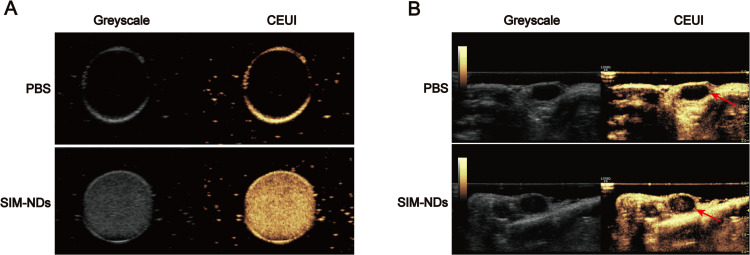
Results: SIM-NDs exhibited excellent pH- and ultrasound-responsive drug release and noticeable ultrasonographic imaging ability, also showing good biocompatibility and biosafety. UTMD could promote increased intracellular reactive oxygen species and consume intracellular glutathione. However, SIM-NDs were efficiently internalized into cells under ultrasound irradiation, followed by the rapid release of SIM, which inhibited intracellular mevalonate production, and synergistically downregulated GPX4 expression, thereby promoting ferroptosis. Moreover, this combined treatment demonstrated strong antitumor ability in vitro and in vivo.
Conclusion: The combination of UTMD and SIM-NDs presents a promising avenue for harnessing ferroptosis in the treatment of malignant tumors.
Keywords: ferroptosis; glutathione peroxidase 4; lipid peroxidation; nanodroplets; theranostics; ultrasound-targeted microbubble disruption.
© 2023 Liu et al.
Conflict of interest statement
The authors declare that they have no competing interests.
Figures


Similar articles
-
Simvastatin induced ferroptosis for triple-negative breast cancer therapy.J Nanobiotechnology. 2021 Oct 9;19(1):311. doi: 10.1186/s12951-021-01058-1. J Nanobiotechnology. 2021. PMID: 34627266 Free PMC article.
-
pH-/Redox-Responsive Nanodroplet Combined with Ultrasound-Targeted Microbubble Destruction for the Targeted Treatment of Drug-Resistant Triple Negative Breast Cancer.ACS Appl Mater Interfaces. 2023 Feb 22;15(7):8958-8973. doi: 10.1021/acsami.2c20478. Epub 2023 Feb 9. ACS Appl Mater Interfaces. 2023. PMID: 36757913
-
Simeprevir induces ferroptosis through β-TrCP/Nrf2/GPX4 axis in triple-negative breast cancer cells.Biomed Pharmacother. 2024 Nov;180:117558. doi: 10.1016/j.biopha.2024.117558. Epub 2024 Oct 14. Biomed Pharmacother. 2024. PMID: 39405915
-
Ferroptosis as a promising targeted therapy for triple negative breast cancer.Breast Cancer Res Treat. 2024 Oct;207(3):497-513. doi: 10.1007/s10549-024-07387-7. Epub 2024 Jun 14. Breast Cancer Res Treat. 2024. PMID: 38874688 Review.
-
Ferroptosis induction via targeting metabolic alterations in triple-negative breast cancer.Biomed Pharmacother. 2023 Dec 31;169:115866. doi: 10.1016/j.biopha.2023.115866. Epub 2023 Nov 10. Biomed Pharmacother. 2023. PMID: 37951026 Review.
Cited by
-
Advances in Ultrasound-Targeted Microbubble Destruction (UTMD) for Breast Cancer Therapy.Int J Nanomedicine. 2025 Feb 3;20:1425-1442. doi: 10.2147/IJN.S504363. eCollection 2025. Int J Nanomedicine. 2025. PMID: 39925678 Free PMC article. Review.
-
The Current Status and Future Directions on Nanoparticles for Tumor Molecular Imaging.Int J Nanomedicine. 2024 Sep 14;19:9549-9574. doi: 10.2147/IJN.S484206. eCollection 2024. Int J Nanomedicine. 2024. PMID: 39296941 Free PMC article. Review.
-
Intersection of ferroptosis and nanomaterials brings benefits to breast cancer.Cell Biol Toxicol. 2025 Jul 22;41(1):119. doi: 10.1007/s10565-025-10067-x. Cell Biol Toxicol. 2025. PMID: 40691737 Free PMC article. Review.
-
Iron-Dependent Cell Death: Exploring Ferroptosis as a Unique Target in Triple-Negative Breast Cancer Management.Cancer Manag Res. 2025 Mar 19;17:625-637. doi: 10.2147/CMAR.S503932. eCollection 2025. Cancer Manag Res. 2025. PMID: 40124838 Free PMC article. Review.
References
MeSH terms
LinkOut - more resources
Full Text Sources
Research Materials
Miscellaneous
