. 2023 May 8;2(5):e0000240.
doi: 10.1371/journal.pdig.0000240. eCollection 2023 May.

Archetypal analysis of longitudinal visual fields for idiopathic intracranial hypertension patients presenting in a clinic setting

Affiliations

Archetypal analysis of longitudinal visual fields for idiopathic intracranial hypertension patients presenting in a clinic setting

Joseph Branco et al. PLOS Digit Health. .

Abstract

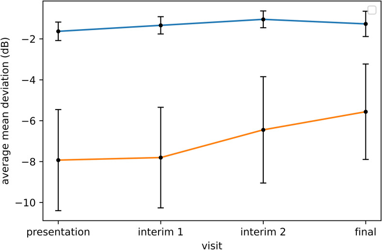
We previously applied archetypal analysis (AA) using visual fields (VF) from the Idiopathic Intracranial Hypertension Treatment Trial (IIHTT) to derive a model, which quantified patterns (or archetypes [ATs] of VF loss), anticipated recovery, and identified residual VF deficits. We hypothesized that AA could produce similar results using IIH VFs collected in clinical practice. We applied AA to 803 VFs from 235 eyes with IIH from an outpatient neuro-ophthalmology clinic and created a clinic-derived model of ATs, with the relative weight (RW) and average total deviation (TD) for each AT. We also created a combined-derived model from an input dataset containing the clinic VFs and 2862 VFs from the IIHTT. We used both models to decompose clinic VF into ATs of varying percent weight (PW), correlated presentation AT PW with mean deviation (MD), and evaluated final visit VFs considered "normal" by MD ≥ -2.00 dB for residual abnormal ATs. The 14-AT clinic-derived and combined-derived models revealed similar patterns of VF loss previously identified in the IIHTT model. AT1 (a normal pattern) was most prevalent in both models (RW = 51.8% for clinic-derived; 35.4% for combined-derived). Presentation AT1 PW correlated with final visit MD (r = 0.82, p < 0.001 for the clinic-derived model; r = 0.59, p < 0.001 for the combined-derived model). Both models showed ATs with similar patterns of regional VF loss. The most common patterns of VF loss in "normal" final visit VFs using each model were clinic-derived AT2 (mild global depression with enlarged blind spot; 44/125 VFs; 34%) and combined-derived AT2 (near-normal; 93/149 VFs; 62%). AA provides quantitative values for IIH-related patterns of VF loss that can be used to monitor VF changes in a clinic setting. Presentation AT1 PW is associated with the degree of VF recovery. AA identifies residual VF deficits not otherwise indicated by MD.


Conflict of interest statement

The authors have declared that no competing interests exist.

Figures

Fig 1
Fig 1. 14-archetype (AT) model derived from the neuro-ophthalmology clinic dataset.
ATs are shown in descending order of relative weight (RW; expressed as a percent), representing their amount of representation within the dataset. The scale (bottom) denotes average total deviation (TD¯) values (range -41 to 7 dB). Each AT pattern is shown with its corresponding TD¯ and RW value.
Fig 2
Fig 2. Average mean deviation (dB) over time for eyes with presentation clinic-derived archetype-1 (AT1) percent weight (PWs) above (blue line) and below (orange line) the mean, showing 95% confidence intervals at each time point.
Fig 3
Fig 3
Case example of archetype (AT) decomposition of visual fields (VFs) from an eye with mild loss at presentation and worsening of loss at two and three weeks (top to bottom). Decompositions are shown using the clinic-derived model (left of grayscale VF), combined-derived model (upper right of grayscale VF) and Idiopathic Intracranial Hypertension Treatment Trial (IIHTT)-derived model (lower right of grayscale VF). All three models identify an enlarged blind spot at presentation. Only the clinic-derived and combined-derived AT models identify a pattern of global loss beginning at two weeks with worsening at three weeks.
Fig 4
Fig 4. Case example of archetype (AT) decomposition of visual fields (VFs) from an eye with severe loss at presentation and change over time.
Decompositions are shown using the clinic-derived model (left of grayscale VF) and combined-derived model (right of grayscale VF). Both models show similar patterns.
Fig 5
Fig 5. Case example of archetype (AT) decomposition of VFs from an eye with mild loss at presentation and residual deficits despite a mean deviation (MD) ≥ -2.00 dB at final visit.
Decompositions are shown using the clinic-derived model (left of grayscale visual field [VF]) and combined-derived model (right of grayscale VF). The combined-derived model also revealed an inferior nasal deficit, but with a percent weight (PW) of 8% (below the cutoff to be considered meaningful).

References

    1. Wall M, Kupersmith MJ, Kieburtz KD, Corbett JJ, Feldon SE, Friedman DI, et al.. The idiopathic intracranial hypertension treatment trial: clinical profile at baseline. JAMA Neurol. 2014. Jun;71(6):693–701. doi: 10.1001/jamaneurol.2014.133 - DOI - PMC - PubMed
    1. Keltner JL, Johnson CA, Cello KE, Wall M, NORDIC Idiopathic Intracranial Hypertension Study Group. Baseline visual field findings in the Idiopathic Intracranial Hypertension Treatment Trial (IIHTT). Invest Ophthalmol Vis Sci. 2014;55(5):3200–3207. - PMC - PubMed
    1. Wall M, Johnson CA, Cello KE, Zamba KD, McDermott MP, Keltner JL, et al.. Visual field outcomes for the Idiopathic Intracranial Hypertension Treatment Trial (IIHTT). Invest Ophthalmol Vis Sci. 2016;57(3):805–812. doi: 10.1167/iovs.15-18626 - DOI - PMC - PubMed
    1. Doshi H, Solli E, Elze T, Pasquale LR, Wall M, Kupersmith MJ. Unsupervised Machine Learning Shows Change in Visual Field Loss in the Idiopathic Intracranial Hypertension Treatment Trial. Ophthalmology. 2022;129(8):903–911. doi: 10.1016/j.ophtha.2022.03.027 - DOI - PubMed
    1. Elze T, Pasquale LR, Shen LQ, Chen TC, Wiggs JL, Bex PJ. Patterns of functional vision loss in glaucoma determined with archetypal analysis. J R Soc Interface. 2015;12(103):20141118. doi: 10.1098/rsif.2014.1118 - DOI - PMC - PubMed

LinkOut - more resources