

Shoe configuration effects on third phalanx and capsule motion of unaffected and laminitic equine hooves in-situ
- PMID: 37155654
- PMCID: PMC10166494
- DOI: 10.1371/journal.pone.0285475
Shoe configuration effects on third phalanx and capsule motion of unaffected and laminitic equine hooves in-situ
Abstract
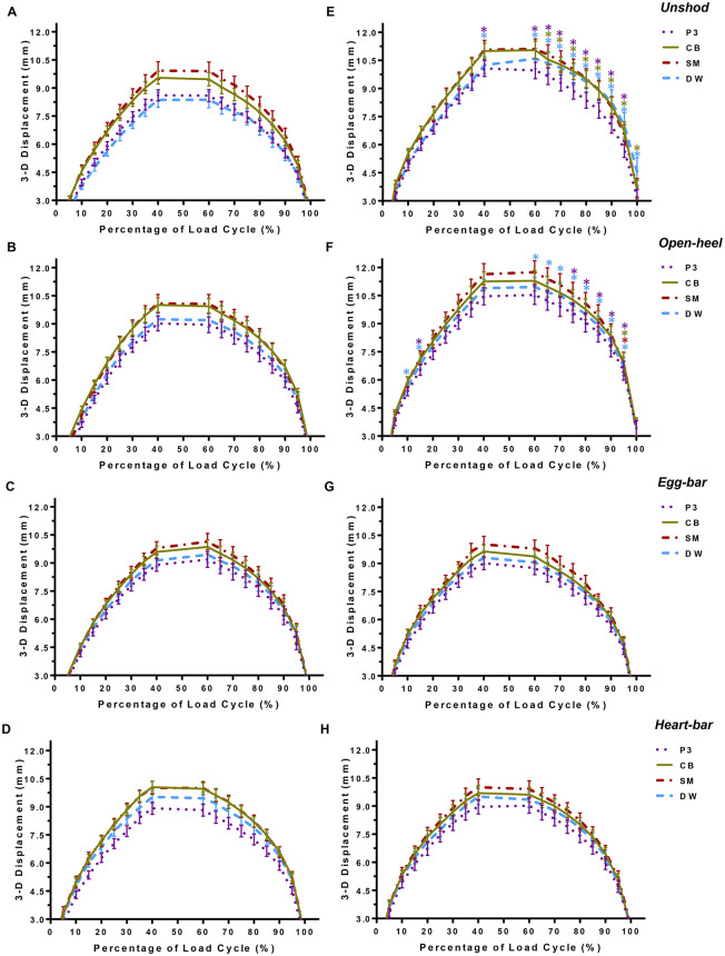
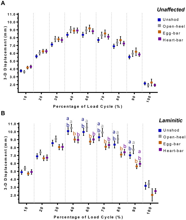
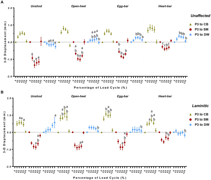
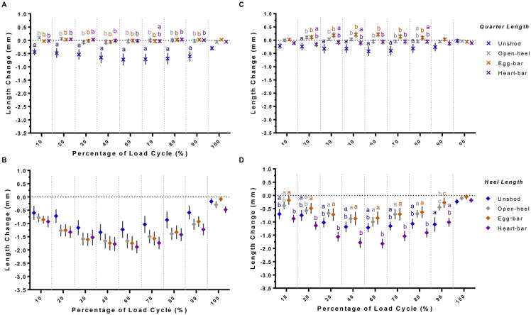
Equine shoes provide hoof protection and support weakened or damaged hoof tissues. Two hypotheses were tested in this study: 1) motion of the third phalanx (P3) and hoof wall deformation are greater in laminitic versus unaffected hooves regardless of shoe type; 2) P3 displacement and hoof wall deformation are greatest while unshod (US), less with open-heel (OH), then egg-bar (EB) shoes, and least with heart-bar (HB) shoes for both hoof conditions. Distal forelimbs (8/condition) were subjected to compressive forces (1.0x10²-5.5x10³ N) while a real-time motion detection system recorded markers on P3 and the hoof wall coronary band, vertical midpoint, and solar margin. Magnitude and direction of P3 displacement and changes in proximal and distal hemi-circumference, quarter and heel height and proximal and distal heel width were quantified. Hoof condition and shoe effects were assessed with 2-way ANOVA (p<0.05). P3 displacement was greater in laminitic hooves when US or with OH, and EB and HB reduced P3 displacement in laminitic hooves. P3 displacement was similar among shoes in unaffected hooves and greatest in laminitic hooves with OH, then US, EB and HB. EB and HB increased P3 displacement from the dorsal wall in unaffected hooves and decreased it in laminitic hooves. OH and EB increased P3 motion from the coronary band in laminitic hooves, and HB decreased P3 motion toward the solar margin in unaffected and laminitic hooves. In laminitic hooves, HB reduced distal hemi-circumference and quarter deformation and increased heel deformation and expansion. Proximal hemi-circumference constriction was inversely related to proximal heel expansion with and without shoes. Overall, shoe configuration alters hoof deformation distinctly between unaffected and laminitic hooves, and HB provided the greatest P3 stability in laminitic hooves. These unique results about P3 motion and hoof deformation in laminitic and unaffected hooves inform shoe selection and design.
Copyright: © 2023 Aoun et al. This is an open access article distributed under the terms of the Creative Commons Attribution License, which permits unrestricted use, distribution, and reproduction in any medium, provided the original author and source are credited.
Conflict of interest statement
The authors have declared that no competing interests exist.
Figures


Similar articles
-
Shoe configuration effects on equine forelimb gait kinetics at a walk.PeerJ. 2025 Feb 26;13:e18940. doi: 10.7717/peerj.18940. eCollection 2025. PeerJ. 2025. PMID: 40028219 Free PMC article.
-
The effect of frog pressure and downward vertical load on hoof wall weight-bearing and third phalanx displacement in the horse--an in vitro study.J S Afr Vet Assoc. 2001 Dec;72(4):217-27. doi: 10.4102/jsava.v72i4.656. J S Afr Vet Assoc. 2001. PMID: 12219918
-
In vitro transmission and attenuation of impact vibrations in the distal forelimb.Equine Vet J Suppl. 1999 Jul;(30):245-8. doi: 10.1111/j.2042-3306.1999.tb05227.x. Equine Vet J Suppl. 1999. PMID: 10659261
-
Chronic laminitis: strategic hoof wall resection.Vet Clin North Am Equine Pract. 2010 Apr;26(1):197-205. doi: 10.1016/j.cveq.2009.12.009. Vet Clin North Am Equine Pract. 2010. PMID: 20381747 Review.
-
Horseshoe effects on equine gait-A systematic scoping review.Vet Surg. 2025 Jan;54(1):31-51. doi: 10.1111/vsu.14162. Epub 2024 Sep 15. Vet Surg. 2025. PMID: 39278729 Free PMC article.
Cited by
-
Shoe configuration effects on equine forelimb gait kinetics at a walk.PeerJ. 2025 Feb 26;13:e18940. doi: 10.7717/peerj.18940. eCollection 2025. PeerJ. 2025. PMID: 40028219 Free PMC article.
-
Investigating Associations between Horse Hoof Conformation and Presence of Lameness.Animals (Basel). 2024 Sep 17;14(18):2697. doi: 10.3390/ani14182697. Animals (Basel). 2024. PMID: 39335286 Free PMC article.
-
Association between radiographic equine distal phalanx characteristics and absence, presence and type of horseshoes.Front Vet Sci. 2025 Jul 25;12:1598038. doi: 10.3389/fvets.2025.1598038. eCollection 2025. Front Vet Sci. 2025. PMID: 40786980 Free PMC article.
References
-
- Redden RF, editor Shoeing the laminitic horse. 43rd Annual Convention of the American Association of Equine Practitioners; 1997; 43:356–359.
-
- Pollitt CC. Equine laminitis. Clinical Techniques in equine practice. 2004;3(1):34–44. doi: 10.1053/j.ctep.2004.07.003 - DOI
-
- Dorn C, Garner HE, Coffman JR, Hahn AW, Tritschler L. Castration and other factors affecting the risk of equine laminitis. The Cornell veterinarian. 1975;65(1):57–64. - PubMed
-
- Treiber KH, Kronfeld DS, Hess TM, Byrd BM, Splan RK, Staniar WB. Evaluation of genetic and metabolic predispositions and nutritional risk factors for pasture-associated laminitis in ponies. Journal of the American Veterinary Medical Association. 2006;228(10):1538–1545. doi: 10.2460/javma.228.10.1538 - DOI - PubMed
-
- Johnson PJ, Messer NT, Slight SH, Wiedmeyer C, Buff P, Ganjam VK. Endocrinopathic laminitis in the horse. Clinical Techniques in Equine Practice. 2004;3(1):45–56. doi: 10.1053/j.ctep.2004.07.004 - DOI
Publication types
MeSH terms
LinkOut - more resources
Full Text Sources
