. 2023 May 16;120(20):e2219644120.
doi: 10.1073/pnas.2219644120. Epub 2023 May 8.

Upregulation of the ERRγ-VDAC1 axis underlies the molecular pathogenesis of pancreatitis

Affiliations

Upregulation of the ERRγ-VDAC1 axis underlies the molecular pathogenesis of pancreatitis

Dipanjan Chanda et al. Proc Natl Acad Sci U S A. .

Abstract

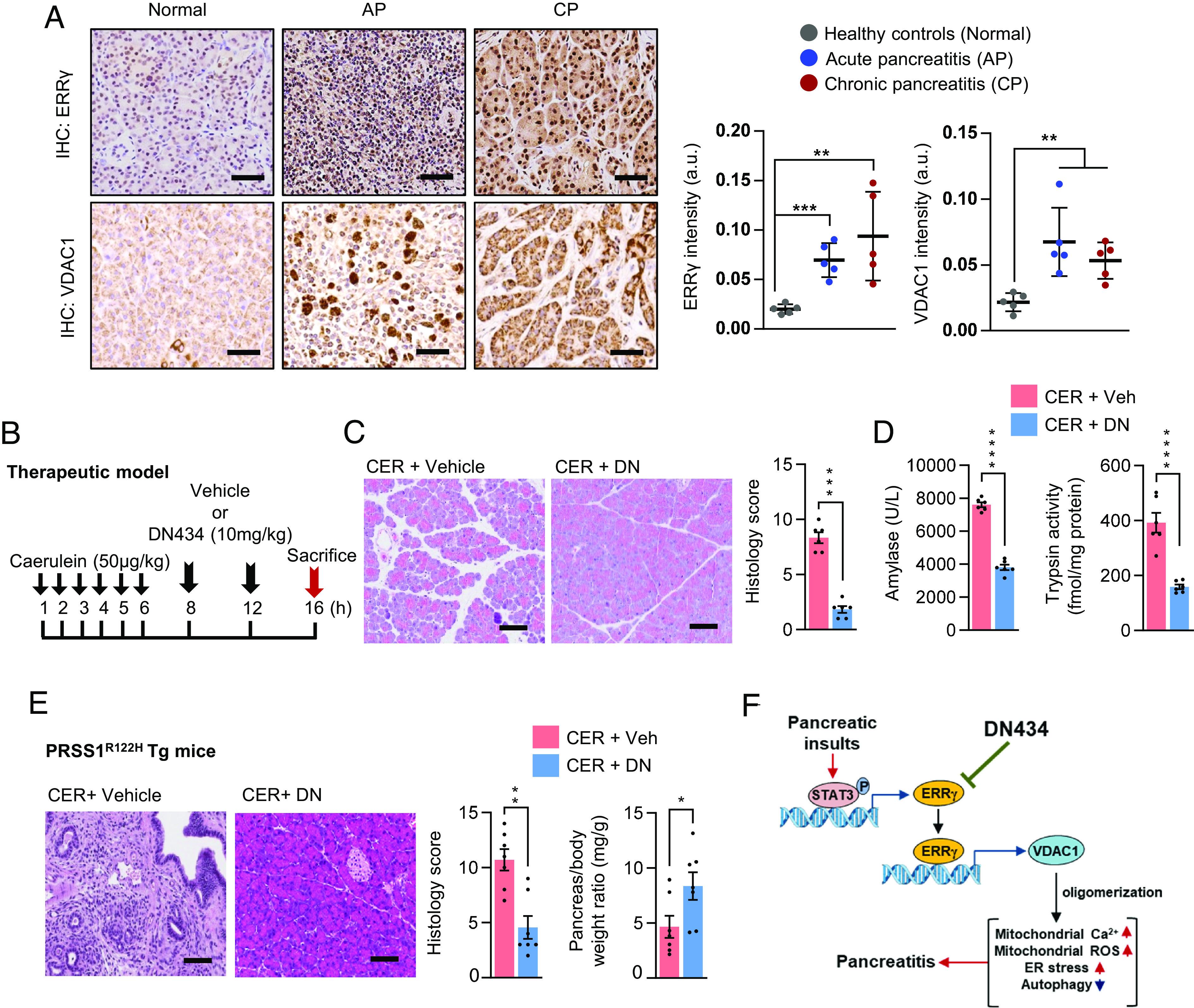
Emerging evidence suggest that transcription factors play multiple roles in the development of pancreatitis, a necroinflammatory condition lacking specific therapy. Estrogen-related receptor γ (ERRγ), a pleiotropic transcription factor, has been reported to play a vital role in pancreatic acinar cell (PAC) homeostasis. However, the role of ERRγ in PAC dysfunction remains hitherto unknown. Here, we demonstrated in both mice models and human cohorts that pancreatitis is associated with an increase in ERRγ gene expression via activation of STAT3. Acinar-specific ERRγ haploinsufficiency or pharmacological inhibition of ERRγ significantly impaired the progression of pancreatitis both in vitro and in vivo. Using systematic transcriptomic analysis, we identified that voltage-dependent anion channel 1 (VDAC1) acts as a molecular mediator of ERRγ. Mechanistically, we showed that induction of ERRγ in cultured acinar cells and mouse pancreata enhanced VDAC1 expression by directly binding to specific site of the Vdac1 gene promoter and resulted in VDAC1 oligomerization. Notably, VDAC1, whose expression and oligomerization were dependent on ERRγ, modulates mitochondrial Ca2+ and ROS levels. Inhibition of the ERRγ-VDAC1 axis could alleviate mitochondrial Ca2+ accumulation, ROS formation and inhibit progression of pancreatitis. Using two different mouse models of pancreatitis, we showed that pharmacological blockade of ERRγ-VDAC1 pathway has therapeutic benefits in mitigating progression of pancreatitis. Likewise, using PRSS1R122H-Tg mice to mimic human hereditary pancreatitis, we demonstrated that ERRγ inhibitor also alleviated pancreatitis. Our findings highlight the importance of ERRγ in pancreatitis progression and suggests its therapeutic intervention for prevention and treatment of pancreatitis.

Keywords: ERRγ; VDAC1; mitochondrial Ca2+; nuclear receptor; pancreatitis.


Conflict of interest statement

H.-Y.J., J.A. and I.-K.L. are board members of NovMetaPharma. All other authors declare that they have no competing interests.

Figures

Fig. 1.
Fig. 1.
ERRγ is a molecular regulator of pancreatitis. (A) Representative H&E images and histology scoring of the pancreas from saline (Sal) and caerulein hyperstimulation (CER) pancreatitis conditions. Animals were killed 16 h after the first saline or caerulein injection. (B) Serum amylase level and intrapancreatic trypsin activity. (C) Pancreatic Errγ relative mRNA level (Left) and immunoblot of ERRγ (Right) from mice pancreata. (D) Representative IHC staining of ERRγ in pancreas tissues. (n = 3 mice/group; two-sided t test). (E) Serum amylase level, intrapancreatic trypsin activity, pancreatic Errγ relative mRNA level and immunoblot of ERRγ from mice pancreata from vehicle and TLCS pancreatitis conditions. Animals were killed 16 h after the vehicle or TLCS injection. (n = 3 to 4 mice/group; two-sided t test). (F) Serum ALT, serum amylase level, and immunoblot of ERRγ from mice pancreata from pair-fed (PF) and EtOH-diet fed (AIP) alcohol-induced pancreatitis conditions. Animals were killed 8 h after the acute alcohol binge. (n = 3 to 6 mice/group; two-sided t test). Results are representative of those from two independent in vivo experiments. Data represent mean ± SEM. *< 0.05, **P < 0.01, ***P <0.001 and ****P < 0.0001. (Scale bars, 50 μm). (See also SI Appendix, Fig. S1).
Fig. 2.
Fig. 2.
Acinar cell-specific Errγ haploinsufficiency retards the progression of pancreatitis. (A) Analysis of ERRγ mRNA expression and protein level in 266-6 cells following knockdown of Errγ using targeted siRNA (siErrγ) (two-tailed t test). (B) LDH release, Rhod2-AM fluorescence, and MitoSOX fluorescence in 266-6 cells following treatment with caerulein in the presence or absence of siErrγ as indicated (two-way ANOVA analysis). (C) The pancreas-specific haplo-insufficient Errγ knockout line (acErrγ+/) was induced by crossing C57BL6/J (B6) mice with Errγf/f mice to initially generate Errγ+/f, followed by intraperitoneal infusion of AAV8-Ela1-iCre into Errγ+/f mice to specifically, but partially, delete Errγ in the whole pancreas. Representative H&E images of the pancreas from Errγ+/f and acErrγ+/ mice. (D) Pancreas weight to body weight ratio of mice in C. (E) Errγ relative mRNA level (Left) and immunoblots for AMY2, ERRγ and CRE from the pancreas and liver of mice in C. (n = 4 mice/group; two-sided t test). (F) Representative H&E images and histology scoring of the pancreas from saline (Sal) and caerulein hyperstimulation (CER) pancreatitis conditions in Errγ+/f and acErrγ+/ mice. Animals were killed 16 h after the first saline or caerulein injection. (G) Serum amylase level and intrapancreatic trypsin activity of mice in F. (H) Immunoblots for ER stress (phospho- and total-eIF2α), and autophagic flux impairment markers (p62) from the pancreas of mice in F. (I) Pancreatic inflammation (Tnfα and Il6), oxidative stress (Hmox1), and ER stress (Chop) markers of mice in F. (n = 3 to 5 mice/group; two-way ANOVA analysis). Results in 266-6 cells (A and B) are representative of those from two to three independent experiments. Results (CI) are representative of those from two independent in vivo experiments. Data represent mean ± SEM. *P < 0.05, **< 0.01, ***P < 0.001 and ****P < 0.0001 n.s. not significant. (Scale bars, 50 μm). (See also SI Appendix, Fig. S2).
Fig. 3.
Fig. 3.
Pharmacological inhibition of ERRγ prevents the development of pancreatitis. (A) Scheme for preventive caerulein hyperstimulation (CER) pancreatitis model in mice. Experimental small molecule inhibitor (DN434; abbreviated as DN; 20 mg/kg) for ERRγ was administered prophylactically, 24 h and 1 h prior to the first caerulein injection. Animals were killed 16 h after the first saline or caerulein injection. (B) Immunoblot for ERRγ from the pancreas of mice in A. (C) Representative H&E images and histology scoring of the pancreas from mice in A. (D) Serum amylase level and intrapancreatic trypsin activity of mice in A. (E) Pancreatic inflammation (Tnfα and Il6), oxidative stress (Hmox1), and ER stress (Chop) markers of mice in A. (F) Representative IHC staining of p62/SQSTM1 of the pancreas from mice in A. (G) Representative EM images (and dotted Inset) of the pancreas from mice in A. Red arrows: ER. (Scale bar, 2 μm). (n = 4 to 6 mice/group; two-way ANOVA analysis). Results are representative of those from two independent in vivo experiments. Data represent mean ± SEM. **P < 0.01, ***P < 0.001 and ****P < 0.0001. ROI; region of interest. (Scale bars, 50 μm). (See also SI Appendix, Fig. S3).
Fig. 4.
Fig. 4.
VDAC1 acts as an effector gene of ERRγ in promoting pancreatitis. (A) Total RNA was isolated from 1° acini cells transduced with adenoviral vectors (Ad) overexpressing GFP or ERRγ for 24 h and analyzed by RNAseq (Top). A volcano plot showing genome-wide changes in mRNA level (Bottom). (B) Heat map depicting the differential expression of genes involved in mitochondrial calcium transport. (C) Analysis of VDAC1 mRNA and protein expression in primary acinar cells following treatment with Ad-ERRγ or Ad-GFP overexpression (10 MOI) for 24 h (two-tailed t test). (D) A putative ERRγ-response element (ERRE) in Vdac1 gene promoter (Left). In vivo chromatin immunoprecipitation (ChIP)-qPCR analysis of ERRγ binding to Vdac1 gene promoter of pancreas harvested from saline (Sal) and caerulein hyperstimulation (CER) pancreatitis conditions (n = 3 mice/group; two-tailed t test). (E) Analysis of VDAC1 mRNA and immunoblot of VDAC1 oligomerization from the pancreas of Errγ+/f and acErrγ+/ mice (n = 3 to 5 mice/group; two-way ANOVA analysis). (F) Analysis of VDAC1 mRNA and immunoblot of VDAC1 oligomerization from the pancreas of preventive caerulein hyperstimulation (CER) pancreatitis model (n = 4 to 6 mice/group; two-way ANOVA analysis). (G) Representative H&E images and histology scoring from the pancreas of mice of preventive caerulein hyperstimulation (CER) pancreatitis model with VDAC1 oligomerization inhibitor, VBIT-12 (20 mg/kg). Animals were killed 16 h after the first saline or caerulein injection. Experimental scheme of this model is described in SI Appendix, Fig. S4D. (H) Immunoblot of VDAC1 oligomerization from the pancreas of mice in G. (I) Serum amylase level and intrapancreatic trypsin activity of mice in (G). (n = 3 to 5 mice/group; two-way ANOVA analysis). Results are representative of those from two independent experiments. Data represent mean ± SEM. ***P < 0.001 and ****P < 0.0001. (Scale bars, 50 μm). (See also SI Appendix, Fig. S4).
Fig. 5.
Fig. 5.
ERRγ inhibitor is an effective experimental therapeutic in treating pancreatitis. (A) Representative IHC staining of ERRγ and VDAC1 in pancreatic tissue from patients and quantification of the samples (n = 5 subjects/group). (B) Scheme of pancreatitis induction (CER) and treatment. Pancreatitis was induced by caerulein, and therapeutic drug (DN) was administered twice, 7 and 11 h after the first caerulein injection. (C) Representative H&E images and histology scoring of the pancreas from mice in B. (D) Serum amylase level and intrapancreatic trypsin activity of mice in B. (n = 6 mice/group; two-tailed t -test) (E) Pancreatitis induction (CER) and treatment in PRSS1R122H mice. Pancreatitis was induced by caerulein, and therapeutic drug (DN) was administered 5 h after the first caerulein injection. Vehicle or DN were given twice daily (10 mg/kg; b.i.d) for the next 7 d. Representative H&E images, histology scoring and pancreas weight to body weight ratio of mice after 7 d of treatment. (n = 7 mice/group; two-tailed t test). (F) Schematic representation of the molecular regulatory role of the ERRγ–VDAC1 axis that contributes toward the pathogenesis of pancreatitis. Results are representative of those from two independent in vivo experiments. Data represent mean ± SEM. *P < 0.05, **< 0.01, ***< 0.001 and ****P < 0.0001. (Scale bars, 50 μm). (See also SI Appendix, Fig. S5 and Table S1).

References

    1. Forsmark C. E., Vege S. S., Wilcox C. M., Acute pancreatitis. N. Engl. J. Med. 375, 1972–1981 (2016). - PubMed
    1. Peng S., et al. , Galactose protects against cell damage in mouse models of acute pancreatitis. J. Clin. Invest. 128, 3769–3778 (2018). - PMC - PubMed
    1. Petersen O. H., Sutton R., Ca2+ signalling and pancreatitis: Effects of alcohol, bile and coffee Trends Pharmacol. Sci. 27, 113–120 (2006). - PubMed
    1. Wan M. H., et al. , Review of experimental animal models of biliary acute pancreatitis and recent advances in basic research. HPB (Oxford) 14, 73–81 (2012). - PMC - PubMed
    1. Komatsu M., et al. , The selective autophagy substrate p62 activates the stress responsive transcription factor Nrf2 through inactivation of Keap1. Nat. Cell Biol. 12, 213–223 (2010). - PubMed

Publication types

LinkOut - more resources